INTRODUCTION
Knee pain occurs in approximately 50% of musculoskeletal injuries and is particularly common in sports injuries and among individuals with repeated sports activity.1 The reported incidence of lower limb injury in sprint athletes is approximately 19%–75%, among which, knee pain accounts for approximately 9%–50%.2 Various factors may contribute to knee pain, including a reduction in the flexibility of ligament laxity, as well as quadriceps weakness and hypermobile patella and tensor fascia lata (TFL).3 Notably, reduced TFL flexibility shows a correlation with knee pain.4,5 Patellofemoral pain syndrome (PFPS) accounts for approximately 25%–40% of all knee pain types, and its cause includes shortened TFL.4,6
The gluteus medius and the TFL prevent contralateral pelvic drop during one-leg standing posture, thereby playing a key role in pelvic stabilization during gait.7 However, in mid-stance phase, the TFL muscle is more prone to shortness or stiffness as it is more dominantly and repeatedly used compared to the gluteus medius, with a consequent role in excessive internal rotation of the hip joint.8 In particular, patients with PFPS may experience knee pain during knee flexion, where the shortened TFL induces patella lateral glide and increases lateral condyle friction between the patella and femur.9,10 According to Noehren and his team, the level of TFL shortening was greater in sprint athletes with iliotibial band syndrome than in the control group without knee pain.11 In another study, ballet dancers with knee pain had a greater level of TFL shortening than the control group without knee pain.5
Many studies have investigated interventions such as stretching, instrument-assisted soft tissue mobilization (IASTM), proprioceptive neuromuscular facilitation (PNF), and massaging, with the aim to increase the flexibility and length of the shortened and stiffened TFL.12-14 TFL stretching has been reported to increase the hip joint range of motion (ROM) and decrease knee pain.15 In the study by Park et al., where 30 adults with shortened TFL received active stretching (AS) in sidelying and standing positions, the hip joint adduction ROM showed a mean increase of 2.3° and 4.1° in the standing position group and the sidelying position group, respectively.16
IASTM, involving the skin massage using a stainless steel device, is another method of intervention to reduce muscle stiffness and increase the ROM.14 In the study by Smuts, where IASTM was applied to healthy adults with shortened TFL, the hip joint adduction ROM was reported to have increased by 4° in comparison to the control.17 Lee et al. performed releasing muscle with a Graston and muscle energy technique (MET) to 30 individuals with shortened hamstring and compared the knee joint extension ROM.18 The Graston group showed a mean increase of 10.2° in knee joint extension ROM with Graston treatment applied to the hamstring muscle. The MET group showed a mean increase of 10° in the knee joint extension ROM after five sets of hamstring muscle contraction. Thus, the knee joint extension ROM was shown to have increased in both groups.18
Myoton has been widely used to measure the mechanical properties of muscles.14,19 Myoton allows easy and rapid measurements of the mechanical properties of muscles, including stiffness, frequency, and decrement in varying positions.20 The stiffness measured by Myoton is the resistance against external forces that distort the muscle shape; it is measured as the force per unit length, in which increasing values indicate increasing levels of stiffness.21 In Kang and Lee, 30 healthy athletes performed IASTM or active calf stretching on the gastrocnemius muscle, and when the stiffness was compared between the two groups, the IASTM group showed a greater reduction than the active calf stretching group.14 Likewise, in a study where female office workers received passive stretching on the sternocleidomastoid, upper trapezius, suboccipitalis, and pectoralis minor muscles, the muscle stiffness was significantly reduced in the stretching group compared to the control.22
In many studies, stretching and IASTM have been suggested to increase the flexibility of shortened muscles, such as the TFL and hamstring, in patients with knee pain. While these studies reported significant improvements in ROM and pain reduction,12,23 no previous study has directly compared the hip joint ROM and muscle stiffness in patients after applying AS and IASTM to a shortened TFL. Thus, the purpose of this study was to determine the hip joint ROM and TFL stiffness after applying AS and IASTM to subjects with a shortened TFL.
METHODS
Twenty subjects with shortened TFL and without abnormality in the lower extremity with respect to orthopedic and neurological surgery were selected for this study. The Ober test was used to select the subjects with shortened TFL capable of hip adduction at 0° of the femur parallel to the ground.24 The subjects were randomized between the AS group (n=10) and the IASTM group (n=10). All subjects were given detailed explanations regarding the purpose and characteristics of the study prior to signing the informed consent form. The study was approved by the Ethics and Research Committee of Daegu Health College(#DHCIRB-2022-03-0001). The general characteristics of the subjects are summarized in Table 1.
An easily accessible and economic smartphone with an internal tilt detector was used to record the ROM. The Goniometer Pro (5fuf5 CO, Stephanskirchen, Germany), tiltmeter application software, which has outstanding reliability (intraclass correlation coefficient [ICC]: 0.99), was installed on the smartphone for ROM measurements.25
The Dr. YOU STM was used to apply the IASTM for increasing the ROM and decreasing the muscle tension. It is made of stainless steel and allows easy access to the parts of the body that are difficult to access with massage.
The Myoton device was applied to stimulate the skin surface with its a small cylindrical probe to provide 0.40 N stimulation for 15 ms. It collected data of muscle stiffness from muscle vibration and acceleration.26
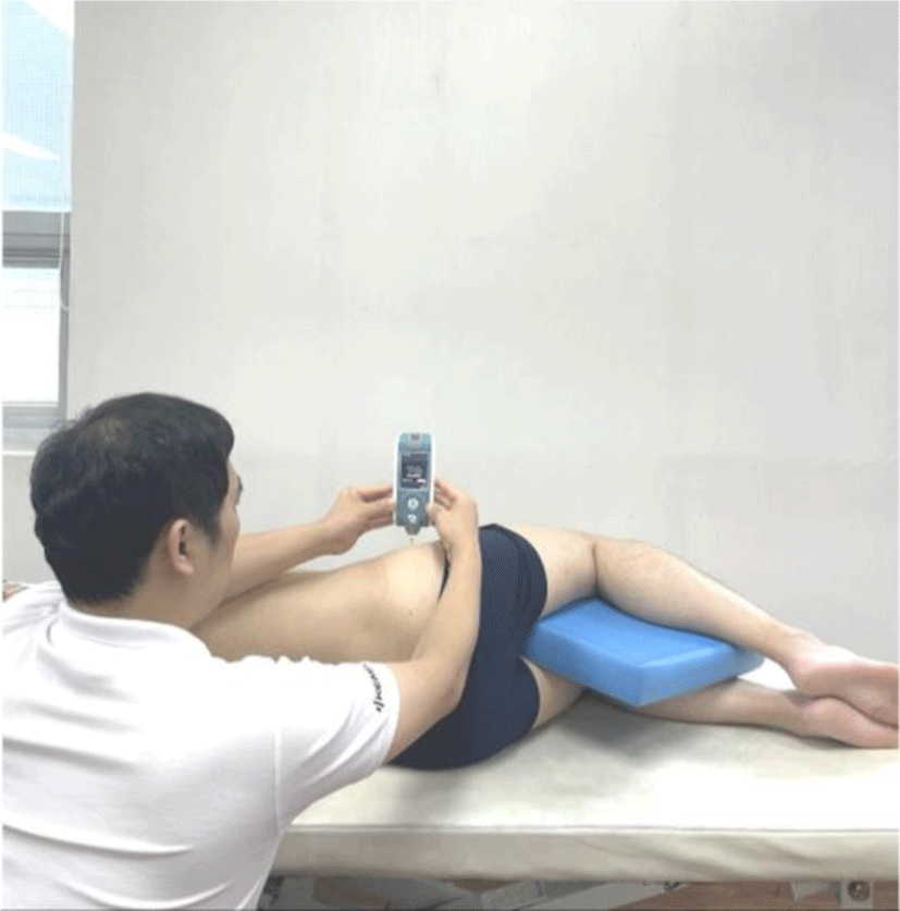
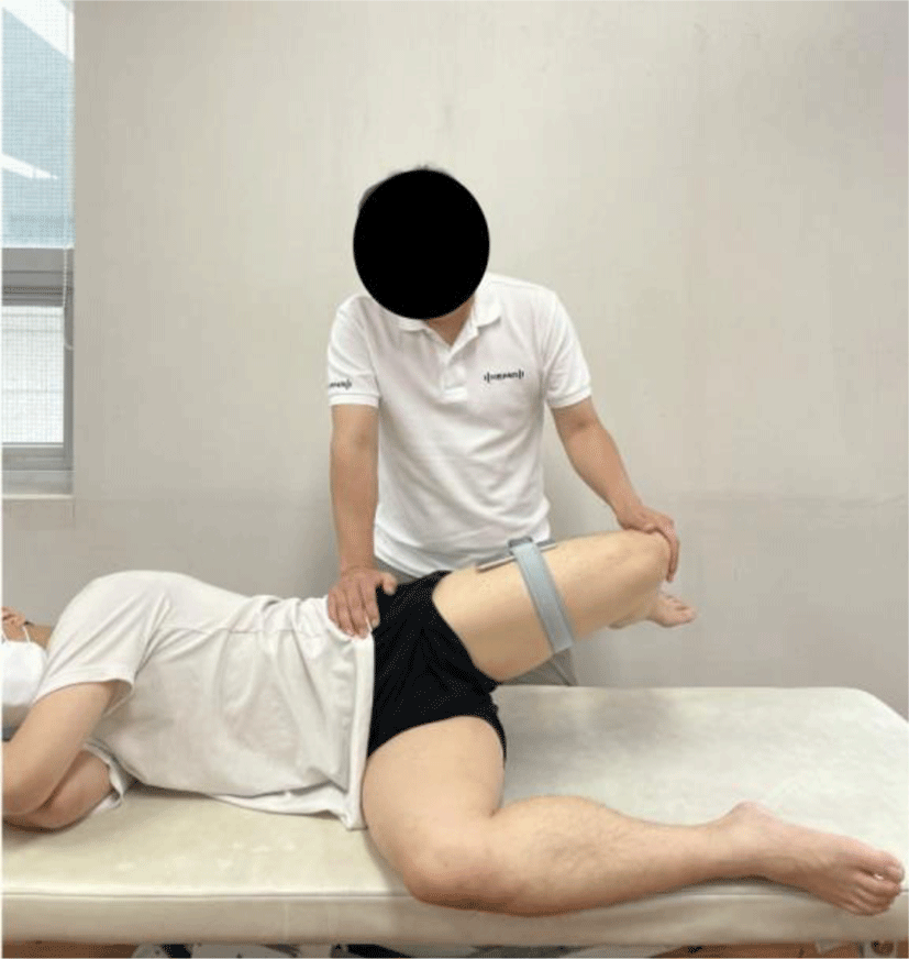
The TFL flexibility of each subject was measured applying the Ober test. The subjects with shorter TFL were selected for the Ober test to divide the subjects into two groups. The reliability of the Ober test was excellent, with an ICC of 0.90.27 To measure the hip joint adduction ROM, the smartphone was fixed using a strap at the point between the knee and the greater trochanter. The hip adduction ROM was set to 0° such that the femur was horizontally level in parallel to the ground. The hip adduction was recorded as a positive value and the hip abduction was recorded as a negative value.27 The Ober test was performed as the subject lay on the table in a sidelying position with the target leg on the top. The leg on the lower side was placed in the flexion of the hip and knee joints so that the lumbar spine could be maintained flat while the top leg was being measured. The investigator used one hand to maintain the pelvis in a fixed state and monitor the movement simultaneously, while using the other hand to hold the knee to maintain the target leg in a parallel line to the trunk through hip joint extension towards adduction. At the onset of measurement, the investigator slowly pulled down the leg in the direction of gravity and then signaled “finish” at the first indication of the movement of the subject’s pelvis (Figure 1). The measurement was taken using a smartphone. The endpoint of the Ober test was determined based on the feeling of discomfort or tightness in the TFL. Each leg was subjected to two tests and the mean was recorded. The subjects were given a 1 min rest between each test. Afterwards, the TFL stiffness was immediately measured using the Myoton. The subject was placed in a sidelying position, with their hip and knee joints in flexion. A cushion was placed between the legs to set the hip joint adduction ROM at 0° and the pelvis in a neutral position. The probe of the Myoton was placed in a vertical position on the surface of the soft tissue. The measurement of the TFL was taken at the point 2 cm in the direction of the anterior superior iliac spine from the greater trochanter of the femur (Figure 2). For repeated measurements, the selected point was marked with a pen. The mean of triplicate measurements was obtained. Next, through lot drawing, ten subjects were randomly allocated to each of the AS and IASTM groups. After the two groups performed the respective interventions, the immediate change of hip ROM was examined by the Ober test and of muscle stiffness by Myoton. The post-intervention measurements were taken immediately without rest to avoid the effect of the TFL extensibility. The interventions were performed by two experts, each with more than 10 years’ experience, in each corresponding physical therapy room. The investigators were blinded to the values being recorded.

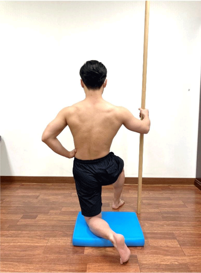
For TFL stretching, the subjects were instructed to take a kneeling posture with both knees at 90° flexion. The leg on the stretching side had the knee touching the floor, whereas the other leg had the knee in 90° flexion with the sole touching the ground. A cushion was placed below the knee on the stretching side to lower the pressure on the patella. Extension, adduction, and external rotation were applied to the hip joint on the stretching side. The subjects held a bar with their hand on the other side to maintain the balance. The stretching continued for 30s as the subject focused on the TFL stretching sensation (Figure 3). Ten sets were performed in total, with a 30s rest between each set.28
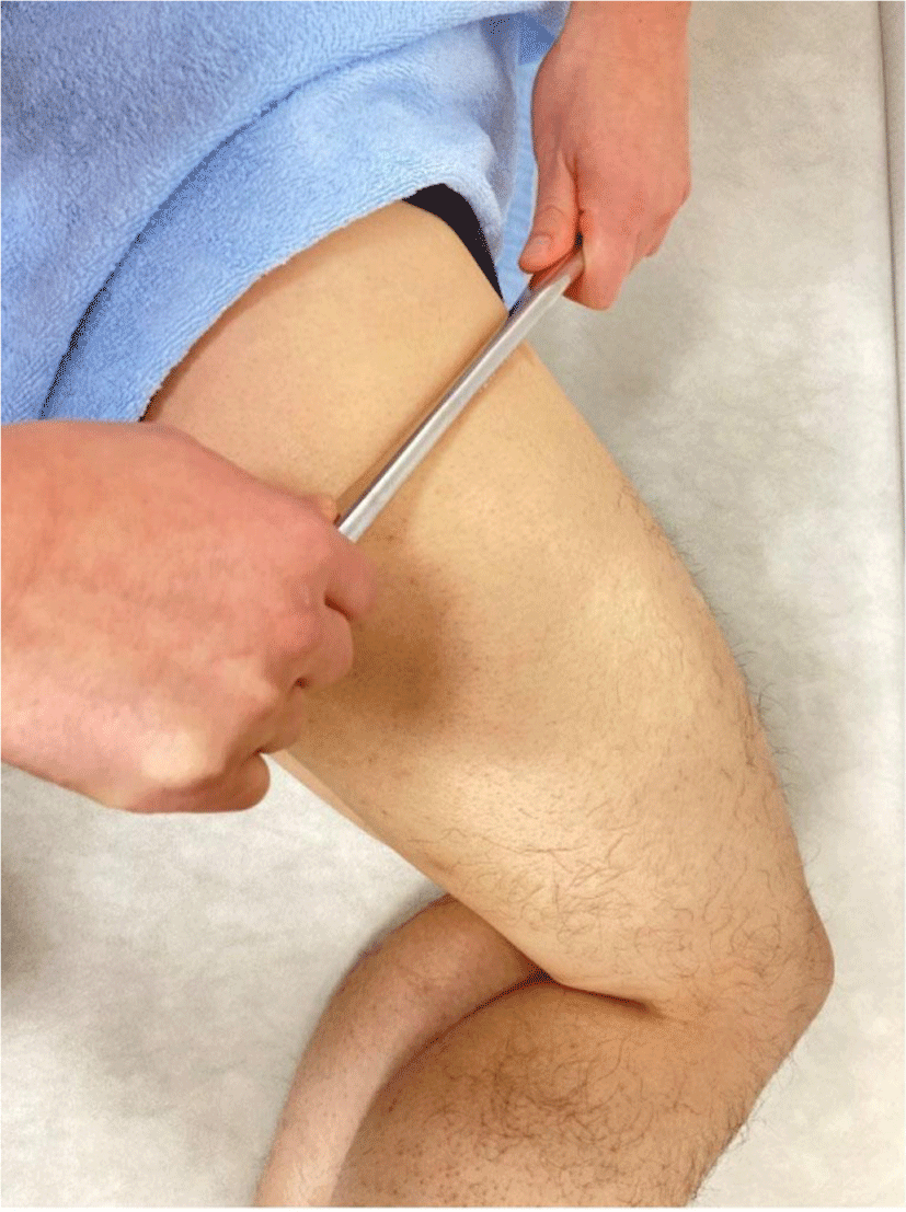
For the IASTM of the TFL, the leg with the shortened TFL was placed on top, with the subject in a sidelying position, with both legs maintained at 45° hip joint flexion and 90° knee joint flexion. A skin cream was applied to the area from the greater trochanter of the femur to the TFL to prevent skin damage during the IASTM. The investigator used a device to apply stroke to the TFL in a horizontal direction from the top to the bottom for approximately 5 min (Figure 4).29

The data were analyzed using SPSS (v.18.0 for Windows; SPSS Inc., Chicago, IL, USA). For the general characteristics of subjects, the Kolmogorov–Smirnov test was used to determine the normality distribution, and an independent t-test was performed to test the homogeneity between the AS and IASTM groups regarding the TFL. A paired t-test was performed to analyze the within-group variation in the hip joint ROM and TFL stiffness between the pre and post-intervention, and an independent t-test was performed to compare the between-group variation. The statistical significance level was set at p<.05.
RESULTS
In the within-group comparison, the hip joint adduction ROM after the intervention showed a significant increase in both AS and IASTM groups, whereas the TFL stiffness showed no significant difference (Table 2). In the between-group comparison, the increase in hip joint adduction ROM was greater for the AS group than for the IASTM group (Table 3).
| Group | AS | IASTM | t | p |
|---|---|---|---|---|
| Hip adduction | 7.63±1.96 | 3.66±1.61 | 5.31 | .00* |
DISCUSSION
This study was conducted to investigate the changes in hip joint adduction ROM and muscle stiffness to compare the effects of AS and IASTM in subjects with shortened TFL. The result of study demonstrated that the hip adduction ROM of the subjects with TFL shortness increased immediately in both AS group and IASTM group. And the greater increase of the hip adduction ROM was revealed in AS group than IASTM group.
In the within-group comparison before and after the intervention, the hip joint adduction ROM increased significantly after the intervention in both the AS and IASTM groups, while the TFL stiffness showed a slight decrease but no significant difference. Rihvk et al. compared stretching group which performed three sets of 30s stretches per set and PNF group which performed five sets of 5s contractions and 1-s relaxations per set to the hamstring muscle in healthy adult subjects and compared the immediate changes in muscle properties.19 The hamstring stiffness before and after intervention showed no significant variation.19 This was in agreement with the current results, where the muscle stiffness was not changed significantly after stretching of the TFL muscle.
In the between-group comparison, the hip joint adduction ROM showed a greater increase of 3.9° in the AS group than in the IASTM group, indicating a significant difference in the variation of hip joint adduction ROM before and after the intervention. Osailan et al. applied stretching and IASTM to the hamstring muscle in adult males with shortened hamstring and compared the changes in hip flexion active ROM after the intervention.30 The hip flexion ROM showed a significant increase in both groups and the IASTM group showed a 2° mean increase of active hip flexion ROM in the stretching group than in the IASTM group.30 This result agreed with the current studies, where the increase in ROM was greater in the AS group compared to the IASTM group. Thus, while the AS was shown to be more effective in causing an immediate increase of muscle flexibility in subjects with TFL shortening, no significant change in TFL stiffness could be obtained through short-term intervention. In a previous study, it was concluded that the ROM could be increased through stretching, with an increase in stretch tolerance rather than a change in the mechanical properties of the muscle.31 The attempt of repeated stretching could have an effect on the flexibility through an increase in stretch tolerance.32 In line with previous studies, the increase in hip joint adduction ROM following TFL stretching in this study is presumed to be the result of the increase in stretch tolerance.
Although the TFL stiffness was not significantly reduced, both the IASTM and AS groups showed increases in hip joint adduction ROM. Several theories have been proposed to account for the effect of IASTM in increasing the joint ROM. First, the IASTM may increase the ROM by reducing the sliding resistance between the muscle and the fascia.33 In addition, the friction caused by the device may reduce the viscosity of the tissue to induce tissue softening.34 While these theories on IASTM may independently or unitedly suggest a potential improvement in joint ROM, the scientific evidence to support such claims remains insufficient. In a study by Kang and Lee, thirty healthy athletes performed IASTM on the gastrocnemius for 60s, and the Myoton measurements showed a reduction in muscle stiffness by 46 N/m.14 However, in this study, the reduction in TFL stiffness following the IASTM was not significant. This difference may be due to the fact that, the IASTM in the study of Kang and Lee was applied to the gastrocnemius rather than the TFL and to the muscle without shortening rather than the shortened muscle, in contrast to the current study. Thus it is considered that it is difficult to compare the two studies directly.
There are several limitations to this study. First, the results should not be taken as reflecting long-term effects as only the immediate effects of the AS and IASTM interventions were compared. Second, all subjects were male young adults; hence, the results may vary from those obtained for female younger adults or subjects of other age groups. Third, the AS in this study was performed with the subjects in a kneeling position, and hence, the results should not be generalized to the AS applied in other positions. Future studies should be conducted to complement these limitations to evaluate the long-term effects of various intervention methods.
CONCLUSIONS
Our findings verified the immediate effects of the AS and IASTM in increasing the hip joint adduction ROM in subjects with shortened TFL, although the TFL stiffness was not reduced. Importantly, the increase in hip joint adduction ROM was increased to a greater extent by the AS compared to the IASTM. Therefore, our findings suggest that the AS is more effective than the IASTM to immediately increase the flexibility of the shortened TFL.


