


INTRODUCTION
Leg length discrepancy (LLD) is a very common musculoskeletal problem, occurring in up to 70% of the general population.1-2 LLD affects the muscle tissue involved in walking movement and posture, increases pressure and moments within the lower limb joints during gait, and creates more tension at musculoskeletal junction points.3-5 LLD is categorized into two primary types: structural (anatomic),6-7 where the actual bone length differs due to various related pathologic conditions, and functional,8-9 where the both legs appear uneven due to mechanical or postural issues. Although LLD is very common, occurring in up to 70% of the general population, and is associated with various musculoskeletal problems, some previous reports have suggested that LLD of approximately 2 cm did not negatively affect walking or running activities.10-11 Otherwise, some studies reported that LLD of less than 2 cm was significantly associated with various musculoskeletal dysfunctions.12-13 In addition, LLDs greater than 1 cm but less than 2 cm are also known to be associated with lower back pain, pelvic and knee pain, and scoliosis.1,13
Considering the closed kinematic chain of the lumbopelvic structures and lower extremities during gait, it is necessary to investigate the kinesiologic influences that occur in the lower joints and pelvic segments in individuals with LLD.14-15 Particularly, abnormal biomechanical influences that develop in the lumbopelvic segment and coxal joints during gait or performing such daily activities can be major factors that elicit related musculoskeletal dysfunctions such as low back pain and decrease the quality of life.12,16 Reliable and objective investigation of the kinematic influences in individuals with LLD on the lumbopelvic structures are essential for the treatment and prevention of related musculoskeletal disorders such as low back pain. Most of the previous LLD studies related to the musculoskeletal problems were verified using variables such as the range of motion of the lower extremity joints during walking, joint moments, ground reaction force, active walking test, and center of gravity shift as outcome measures.17-18 Few studies, however, have verified the influences of orthotic correction of LLD on the kinematic characteristics of the three-dimensional (3D) lumbopelvic movement during gait in individuals with LLD applying various insole orthoses including a customized 3D-printing insole orthosis fabricated based on 3D scanning technology.15,17 Therefore, the aim of this study was to verify the influences of various insole orthoses on 3D pelvic motion in individuals with LLD during gait. It was hypothesized that when wearing a 3D-printed insole orthosis based on high technology system, the participants would walk with greater stability of lumbopelvic segment during gait compared to the other insole orthosis conditions. It was also hypothesized that when wearing the general shoes during gait, the participants would walk with increased pelvic perturbation compared to those wearing shoes with insole orthosis conditions.
METHOD
Twenty-two individuals (14 males, 8 females) with LLD participated in this study. Before LLD evaluation was executed, the participants received a questionnaire that included general health status and some musculoskeletal postural problems such as scoliosis or low back pain. The survey results showed that the participants’ overall health was relatively good, and there were no musculoskeletal dysfunctions that could prevent them from performing the experimental procedures, including the free walking test. The selection criteria for participants were those who had a leg length difference of 5 mm or more between the two lower extremities, no underlying musculoskeletal or neurological diseases that interfered with free walking, and were able to walk comfortably and at a free speed on a 6 m laboratory walkway. LLD evaluation was performed using a tape measure between the anterior superior iliac spine and the medial malleolus in a supine position.13,19 When measuring with a tape measure, measurement errors of 1 to 10 mm may occur depending on the measurer’s skill level, measuring posture, and pelvic tilt.1,13 To minimize these errors, the measurements were performed by a clinician with at least 15 years of clinical experience. The mean and standard deviation of LLD level of the participants measured using a tapeline was 8.4±2.1 mm. Those with lower extremity osteoarthritis or neurological deficits affecting free walking or taking neurological medications were excluded from the study. All participants understood the general experimental process and study purpose, provided written informed consent, and voluntarily participated in the study. The Institutional Review Board of Jeonju University approved the study design (jjIRB-170615-HR-2017-0609).
First, to produce a 3D-printed insole, a digital image of each participant’s foot and sole needs to be obtained. The acquisition of digital foot outline silhouette was performed using a 3D scanner device (MediACE Scan, MS320F, RealDimension Inc., Korea) (Figure 1A), with its moderate to high reliability demonstrated and reported by previous study.20 The scanner device consisted of a scan body, a scanner arm, a transparent foot platform, and a seat. Two USB cables connected between the scanner and main computer were used to operate the scanner itself and to transfer the acquired foot and sole data to the computer-aided design (CAD) software system (MediACE3D V1, RealDimension Inc., Korea).
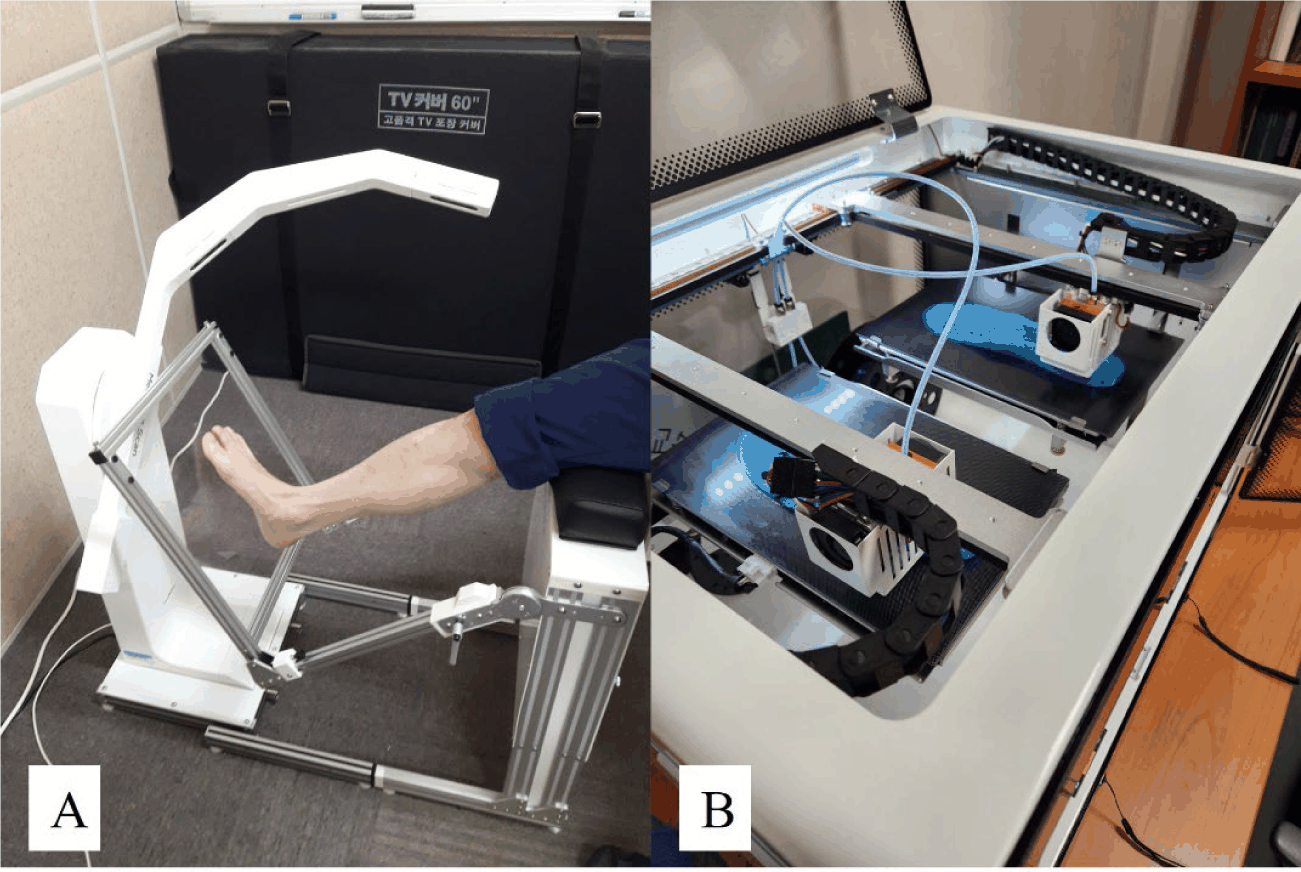
The final virtual 3D-printed insole was completed through the CAD program based on the acquired foot and plantar sole silhouette models using the 3D scanner device according to each participant LLD correction. The customized 3D-printed insole orthosis (C3DIO) was designed to reflect each participant’s foot shape and LLD characteristics. The basic C3DIO bottom thickness was designed to be 3 mm, and the right or left insole bottom was modified to a height that compensates for the LLD level of each participant. The 3D printer device (iSun3D Flx2, eSUN Industrial Co., Shenzhen, China) and its operation software (Simplify3D V4.0.1, Cincinnati, OH, USA) were used to obtain the completed final C3DIO experimental materials (Figure 1B). The 3D printer is composed of two 0.8 mm ejection nozzle, two printing flatforms, and two 3.5-inch operation digital panels, so it was able to print both left and right foot orthoses simultaneously. The thermoplastic polyurethane (TPU) (TPU-flexible 95A, Cubicon Co., Sungnam, Korea) was used as a printing filament material to fabricate the C3DIO for LLD modification. The thickness adjustment insole orthosis (TAIO) was made based on a raised LLD-corrected insole (Cubio, JKGOLD, Korea) according to each participant LLD level and was corrected to fit the shoes. The general shoe insole (GSI) condition means normal walking shoes without any orthotic insole inside of the shoes.
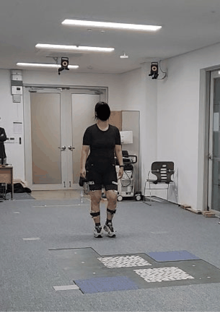
To verify the pelvic kinematics, 40 reflective markers attached to the participants’ pelvic segment and the lower extremity landmarks based on the six-degrees-of-freedom (6DOF) model.21 A Vicon Motion Analysis System (Vicon Inc., Oxford, England) with six cameras (T10) operating at a 100 Hz sampling rate was used to acquire 3D pelvic kinematic data in three different insole orthosis conditions during gait. The general shoes (S260, Supercomet Co., Gimhae, Korea) were provided to equip with each experimental insole condition according to each subject’s foot size. Experimental walking trials under three different insole orthotic conditions were randomly executed to verify the 3D maximal pelvic movements during gait (Figure 2). In each orthotic condition, the participants were asked to free walk a 10 m walkway with a comfortable speed in the gait laboratory room. A total of 8 successful walking trials were used in the final analysis in each experimental condition. A Nexus 1.8.5 software (Vicon Inc., Oxford, UK) was used to process the kinematic data of the pelvic segment in 3D space obtained the Vicon system. The proceeded pelvic kinematic data was exported as c3d files and transmitted to a Visual3D program (C-Motion, Rockville, MD, USA) for statistical analysis and final reports. The Visual3D software reported virtual musculoskeletal models based on attached reflective markers and analyzed the maximal 3D pelvic kinematic data while walking.
All analysed pelvic kinematic variables were satisfied with normal distribution and parametric test. Repeated-measures analysis of variance (ANOVA) with Bonferroni’s correction was used to analyse the kinematic pelvic motions based on three different insole orthotic conditions and foot sides during gait. When the main effect was found to be significant by ANOVA results, the post-hoc test was performed the pairwise comparison between the orthotic conditions to confirm significance. All statistical analyses were confirmed using SPSS version 26.0 (IBM Corp., Armonk, NY, USA). Statistical significance was set at an α level of 0.05.
RESULTS
Table 1 presented the general characteristics of the participants and the average walking parameters. There was significant difference in walking speed between GSI and C3DIO conditions (p<0.05), on the other hand, there were no significant differences in step length, and step width among the insole orthoses conditions (p>0.05) (Table 1). All maximal pelvic motion variables obtained during free walking trials for ANOVA analysis were satisfied with the parametric test and Mauchly’s assumption of sphericity.
There were significant differences in some maximal pelvic kinematic variables between the insole orthosis conditions during free walking (Table 2). The maximal elevation (F(2,42)=6.357, p=0.006), maximal depression (F(2,42)=8.467, p=0.002), and total maximal pelvic motion in the frontal plane (F(2,42)=7.033, p=0.004) of the pelvic segment differed significantly according to shoe insole orthosis conditions (Table 2). Otherwise, there were no significant differences in maximal pelvic motion values between both leg sides (p>0.05) (Table 2). The interactive effects between the insole conditions and the leg sides in all the maximal pelvic kinematic variables were not found (p>0.05) (Table 2). The 3D pelvic kinematic data showed in Table 3 partially support the initial hypothesis of this study. As a result of post-hoc comparisons of maximal pelvic motions, the pelvic maximal elevation and total range in frontal pelvic motion were significantly decreased in the C3DIO condition than in the GSI and TAIO shoe conditions (p<0.05) (Table 3). In addition, all maximal frontal pelvic kinematic variables occurred in GSI condition showed significantly greater changes than in the TAIO shoe condition (p<0.05) (Table 3). However, no significant differences were found in the sagittal and transverse maximal pelvic kinematic motion values among the three different insole orthosis conditions during gait (p>0.05) (Table 3).
DISCUSSION
This study was conducted to examine the effects of pelvic kinematics during walking in individuals with LLD using standard shoes, height-adjustable insoles, and 3D-printed insoles. A 3D motion analysis-based assessment system was used to measure precise 3D pelvic movements during walking. The anteroposterior tilt of the pelvis is known to be very small, ranging approximately 2 to 4° when walking at a free walking pace.22 In this study, under standard shoes condition, the average maximum range of pelvic tilt during walking for all subjects was approximately 2.68°. The maximum pelvic range of motion in the frontal plane was 9.97°, and in the horizontal plane, 11.35°, which were greater than the average pelvic mobility observed in coronal and transverse planes during walking in people with no difference in lower limb length.23 Although the LLD severity of the study participants was mild, with an average discrepancy of less than 1 cm, the values were still different from the range of normal pelvic mobility.
When comparing the three insole orthoses applied during walking, the GSI condition showed significantly greater pelvic movements during walking in the maximum pelvic elevation, maximum pelvic depression, and the total range in the frontal plane than to the other two insole orthotic conditions. These differences were more pronounced in participants with severe LLD. Conversely, there were no significant differences in pelvic ROM in the sagittal and horizontal planes among the three insole orthoses, indicating that LLD during walking did not affect pelvic movement. These results clinically implicate that insole orthotic interventions applied for the clinical correction of LLD can influence coronal pelvic motions. Walsh et al.24 sequentially applied the insole orthoses ranging from 1 cm to 5 cm to seven participants to verify the mechanism of compensatory responses occurring in the pelvic segment and lower extremity joints according to various artificial LLDs. Their study showed a significant increase in pelvic movements in the coronal plane on the long leg side in response to artificial LLD of up to 2.2 cm, and compensatory movements of the knee and ankle joints on the long leg side along with compensation of the coronal plane pelvis were observed for artificial LLD conditions greater than 2.2 cm. The average LLD of the participants in this study was 0.84 cm, and most subjects had a leg length difference of less than 1 cm. In this study, these subject characteristics may explain why significant changes in coronal pelvic kinematic variables were observed only under the TAIO and C3DIO intervention conditions compared to the GSI condition. Therefore, the results of this study suggest that even an LLD of less than 1 cm can cause unnecessary pelvic movements in the coronal plane, which can be a major cause of other musculoskeletal dysfunctions such as low back pain or hip joint problems.2,16,25,26
When the C3DIO condition was applied to correct the participants’ LLD, a significant decrease in maximum pelvic elevation and total coronal pelvic motion was observed compared to the TAIO intervention. These results suggest that the C3DIO condition contributes more to pelvic stability during walking than the TAIO as a clinical corrective intervention for LLD. TAIO was simply manufactured by adjusting the insole height in 3 mm increments, but C3DIO was manufactured by adjusting the height of the entire sole of the foot through personalized adjustment using high-performance 3D scanner equipment. Insoles like the TAIO, which are inserted into general shoes to correct LLD, are not custom-made for individual feet, leading to discomfort, bunions, and pain in the foot and ankle regions due to poor footwear fit.13 Furthermore, a subject survey revealed that the C3DIO, designed and developed for LLD correction in this study, did not only increase insole height but also provided excellent comfort and fit, customized to each participant’s foot and plantar area, making it clinically more useful than the TAIO correction condition. Therefore, clinical interventions for LLD using C3DIO, customized through individual measurements and designs using high-performance, high-tech 3D scanners and CAD program, can overcome the limitations of conventional orthotic interventions, contribute to pelvic movement during gait, improve gait efficiency.
However, this study has the following limitations. Due to difficulties in recruiting sufficient participants, the study was conducted on a general population with relatively mild LLD, applying various insole orthotic conditions. Furthermore, the long and short legs could not be separated into right and left sides, making it difficult to generalize the results to all patients with LLD. Due to measurement and assessment difficulties, we were unable to comprehensively evaluate the kinematic and biomechanical characteristics of the lumbar joints that could influence pelvic kinematics. These limitations include the unnatural gait of the subjects, who had to step on the force plate while walking, the inhomogeneity of the materials used in each orthotic insole, and the inability to adequately assess the impact of foot dominance on pelvic mobility prior to the experiment. Future studies are needed to overcome these study limitations and verify the interrelationships between various insole orthotic interventions on pelvic mobility as well as clinical and biomechanical characteristics of the lumbar spine and lower extremity joints in individuals with various LLD severity levels.
CONCLUSIONS
This study investigated the effects of three different insole orthosis conditions on pelvic kinematics during walking in 22 individuals with LLD. As an orthosis intervention, the C3DIO manufactured method using a high-tech 3D scanner and 3D printing equipment has significant influence on the pelvic stability of the coronal motion plane during free walking in individuals with LLD compared to the mass-produced insole orthosis and general shoe conditions. Abnormal pelvic stability during gait may cause low back pain, arthrosis of the hip joints, and lumbopelvic segment deformities such as sacroiliac pain and instability. The results of this study suggest that the personalized insole orthosis fabricated based on 3D scanning system and 3D printing technology for LLD reduced pelvic motion and subsequently contributed to preventing excessive movement of the pelvic segment during gait.