


INTRODUCTION
Plantar fasciitis (PF) is a common condition affecting the lower extremities. It is characterized by pain at the insertion site of the plantar fascia, particularly at the proximal plantar heel, with symptoms worsening significantly during weight-bearing activities.1 As PF progresses, pain intensifies, leading to reduced weight-bearing capacity in daily tasks.1 However, its exact etiology remains unclear.
Previous studies have identified several risk factors for PF, including weakness of the abductor hallucis (AbdH) muscle, shortened calf muscles, weakened tibialis posterior, an imbalance between intrinsic and extrinsic foot muscles, and excessive foot eversion.2 Notably, shortened calf muscles are a key contributor to restricted ankle dorsiflexion. Reduced dorsiflexion leads to excessive foot pronation and increased stress on the plantar fascia during weight-bearing movements such as walking and squatting.3 Additionally, recent research suggests that PF is influenced by weakness in the hip abductors and external rotators.4,5 Deficits in these muscles groups contribute to internal rotation and adduction of the hip joint during daily activities, further promoting foot pronation.6
Various physical therapy interventions have been investigated for the conservative management of PF, including foot orthoses, muscle stretching and strengthening, taping, and manual therapy.7,8,9 Some studies have found that foot orthoses effectively correct foot alignment and alleviate symptoms associated with excessive foot pronation; however, they may also contribute to intrinsic muscle atrophy.10 Sharma et al. reported symptom improvement in PF patients who performed plantar fascia and gastrocnemius stretching three times daily for 30 s each over 8 weeks.11 However, despite extensive research on the effects of stretching for PF, evidence supporting its therapeutic effectiveness remains limited. To strengthen the intrinsic foot muscles in PF patients with foot pronation, clinicians have incorporated low-load exercises such as short foot exercises.12
Sahrmann classified foot pronation, supination, and insufficient dorsiflexion syndrome based on the Movement System Impairment (MSI) approach.13 In particular, this classification system focuses on identifying abnormal alignment and movement patterns that contribute to foot pain and assessing functional changes. Foot pronation syndrome is characterized by a flattened arch, restricted ankle dorsiflexion, and PF. However, no studies have yet applied the MSI approach specifically to patients who had foot pronation syndrome with PF. Previous rehabilitation reports for PF and pronation have primarily emphasized stretching, orthoses, or isolated muscle training, with limited integration across multiple functional domains.7,8 In contrast, the present case introduces a novel MSI-based intervention protocol that integrates alignment correction, movement retraining, and targeted strengthening of both hip and foot musculature, thereby establishing an innovative, multi-domain framework for comprehensive assessment and intervention. This study hypothesized that correcting abnormal foot alignment and movement patterns would reduce pain and eliminate the underlying cause of tissue irritation. Therefore, the aim was to investigate the treatment and prognosis of the MSI approach in patients with foot pronation syndrome and PF-related foot pain.
CASE HISTORY
A 30-year-old man (height: 174.5 cm, weight: 72.4 kg, and body mass index: 23.8) visited W Medical Center in Daegu due to left plantar heel pain. He was a hairstylist and is currently unemployed, typically engaging in standing or walking activities for 6 h per day. He had been experiencing foot pain for 2 months, which was most severe with the first step in the morning, with a visual analogue scale (VAS) score of 7/10, and less intense in the afternoon (VAS: 5/10), particularly after walking for more than 10 min. A physician diagnosed him with PF and prescribed physical therapy. PF was diagnosed based on tenderness at the medial calcaneal tuberosity and pain elicited with the first step during weight-bearing. This study was approved by the Institutional Review Board in Daegu Health College (DHCIRB-2025-03-0001).
PHYSICAL EXAMINATION
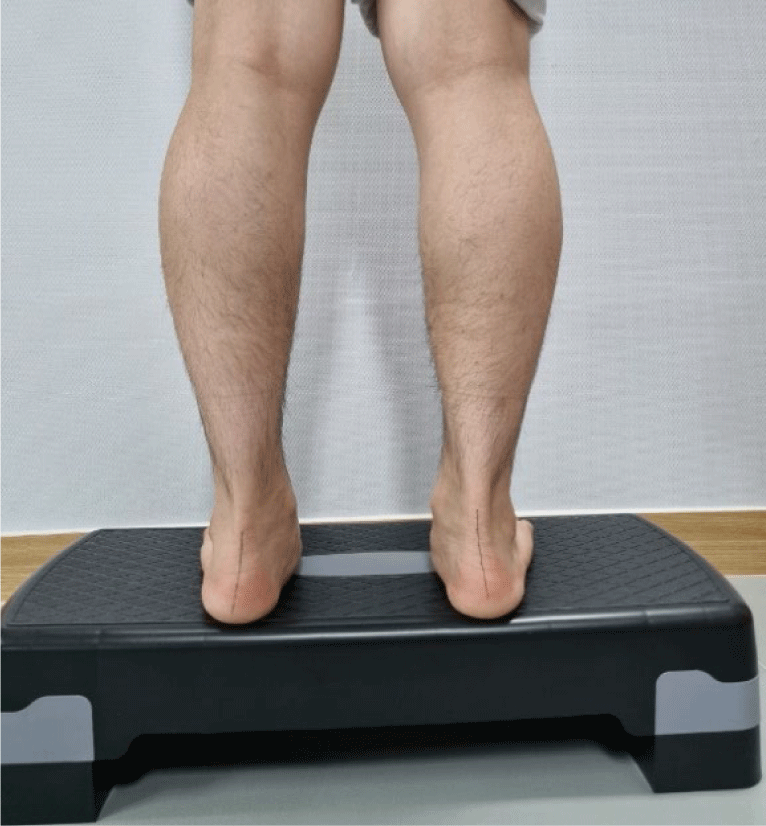
The examiner performed tests in various postures based on MSI principles. In the standing position, the patient exhibited an everted calcaneus on both sides and a lower-than-normal arch. Additionally, the internal rotation of the hip joint and the lateral rotation of the knee joint were observed. Foot alignment was assessed using both the resting calcaneal stance position (RCSP) and navicular drop (ND) tests. For RCSP measurement, the examiner first marked a bisecting line on the patient’s calcaneus while he was in a prone position. The patient was then instructed to stand in a comfortable position on a 15-cm-high step box.14 Using a smartphone, the examiner measured RCSP, which showed 7.7° of calcaneal eversion on the left side (Figure 1). A previous study reported that the RCSP test for a pronated foot has a sensitivity of 67% and a specificity of 85%.15
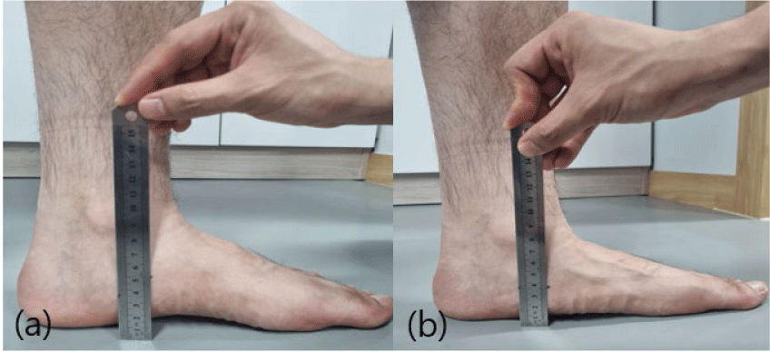
For the ND test, the patient was seated on a chair with feet flat on the floor, while the hips, knees, and ankles were positioned at approximately 90°, and the tibia remained perpendicular to the floor. The examiner used their thumb and index finger to palpate both the medial and lateral aspects of the talus. The talus was then positioned centrally to determine the subtalar joint’s neutral position. The examiner palpated the navicular tuberosity and marked it with a pen. The height from the floor to the navicular tuberosity was first measured in the seated position using a ruler (Figure 2a). The measurement was then repeated in the standing position (Figure 2b). The ND value was recorded as the difference in navicular height between sitting and standing (Table 1).16 A previous study reported high intrarater reliability (0.73) and interrater reliability (0.83) for the ND test.17
5/5, normal strength; 4+/5, able to hold against moderate to strong resistance; 4/5, able to hold against moderate resistance; 4-/5, able to hold against slight to moderate resistance; 3+/5, able to hold against minimal resistance; 3/5, able to hold against gravity but not against additional minimal resistance applied manually; 2+/5, moves through partial range of motion against gravity.
For ankle dorsiflexion range of motion (ROM) measurement, the examiner used a goniometer application on a Samsung Galaxy smartphone. The smartphone screen was aligned parallel to the patient’s fibula, and the touch screen was used to activate the “set” button to determine the fixation axis. The patient was instructed to perform maximal ankle dorsiflexion while in a long sitting position. The examiner then positioned the smartphone parallel to the patient’s fifth metatarsal bone and pressed the “stop” button to record the measurement (Figure 3a). A previous study demonstrated that measuring dorsiflexion ROM with a smartphone has high intrarater reliability (0.91) and interrater reliability (0.89).18
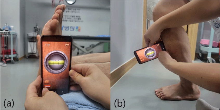
The weight-bearing lunge test (WBLT) was performed to measure the patient’s dorsiflexion ROM in a weight-bearing position. The patient aligned his heel in alignment with the second toe and bent his knee while ensuring that the heel remained in contact with the floor and subtalar pronation was minimized.19 For measurement, the “iHandy Level” app, a digital inclinometer, was used on a smartphone. The device was positioned 15 cm below the tibial tuberosity (Figure 3b). A previous study reported high intrarater reliability (0.97) and interrater reliability (0.76) for dorsiflexion ROM measurement using this smartphone application in the WBLT.20 Each measurement was performed three times, with a 1-min rest between trials. The average of the three measurements was used for data analysis.
All measurements were standardized to ensure reliability. RCSP, ND, ankle dorsiflexion ROM, and WBLT were conducted using a Samsung Galaxy S22 smartphone (Samsung Electronics, Seoul, Korea) with the iHandy Level (v4.6.2) and a goniometer app (v3.1.0). Before each session, the device was calibrated on a leveled surface according to the manufacturer’s instructions. The examiner was a licensed physical therapist with 10 years of clinical experience and specific training in MSI-based assessment. All tests were performed barefoot on a flat, non-slip floor. For RCSP, ND, and dorsiflexion ROM, three trials were performed, and mean values were calculated for analysis. Each measurement was performed three times, with a 1-min rest between trials. Camera alignment, inclinometer placement, and axis markings were standardized using anatomical landmarks, with trial structure identical across all sessions.
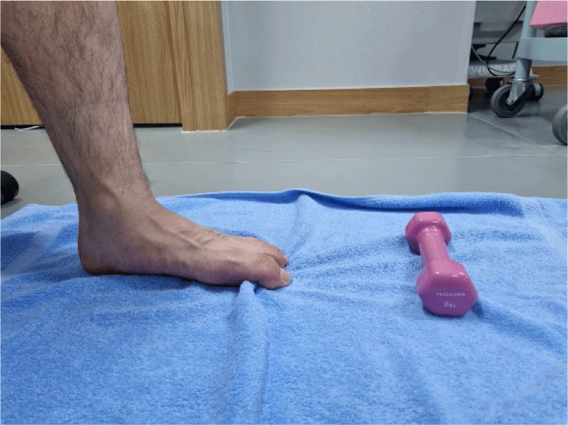
Muscle performance impairment was evaluated using the one-leg heel lift and short foot exercises. Muscle performance was assessed by the maximum number of one-leg heel raises performed until fatigue, defined as failure to achieve 50% of the initial lift height. Short foot exercise performance was evaluated by the patient’s ability to maintain metatarsophalangeal flexion without compensatory interphalangeal flexion. The patient was able to complete 10 repetitions of the one-leg heel lift before experiencing muscle fatigue, with a progressive decrease in heel lift height as repetitions continued (Table 1). The patient was instructed to pull a towel under the foot by flexing the metatarsophalangeal joints. However, compensatory flexion of the interphalangeal joints was observed (Figure 4).
Manual muscle testing was conducted in accordance with Kendall, revealing weakness of the posterior fibers of gluteus medius and tibialis posterior (Table 1).21 Muscle strength was graded using the manual muscle testing scale, where 3 indicates active movement against gravity, 4 indicates movement against moderate resistance, and 5 indicates full strength.21
DIAGNOSIS
For movement analysis, the patient performed a small knee bend and gait assessment. During the small knee bend, calcaneal eversion increased, and foot arch height decreased. VAS score increased to 6/10. However, after applying arch support, the VAS decreased to 1/10. During gait assessment, the patient reported a VAS score of 6/10, which reduced to 2/10 after applying taping to limit foot pronation. Taping and arch support were applied only during the initial assessment phase to evaluate pain modulation and were not included in the 6-week intervention protocol.
Based on the examination findings, the patient was diagnosed with foot pronation syndrome with PF according to the MSI classification (Table 2).13 Since the test results aligned with foot pronation syndrome, the MSI approach was applied for treatment. The primary objectives were to alleviate pain, restore foot alignment, normalize movement patterns, and enhance muscle performance.
TREATMENT
The patient underwent physical therapy three times per week for 6 weeks, following the MSI classification-based diagnosis. He visited the physical therapy center once a week for manual correction of alignment and reassessment of movement impairments, while also performing targeted exercises to improve muscle function. The remaining two sessions per week were conducted as home-based training (Table 3). As symptoms improved, the exercise load was progressively increased.
Exercise progression followed predefined rules. Resistance or volume was increased by approximately 10–20% weekly if no compensatory patterns were observed and if the patient reported a VAS ≤3 during or after exercise. If cramps or compensatory movements appeared, the exercise was regressed to the previous level. The patient maintained a weekly log, and adherence was reviewed during each clinical visit. Missed home sessions were addressed through therapist check-ins and adjusted scheduling to ensure continuity.
The patient was instructed to perform each exercise within a pain-free range while avoiding compensatory movements. He was advised to stop exercising if pain increased and to contact the physical therapist if he had any questions during home training.
To increase ankle dorsiflexion ROM in the initial phase, calf muscle stretching was performed in a long sitting position (non-weight-bearing). The exercise was performed three times per day, with three repetitions of 30s each. To strengthen the foot intrinsic muscles, the patient performed the short foot exercise and the toes spread-out exercise. For the short foot exercise, the patient sat on a chair with a towel placed on the floor. A 1-kg dumbbell was positioned at one end of the towel, while the patient’s foot was placed at the other. Using metatarsophalangeal flexion, he was instructed to gently pull the towel along the floor.3,10 For the toes spread-out exercise, the patient sat on a chair with his knees and feet positioned vertically and was guided to actively spread his toes apart. The patient maintained all toes in an extended position while keeping the metatarsals and heel in contact with the floor. He then abducted the little toe laterally and pressed it onto the ground, followed by abducting the big toe medially and pressing it down. The patient sustained pressure on both the little toe and big toe against the ground for 10s, ensuring they did not lift. This exercise was performed in 3 sets of 10 repetitions.22 To strengthen the tibialis posterior, an extrinsic foot muscle and inverter, an elastic band was wrapped from the navicular bone to the first metatarsal head, maintaining a 45° incline between the foot and the floor. Resistance was applied in the directions of ankle eversion and abduction. The patient actively resisted by maintaining ankle inversion and adduction while contracting the tibialis posterior. This exercise was performed in 3 sets of 10 repetitions, with a 2–3 min rest between sets. Over the 6 weeks, the number of sets or band resistance was progressively increased.23 Additionally, to strengthen the posterior fibers of the gluteus medius, the patient performed an exercise in a side-lying position. The bottom leg was positioned with the hip and knee flexed at 60°, while the exercising leg was placed in slight hip extension and external rotation. The patient was instructed to perform hip abduction to the maximum possible range and then slowly return to the starting position. This exercise was performed in 3 sets of 10 repetitions, 3 times per week.3,24
At the second visit, the patient reported a slight reduction in pain intensity when taking his first step in the morning. He also noted decreased plantar heel pain in the afternoon after starting work in the morning. However, the patient experienced difficulty controlling compensatory movements when performing exercises independently at home. During the short foot exercise in a sitting position, the patient complained that it was difficult to flex only the metatarsophalangeal joint without also flexing the toes and experienced mild cramping after seven repetitions. To address this, the patient was advised to reduce the dumbbell resistance to 0.5 kg and decrease the exercise volume from 3 sets of 10 repetitions to 2 sets of 10 repetitions, ensuring the exercise could be performed without causing cramps. Following this adjustment, the patient was able to perform the short foot exercise without toe flexion. The examiner emphasized the importance of performing all exercises at an appropriate intensity.
The patient reported a gradual decrease in foot pain intensity, significant improvement in morning pain, and increased ease in completing all exercise sets. The exercises were no longer challenging as before. Calf stretching was progressed from a long sitting position to a standing position. The patient was instructed to face a wall, position the treated leg behind the unaffected leg, and bend the front knee forward until feeling a tolerable stretch in the calf muscles of the treated leg.25 To maintain balance and prevent the heel of the treated leg from lifting off the floor, the patient placed both hands against the wall. Stretching was performed 10 times for 30s each, with 30s rest intervals between stretches.26 The patient was also directed to transition the short foot exercise and toes spread-out exercise from a sitting to a standing position and to increase the holding time from 5 to 10s. The positional change required greater focus to prevent compensatory movements. The patient was first instructed to sustain the exercise in a sitting position for 10s and, if no compensatory movement occurred, to progress to performing it in a standing position for 5s. For the gluteus medius exercise, the holding time was increased to 5s, and the number of repetitions was raised from 10 to 12. If the patient’s pain continued to improve and there was no discomfort during daily activities, it was decided that the exercise load would be increased in the next session.
The patient reported no pain or discomfort during most daily activities, except for occasional discomfort in the plantar heel when taking the first step in the morning and while working. The primary goal of the fourth session was to enhance exercise performance by increasing the exercise load. The patient noted a reduced pulling sensation during calf muscle stretching. Additionally, he was able to maintain the short foot exercise and toes spread-out exercise in a standing position for 5s without compensatory movements. To increase the exercise load, the patient was instructed to perform 3 sets of 15 repetitions, holding each repetition for 10s. For the tibialis posterior exercise, as the patient could perform it without compensation in a sitting position, the exercise was progressed to a standing heel raise. To prevent excessive activation of the gastrocnemius and soleus, a modified adduction heel raise was introduced.27 A tennis ball was placed between the medial sides of the heels, and the patient was instructed to press the ball firmly while performing the heel raise to further strengthen the tibialis posterior muscle.
Since the gluteus medius strengthening exercise could be performed without compensation in a side-lying position, it was progressed to a standing position. As the exercise load gradually increased with symptom improvement, additional attention was given to preventing compensatory movements.
OUTCOME
The patient’s plantar heel pain improved significantly, with the VAS score decreasing from 7/10 to 0/10 during the first step in the morning and from 5/10 to 0/10 after prolonged work. RCSP decreased from 7.7° to 4.7° on the left, while the ND test result improved from 16 mm to 8 mm. Active dorsiflexion ROM increased from 1.5° to 13.7°, and WBLT improved from 35.3° to 64.8°. The strength grades of the gluteus medius and tibialis posterior muscles increased from 3/5 to 4/5 and from 3+/5 to 4+/5, respectively. The number of single heel raises increased from 10 to 26 (Table 1). These improvements were closely linked to the MSI-based intervention strategy, which addressed abnormal alignment, reduced excessive pronation, and enhanced hip and foot muscle performance.
DISCUSSION
This study investigated the diagnosis and treatment of a patient with PF based on the MSI approach. Unlike general exercise therapy, this case emphasized a multi-domain MSI strategy incorporating alignment correction, movement retraining, and targeted strengthening of the gluteus and tibialis posterior. The reduction in VAS from 7 to 0 exceeded the minimal clinically important difference reported for plantar heel pain, confirming clinical relevance. The improvements in RCSP (7.7° → 4.7°), ND (16 mm → 8 mm), and dorsiflexion ROM (1.5° → 13.7° active; 35.3° → 64.8° WBLT) were clinically meaningful, as they fall within ranges associated with reduced pronation and improved functional stability. The increase in dorsiflexion ROM was directly related to improved performance in weight-bearing activities such as squatting and walking without pain. Furthermore, strengthened tibialis posterior and gluteus medius muscles provided enhanced dynamic arch support, reducing excessive pronation during gait. Changes in navicular height also contributed to improved medial longitudinal arch stability and pain-free movement.
The patient’s left calcaneal alignment showed valgus at 7.7°, and ND measured 16 mm, indicating a lower-than-normal arch. These factors may have contributed to excessive subtalar pronation during weight-bearing activities such as walking, squatting, and single-leg standing, increasing tensile stress on the plantar fascia. To address compensatory subtalar pronation, the patient performed short foot and toes spread exercises. As the patient’s exercise performance improved, the difficulty was progressively increased by transitioning from a sitting to a standing position. After 6 weeks of training, the patient’s RCSP improved from 7.7° to 4.7° on the left, while the left ND measurement decreased from 16 mm to 8 mm. Additionally, pain no longer occurred during weight-bearing activities. The reduction in RCSP and ND following the exercises was believed to have contributed to a decrease in excessive pronation during weight-bearing movements. The immediate pain reduction observed with taping and arch support was attributed to transient mechanical effects during testing, distinct from the long-term improvements achieved through the exercise-based MSI intervention. Brijwasi and Borkar conducted a study on adults with flexible flat feet, dividing them into two groups. Over 6 weeks, a control group of 24 participants performed active dorsiflexion exercises and plantar flexion exercises, while an experimental group of 25 participants performed short foot exercises and gluteal muscle strengthening in addition to the control group’s exercises. The experimental group demonstrated an approximate 4 mm improvement in ND height compared to the control group, along with an increase of about 19° in the medial longitudinal arch (MLA) angle.24 These findings were consistent with those of our study.
The patient’s active ankle dorsiflexion ROM was initially 1.5° in the long sitting position and 35.3° in the WBLT. Restricted dorsiflexion ROM was believed to contribute to increased flexibility in subtalar pronation during weight-bearing activities. To improve dorsiflexion, the patient performed calf stretching for 6 weeks, which resulted in an increase from 1.5° to 13.7°. Additionally, the WBLT angle significantly improved from 35.3° before treatment to 64.8° after treatment. Shashua et al. conducted a study involving 50 subjects with PF, dividing them into two groups of 25.28 The control group received stretching and ultrasound, while the experimental group received additional mobilization of the ankle and midfoot joints for 2 weeks. Following treatment, dorsiflexion ROM in the WBLT increased from 39.8° to 42.0° in the experimental and from 39.6° to 42.6° in the control group. While both groups showed significant improvements, the difference between them was not statistically significant.28 These findings suggest that PF and limited dorsiflexion ROM are associated more with muscle restrictions than joint limitations, which aligns with the results of our study. Limited ankle dorsiflexion has been shown to induce compensatory subtalar pronation during functional activities such as walking and may contribute to dorsiflexion at the midtarsal joint.29 Accordingly, an increase in ankle dorsiflexion ROM is presumed to mitigate tensile stress on the plantar fascia by reducing compensatory movements of adjacent joints during weight-bearing activities.
Before treatment, the patient exhibited weakness in the posterior fibers of the gluteus medius and tibialis posterior. Following 6 weeks of exercise, manual muscle testing revealed an improvement in tibialis posterior strength from 3+/5 to 4+/5, allowing the muscle to withstand moderate resistance, while the strength of the posterior fibers of the gluteus medius increased from 3 to 4. Additionally, pronation compensation was no longer observed during weight-bearing activities, indicating that both the tibialis posterior and gluteus medius were effectively engaged. Alam et al. studied 28 adults with pronated feet, dividing them into two groups of 14. The experimental group performed towel curl exercises, tibialis posterior strengthening, and iliopsoas stretching, while the control group only performed towel curl exercises three times a week for 6 weeks. ND height, muscle activity, and dynamic balance (Y-balance test) were assessed before and after the intervention.23 The within-group analysis showed a more significant improvement in ND height in the experimental group (37.03% decrease) compared to the control group (13.07% decrease), suggesting that extrinsic muscles like the tibialis posterior, in addition to intrinsic foot muscles, play a crucial role in preventing foot pronation. Furthermore, electromyographic activity of the tibialis anterior decreased by 65.67% in the experimental group and 8.2% in the control group. The AbdH muscle activity increased by 74.7% and 26.97% in the experimental and control groups, respectively.23 Choi et al. conducted a study on 32 adults with pes planus to compare muscle activity and the MLA angle during short foot exercises performed with and without isometric hip abduction (IHA) in both standing and sitting postures.30 AbdH activity increased significantly to 64.46% MVIC with IHA and 43.20% MVIC without IHA, while TA activity was notably lower at 6.45% MVIC with IHA and 15.29% MVIC without IHA. The increase in the MLA angle was significantly greater after performing short foot exercises with IHA in a standing position compared to other short foot exercise conditions.30 This highlights the role of the hip abductor in enhancing the activation of intrinsic foot muscles, which should be considered when designing rehabilitation programs for patients with pes planus. These findings were consistent with those of this case study.
This study has several limitations. First, its findings cannot be widely generalized since only a single patient was included, underscoring the need for future research with a larger sample size. Second, follow-up assessments were not conducted beyond the pre- and post-exercise measurements, making it unclear whether the improvements were sustained over time. Third, as the same therapist performed both the assessments and interventions, assessor blinding was not applied. This methodological limitation may impact the external validity of the findings and should be taken into account when interpreting the results. Therefore, further studies are required to determine the long-term effectiveness of the MSI approach in managing foot pronation syndrome with PF.
CONCLUSIONS
This case study evaluated the short-term effectiveness of the MSI approach in diagnosing and treating a patient who had foot pronation syndrome with PF. The patient performed a 6-week exercise program based on the MSI approach to address movement impairment and muscle performance deficits contributing to PF and foot pronation syndrome. These findings suggest that the MSI approach can be an effective clinical intervention for reducing plantar heel pain, improving foot alignment, and enhancing functional performance in patients with foot pronation syndrome accompanied by PF.