


INTRODUCTION
Lumbar disc herniation is one of the most common causes of back pain, occurring mostly in adults between the ages of 30 and 50.1 However, it is rare in adolescents, and the clinical presentation is similar to that seen in adults, but unlike in adults, where it is mostly considered a disc degeneration, it has an unclear etiology.2
A laminotomy, which removes a portion of the vertebral lamina bone to relieve pain and pressure on the spinal cord and nerves, is often used in adolescents and young adults because it has advantages over a laminectomy, which removes most of the lamina, such as restoring normal bone anatomy and preventing epidural compression.3,4
Persistent spinal pain syndrome (PSPS) is the persistence of pain after spinal surgery and may be related to several factors, including surgical technique, preexisting diagnosis, patient health, and psychosocial factors, making it difficult to provide effective treatment.5 In this regard, Daniels et al6 reported that neuromobilization and myofascial release may be recommended for patients with persistent pain and disability after back surgery, and Papalia et al7 reported in a systematic review that high-frequency spinal cord stimulation was effective in improving pain and reducing disability scores in patients with PSPS. However, these studies report a lack of high quality evidence to recommend these interventions and a lack of heterogeneity in population characteristics among the categorized studies, suggesting that further research is needed to confirm the effectiveness of noninvasive interventions in patients with PSPS.
Based on movement system impairment (MSI), Sahrmann categorized low back disorders into lumbar extension, flexion, rotation, extension-rotation, and flexion-rotation syndromes.8 She also explained that even in the presence of idiosyncratic problems such as disc protrusion or spondylolisthesis, correcting impairments in trunk muscle performance helps to eliminate the cause of the problem.8 Despite the benefits for these idiosyncratic problems, no study has applied the MSI approach to patients with persistent spinal pain syndrome. Therefore, this study aimed to diagnose the types of patients with PSPS based on the MSI approach and to describe the treatment and prognosis of PSPS patients according to the identified types.
CASE REPORT
The purpose of this case report is to describe the diagnosis, intervention, and management of a patient with PSPS using Sahrmann's MSI approach and the resulting outcomes.
CASE HISTORY
The patient is a 17-year-old male who had back pain, numbness along the right gluteal region, and paresthesia in the soles of his feet for 6 months, and underwent right L4/5 disc rupture and right laminotomy at another hospital 2 months before visiting our clinic, and even after the surgery, he came to us complaining of back pain and numbness, lower limb muscle weakness, and paresthesia in the soles of his feet. The patient was a student at an industrial high school and often worked standing for more than 1 hour or sat down to read, and although he usually had numbness, he complained of numbness and weakness that made it difficult to change his behavior when standing or sitting for more than 10 minutes. During the first interview, he presented with a limp gait and related that he had been limping for about 3 months. The patient demonstrated excessive dorsiflexion, bending at the waist first, and when asked to perform the movement, the movement was greater at the spine than at the waist and hip joints. The patient also complained of pain when asked to perform the same extension movement. The patient reported a pain rating of 6 out of 10 in the lower back, and a pain rating of 8 when standing for more than 10 minutes or when flexing the lower back (Table 1). This study was approved by the Daegu University Institutional Review Board (1040621-202505-HR-034).
PHYSICAL EXAMINATION
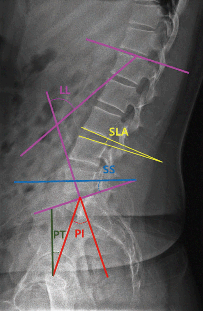
Physical examination was performed to identify motor impairment according to Sahrmann's MSI approach.8 The examination was performed by the first author, a physiotherapist working at the hospital. The examiner performed a muscle manual test of the lower extremity muscles and a passive straight leg raise test (PSLR) and measured the lumbar lordosis angle (LL), sacral slope (SS), pelvic incidence (PI), pelvic tilt (PT), and segment lumbar angle (SLA) with radiographs to check alignment and movement. In addition, the return from forward bending test and prone 10° hip extension with knee flexed were performed to evaluate movement.9,10,11,12 The patient was in pain at the time of initial evaluation, and most tests had difficulty obtaining clear results due to the pain, so the tests had to be performed in the position that was required for treatment. Therefore, the return from forward bending test and prone 10° hip extension with knee flexed tests were performed.
Radiographic measurements were taken using the Jumong General X-ray System (SG HealthCare, Seoul, Republic of Korea) and confirmed using the picture archiving and communication system (PACS) of ViewRex3 (ver 3.0.8.8(1.0), TechHeim Co., Ltd., Seoul, Republic of Korea). LL, SS, PI, and PT were measured before surgery, one month after surgery, and after 12 weeks of intervention (5 months after surgery) to confirm alignment.10,11 Neutral and extension segment lordosis angles (SLA) were measured before surgery, one month after surgery, and after 12 weeks of intervention to confirm the movement of each lumbar segment on radiographs of the patient's perceived neutral and extension positions, and the difference in movement between the extension and neutral positions was determined before surgery and after intervention. One month after surgery, only the neutral position was taken due to pain (Table 2) (Figure 1).12
Measurement of PSLR was performed manually by the examiner in the supine position, with pain during pre-execution and compensation for pain avoidance (Table 1).13
In movement tests, the Return from forward bending test was positive because movement at the low back occurred before the hip joint when returning from forward bending, and the Prone 10° hip extension with knee flexed test was positive because painful hip extension and anterior tilt of the pelvis occurred during hip extension in the prone position (Table 1).8
DIAGNOSIS
The MSI approach was used to confirm the findings of the examination and the patient was diagnosed with lumbar extension syndrome (Table 3).8 Although the patient's examination did not show an anterior pelvic tilt posture, the MSI approach was applied to the patient's therapeutic intervention because the other findings were consistent with lumbar extension syndrome. The main goals of treatment included restoration of normal motion and reduction of pain during movement performance.
TREATMENT
The treatment was performed 18 times over a 12-week period according to the MSI diagnosis, with each session consisting of 15 minutes of superficial heat therapy, interferential wave current therapy, and 5 minutes of deep heat therapy, and 20 minutes of high-frequency heat therapy using the Winback 3SE (Back 3SE, Winback, France) to relax the tense superficial muscles in the lower back area.14 The exercise program was then performed for at least 30 minutes, with the type and intensity of the exercise program depending on the patient's exercise performance and symptom improvement by week, and the exercises performed were filmed by the patient to be repeated at home. The exercise program included Sahrmann's exercises to correct the muscle imbalances and movement disorders identified through the examination (Table 4).8 Patients were instructed to contact the physical therapist immediately if they experienced increased pain or confusion in performing the exercises at home, and the exercises were videotaped so that the physical therapist could review the video and correct the exercises at the next visit.
For movement education on movements that are frequently used in daily life, during the initial treatment, the patient was taught to emphasize the extension of the backbone in sitting and standing positions, the contraction of the latissimus dorsi muscles in response to trunk movements, and the sequence of movements of the hip joints and lumbar vertebrae. At each subsequent visit, the patient's sitting and standing posture was observed and re-educated with the physiotherapist.
First, we tried to strengthen the basic muscular strength needed to perform sitting and standing movements, thereby correcting posture in daily life.
Second, we tried to improve the quality of movement by re-educating the sequence of movements that occur in the patient's functional movements.
OUTCOME
After eighteen treatment sessions, the patient's pain improved positively to a VAS score of 0 on a scale of 1 to 10 at rest and during activity, and he showed improvement in strength in all muscles measured (Table 5). Notably, the patient reported no numbness or weakness while sitting or standing for more than an hour, and no limp while walking. Radiographic measurements showed segmental movement changes with alignment and motion. PSLR showed no neurologic symptoms on either side, and the Return from forward bending test and Prone 10° hip extension with knee flexed test were negative, with positive results compared to baseline (Table 1).
5/5, normal strength; 4+/5, able to hold against moderate to strong resistance; 4/5, able to hold against moderate resistance; 3+/5, able to hold against minimal resistance; 3/5, able to hold against gravity but not against additional minimal resistance applied manually. Abbreviation: N/A, not available.
DISCUSSION
This study reports on the MSI diagnosis and treatment of a patient with PSPS who remained symptomatic after laminotomy, and confirms that the symptoms observed in the patient are consistent with the MSI diagnosis of lumbar extension syndrome. A 12-week program of diagnosis-specific exercises had a positive effect on the patient's symptoms.
Before starting treatment, the patient presented with numbness and back pain along with limping, which was confirmed by PSLR and strength testing, which showed nerve sensitivity and weakness in the key muscles of L4 and L5. The PSLR assessment on the left shows an up slip of the right pelvis, which suggests a pelvic instability as well. Furthermore, the trunk flexion and extension movements were performed to check movement quality, and both flexion and extension showed excessive thoracic spine motion with reduced motion at the lumbar spine and hip joints. When comparing trunk flexion and extension movements in PSLR and standing, pain only occurred in extension in standing. The occurrence of this pain due to posture was confirmed by the direct movement of the lumbar with the compensatory movements of the thoracic spine removed, and the appearance of the lumbar extension during the PSLR process confirmed that it was a lumbar extension syndrome. In lumbar extension syndrome, weakness in the hip extensors and lower abdomen occurs.8 Therefore, we applied abdominal drawing-in maneuver (ADIM) and gluteus maximus squeeze in supine and prone positions with minimal movements to strengthen the hip extensors and lower abdominal muscles, and applied them in various positions as the patient's symptoms improved. After 12 weeks, muscle strength in the lower abdomen and hip extensors increased, and the ADIM and gluteus maximus squeeze exercises applied to improve muscle strength in patients with pain were effective.
Previous studies have shown that ideal lumbar alignment is associated with functional impairment when the PI value is equal to the sum of SS and PT, and when PI minus LL is greater than 11.10 In this study, the sum of SS and PT was not equal to the PI value, but the difference between the pre- and post-intervention values was decreasing, which may indicate a positive outcome in the future. The comparison of the post-intervention values with those measured 1month after surgery shows a further increase in the difference between the sum of SS and PT and the PI value. Although this result may seem negative, the fact that the decrease in PT values in the post-intervention results is greater than the increase in SS values suggests that the effect is due to the pelvis recovering from retroversion. In addition, PI minus LL showed a decrease after the intervention, which may indicate a positive functional impact in the future.
In the case of SLA, an angle closer to zero means that the upper and lower segments are parallel, while a positive angle means that the space between the posterior segments is narrower, and a negative angle means that the space between the anterior segments is narrower.12
The SLA results in the neutral position of this study showed that the upper lumbar spine, such as T12-L1 and L1-2, showed an alignment change from a gradually decreased lordotic curve to a gradually increased lordotic curve, and the L3-4, L4-5, and L5-S1 segments showed a decrease in angle change after MSI intervention and gained intervertebral space. These results are consistent with a decrease in PT values in the neutral position. An increase in PT value indicates pelvic retroversion, which occurs with hip joint extension. However, in this study, the radiographic measurements were taken in a standing position, and these results suggest that the standing position measured before surgery and one month after surgery was actually a state of hip joint extension, and it can be considered that the MSI-based exercise program in this study is moving toward a neutral position rather than a hip-pelvis-hip complex with extension in a standing position. Previous studies have suggested that the normal PT value is less than 15°, and interventions focusing on motor control of the hip joint and lumbar spine are needed in future treatment to reduce the current PT value of 19.8°.10
The difference between the SLA values in the neutral position and in the supination state before and after the intervention showed that the extension movement was evened out in all segments after the intervention compared to before the surgery. In particular, the excessive L4-5 segmental movement in the extension state due to the L4-5 disc rupture, which was suspected to be the main problem of the preoperative back pain, seems to have been reduced by the rehabilitation, which is consistent with the results of increased muscle strength of the muscles dominated by the pain and the segment. Exercises that emphasize segmental movements of the spine improve spinal mobility and reduce overall peri-vertebral muscle tension.15 Trunk flexion and extension using a foam roller, which was the intervention in this study, is designed to recognize the position of the spinal segments and the combined movements of the lumbar-pelvic-hip joint complex through trunk flexion and extension on a foam roller, and it is likely that segmental movements were learned through the application of these exercises.
The results of the return from forward bending test and prone 10° hip extension with knee flexed were negative after intervention. A positive return from forward bending test indicates erector spinae dominance over the hip flexors with hip flexor shortening, while a positive prone 10° hip extension with knee flexed indicates hip flexor shortening and pelvic instability.8 Retchford et al16 reported that the hip external rotators contribute to the stability of the hip and pelvis due to the length and position of the muscles themselves, and Gottschalk et al17 reported that the gluteus medius contributes to the stability of the lumbar-pelvic-hip complex by regulating gait function and pelvic rotation individually, and is an important factor in the stability of the lumbar-pelvic-hip complex. The results of these previous studies provide evidence that the side lying hip abduction and external rotation applied in the present study contributed to pelvic stability and therefore were negative after the intervention. In addition, although it is common to apply stretching to shortened hip flexors, we did not apply stretching to the hip flexors in patients with painful PSPS because we were concerned that stretching the hip flexors would further contribute to back instability.18 In addition, since the patient had a concomitant weakness of the hip flexors, strengthening exercises were necessary, and it is thought that the repetition of controlled movements of the pure hip joint with the stability of the lumbar spine and pelvis secured through backward and forward rocking, hip flexion in upright sitting position, and standing hip extension using a slider applied in this study influenced the strengthening and length change of the muscle.
In this case report, MSI-based testing diagnosed a PSPS patient with a difficult-to-specify lumbar extension syndrome due to a variety of causes and symptoms, and the MSI intervention applied to address the diagnosed problem successfully achieved the therapeutic goal. These results suggest that MSI-based diagnosis may have a broader scope of acceptability in clinical practice.
There are a few limitations to this study. First, the study only included one subject, which makes it difficult to generalize the results. Second, there was no follow-up evaluation, so the long-term sustainability of the results cannot be determined. Third, the study only examined lat view radiographs, so the alignment of the lumbar-pelvic-hip joint complex could not be checked in three dimensions. Therefore, future studies should be designed with a larger sample size and images from different angles to determine the long-term effects of the MSI approach.
CONCLUSIONS
This study describes the MSI-based diagnosis and treat-ment of a patient with PSPS. The patient was diagnosed with lumbar extension syndrome and performed an exercise program designed to treat muscle imbalances and movement disorders. The patient experienced a decrease in low back pain and improvements in strength and functional activity. This report suggests that the clinical application of the MSI approach is effective in the diagnosis and treatment of patients with PSPS.