


INTRODUCTION
The Y-raise exercise (Y) is an exercise performed in a prone position where the arms are lifted overhead to perform shoulder flexion, which contributes to the stability and functional improvement of the shoulder complex.1-3 In the Y, the trapezius is one of the important muscles that work, and among its parts, the lower trapezius (LT) is the most activated. Previous research compared the muscle activation of the LT during the backward rocking arm lift and the backward rocking diagonal arm lift (BRDAL). The research results showed that the muscle activation of the LT was 47.99% in backward rocking arm lift and 63.5% in BRDAL, confirming more efficient LT muscle activation in BRDAL.4 Several previous studies compared the muscle activation of the LT when performing the Y at various shoulder abduction angles (180°, 160°, 125°, 90°, 75°), and the results indicated it was highest at 160° of shoulder abduction.1, 5-7 This is because the direction of the exercise aligns with the fiber direction of the LT.1, 5-8 Therefore, the Y is recommended as an efficient exercise for the LT.
Pectoralis minor (PM) stretching is used to increase the length of the Pm muscle. The Pm is a muscle that attaches to the coracoid process of the scapula from the 3rd to 5th ribs. When it is shortened (or tight), it increases the internal rotation and anterior tilt of the scapula, leading to a rounded shoulder posture.9 Previous studies have confirmed that when Pm stretching is applied, the rounded shoulder posture is improved compared to the pre-treatment baseline. When the Pm is shortened (or tight), the horizontal abduction angle of the shoulder joint decreases.10,11 This is because the length of the sarcomere shortens in the muscle contraction mechanism, and the separation of the actin and myosin cross-bridges is limited. Furthermore, a shortened Pm generates strong passive tension, which restricts the upward rotation, abduction, and posterior tilt of the scapula. When the length of the Pm increases through Pm stretching, it forms an appropriate length-tension relationship that facilitates the movement of the scapula.12 Furthermore, when scapular posterior tilt exercises were applied to subjects with a rounded shoulder posture after Pm stretching, the LT muscle activation significantly increased compared to applying the scapular posterior tilt exercises alone. This is because Pm stretching improved the alignment of the scapula and contributed to the muscle activation of the LT, which is the primary mover for scapular upward rotation.13 Therefore, Pm stretching is recommended as an effective therapeutic method for improving scapular alignment.
Taping is another intervention method used in clinical practice. Taping is classified into elastic and rigid tapes depending on its intended use and elasticity, with rigid tape being primarily used for mechanical restraint.14 On the other hand, elastic tape is divided into Kinesio Taping and Dynamic Taping.15 Kinesio Taping stretches only in the horizontal direction and improves local circulation, reduces swelling, corrects ligament damage, and enhances proprioception. Dynamic Taping, unlike Kinesio Taping, stretches in all directions (horizontal and vertical) and exhibits high elastic resistance and an elongation of over 200%. Furthermore, when the tape is applied, a recoil effect occurs, providing force absorption and support during pre-stretching. This is similar to taping techniques using rigid (non-elastic) tape, resulting in an effect that inhibits muscle activity.14,16 Furthermore, a study that applied Dynamic Taping to patients with tension-type headache and chronic neck pain with forward head posture showed a decrease in the tone of the upper trapezius (UT) and the headache index.16 A comparison was made of the changes in rounded shoulder posture following Dynamic Taping and Kinesio Taping interventions in university students in their 20s. The change in rounded shoulder posture decreased from 5.73 cm to 3.93 cm in the group that received Dynamic Taping, and it decreased from 5.40 cm to 4.66 cm in the group that received Kinesio Taping. This indicates that the group that received Dynamic Taping was more effective in improving postural alignment than the group that received Kinesio Taping.17 Therefore, Dynamic Taping is being suggested as an effective intervention method for improving shoulder posture alignment.14,15 A previous study reported that when the Pm muscle was stretched and relaxed, a viscoelastic response occurred in the muscle-tendon unit, which reduced unnecessary tension in the pectoralis minor and resulted in the improvement of scapular alignment.18 Furthermore, it was reported that when subjects with shoulder pain performed scapular upward rotation while taping was applied to the LT, the muscle activity of all muscles surrounding the shoulder girdle decreased.18 This suggests that the application of taping assists in scapular stability and distributes the load by appropriately positioning the scapula on the thoracic spine.
As can be seen through previous studies, applying Dynamic Taping to subjects can confirm that it reduces the tension of the UT and influences changes in rounded shoulder posture. Additionally, the selective muscle activation of the LT was confirmed after applying Pm stretching and the Y to subjects with a rounded shoulder posture. However, current research trends have not yet investigated the comparison of UT and LT muscle activation during the Y after applying pec minor stretching and taping to the LT. Therefore, this study aims to investigate the effect on the muscle activation of the UT and LT when performing the Y without intervention compared to performing the Y after applying pec minor stretching and LT taping. The research hypothesis was set as follows: First, the YLTPS will show a significantly greater increase in LT muscle activation compared to the Y. Second, the YLTPS will show a significant decrease in UT muscle activation compared to performing the Y. Third, it was hypothesized that the ratio of LT/UT muscle activation would be higher when performing the YLTPS compared to performing the Y.
METHODS
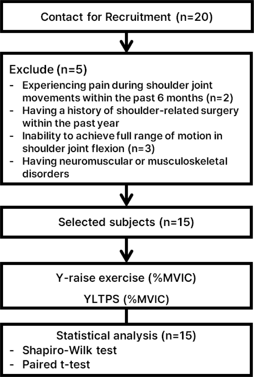
G*Power (version 3.1.2; Franz Faul, University of Kiel, Germany) was used to determine the sample size (number of subjects). Referring to a previous study and utilizing the LT activity from the five pilots in this current study, the required sample size was 15 participants when setting the effect size at 0.78, the significance level (α) at 0.05, and the power (1−β) at 0.8. Healthy adult males (24.3±3.0 years; 175.7±6.3 cm; 72.3±10.3 kg; 23.4±3.0 kg/m) were recruited.19 The exclusion criteria for the study were subjects with: 1) severe shoulder joint pain (8 or more out of 10 on the Visual Analog Scale), 2) a history of surgery/procedure in the shoulder area within the past 3 months, 3) an inability to achieve full shoulder flexion or pain during movement, 4) currently undergoing treatment for cardiovascular, cardiopulmonary, metabolic, or renal diseases, and 5) a prior history of shoulder joint disease (past illness, injury, genetic, or congenital condition).8, 20,21 This study was conducted after receiving approval from the Hoseo University Institutional Review Board [1041231-240708-HR-181]. All participants were fully informed about the study procedures and participated after signing a voluntary consent form (Figure. 1, Table 1).
| Characteristics | Healthy group (n=15) |
|---|---|
| Age (years) | 24.27 ± 2.99 |
| Weight(kg) | 72.33 ± 10.3 |
| Height (cm) | 175.67 ± 6.25 |
| BMI (kg/m2) | 23.42 ± 2.99 |
Surface electromyography (EMG) equipment (Ulitium EMG system, Noraxon, USA) is used to measure muscle activation during exercise performance. The EMG settings were as follows: To analyze the muscle activation of the UT, LT, and serratus anterior, the band-pass filter was set to 10-450 Hz, the sampling rate to 1,024 Hz, and the notch filter to 60 Hz. The collected data was processed using the Root Mean Square (RMS) with a moving window of 50 ms.22 Before attaching the electrodes, the hair at the attachment site was shaved and the area was cleaned with an alcohol swab to minimize skin resistance. Ag/AgCl disposable surface electrodes were attached to the corresponding areas.20 The electrodes were attached to the UT, LT, and serratus anterior (SA) of the subjects according to the guidelines of Criswell. The electrode for the UT was attached after palpating the spot between the C7 spinous process and the acromion of the scapula where the muscle bulked up the most during muscle contraction. The electrode for the LT was attached obliquely to the oblique muscle that bulked up the most during muscle contraction, in the area 5 cm below the spine of the scapula and near the inferomedial border of the scapula. The electrode for the SA was attached medial to the latissimus dorsi at the inferior tip of the scapula in the axillary region. Muscle activation was collected from the subjects' dominant arm, and the first and last 1 second of the signal were removed.20,23
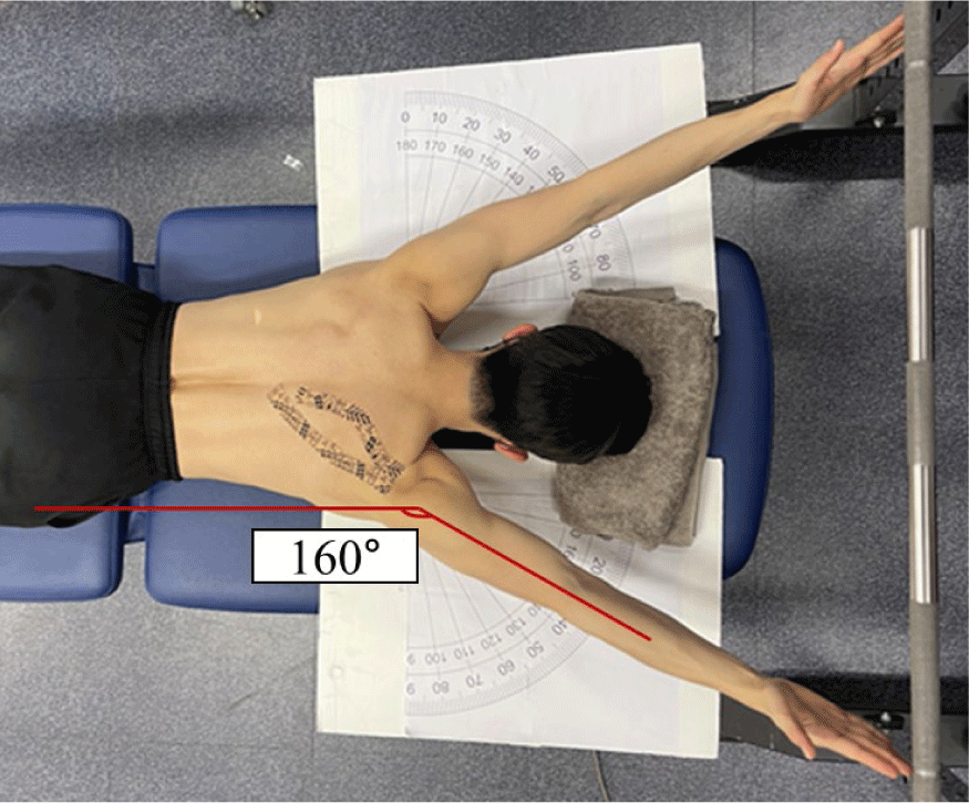
Prior to conducting this study, the examiner thoroughly explained the correct exercise posture to the subjects, and the study proceeded only after the subjects had practiced sufficiently until they were familiar with the exercise methods. The exercise was performed for 5 seconds per repetition, 3 repetitions in each exercise posture, and 1 minute of rest was provided between repetitions. Subjects who failed to perform the exercise with correct posture were excluded. The sequence of exercise progression was as follows: First, the Y was performed without any intervention, followed by the YLTPS. Two minutes of rest was provided between each exercise to minimize muscle fatigue, and the subjects performed the exercise at 5-second intervals according to the metronome speed.20 The subjects were placed prone on a height-adjustable table, and the angle of their shoulder joint was adjusted using a board marked with angles. With the acromion of the scapula as the reference point, the subjects were instructed to position both arms abducted to 160°, and the height of the target bar was set so that the height of the arm lift reached 180° of shoulder flexion. The humerus was rotated externally so that the palm faced the ceiling to prevent limitations caused by anatomical structures. While the subjects performed the exercise by maintaining maximum flexion strength for 5 seconds with their hands touching the target bar, the examiner measured the muscle activation of the UT, LT, and SA.19 After completing the Y, considering muscle fatigue, the subjects were given a 2-minute rest before performing the Pm stretching. In the supine position (lying on the back), the subject was prepared by having their elbows flexed to 90° and their shoulder joints horizontally abducted. The examiner fixed the subject's sternum and held the elbow, then performed the stretching by horizontally abducting the subject's shoulder to the maximum possible range. The stretching was performed for 10 seconds per repetition and repeated 12 times. A 10-second rest period was provided between each stretch.24,25 Afterwards, Dynamic Taping was applied to the LT. The application position for the Dynamic Taping was between the subject's scapular spine and the T7 vertebra, and it was applied with light tension.25,26 Subsequently, the subjects performed the exercise the same way as the Y they had initially (Figure. 2).
To standardize the measured muscle activation values, the Maximum Voluntary Isometric Contraction (%MVIC) was measured for the UT, LT, and serratus anterior. The method for measuring the %MVIC of the UT is as follows: The subject performed shoulder elevation and neck extension while turning their neck away from the dominant side muscle in a sitting position. The examiner applied resistance to the subject's head to induce anterolateral flexion while simultaneously applying downward resistance to the shoulder. For the %MVIC of the LT, the subject was prone (lying on their stomach), and they lifted their arm in a diagonal abduction while performing a posterior tilt of the scapula. The examiner provided downward resistance to the subject's arm. For the %MVIC of the serratus anterior, the subject was measured in a supine position (lying on their back) with the arm flexed to 90°, and the measurement was taken while performing shoulder protraction (abduction of the scapula). The examiner fixed the subject's fist and applied resistance by pushing dorsally (towards the back). The examiner measured the %MVIC for each of the subject's muscles for 5 seconds, and 1 minute of rest was provided between measurements to reduce muscle fatigue.20
All data collected in this study were analyzed using SPSS ver. 20.0 program (IBM Co., Armonk, NY, USA). The normality of the data distribution was confirmed using the Shapiro-Wilk test (p>0.05). The Paired t-test was used to compare the muscle activation of the UT, LT, and SA, as well as the muscle activation ratios of LT/UT and SA/UT. The statistical significance level was set at p<0.05.
RESULTS
There was a significant difference in the muscle activation of the UT and LT between the Y and the YLTPS (p<0.05). The muscle activation of the UT was measured as 39.06% in the Y and 26.84% in the YLTPS, showing a significant decrease for the YLTPS compared to the Y (p<0.05). The muscle activation of the LT was measured as 59.80% in the Y and 67.16% in the YLTPS, showing a significant increase for YLTPS compared to the Y (p<0.05). However, the muscle activation of the SA was measured as 34.75% and 33.74% for the Y and the YLTPS, respectively, and no statistically significant difference was found between the exercises (p>0.05) (Table 2, Figure. 3).
| %MVIC | Mean ± SD | Difference value | t | p | Effect size | |
|---|---|---|---|---|---|---|
| Y | YLTPS | |||||
| UT | 39.06±16.44 | 26.84±14.63 | 12.22 | 3.71 | .00* | 0.78 |
| LT | 59.80±13.21 | 67.16±14.90 | 7.36 | 2.6 | .02* | 0.52 |
| SA | 34.75±16.30 | 33.74±15.26 | 1.01 | .27 | .80 | 0.06 |
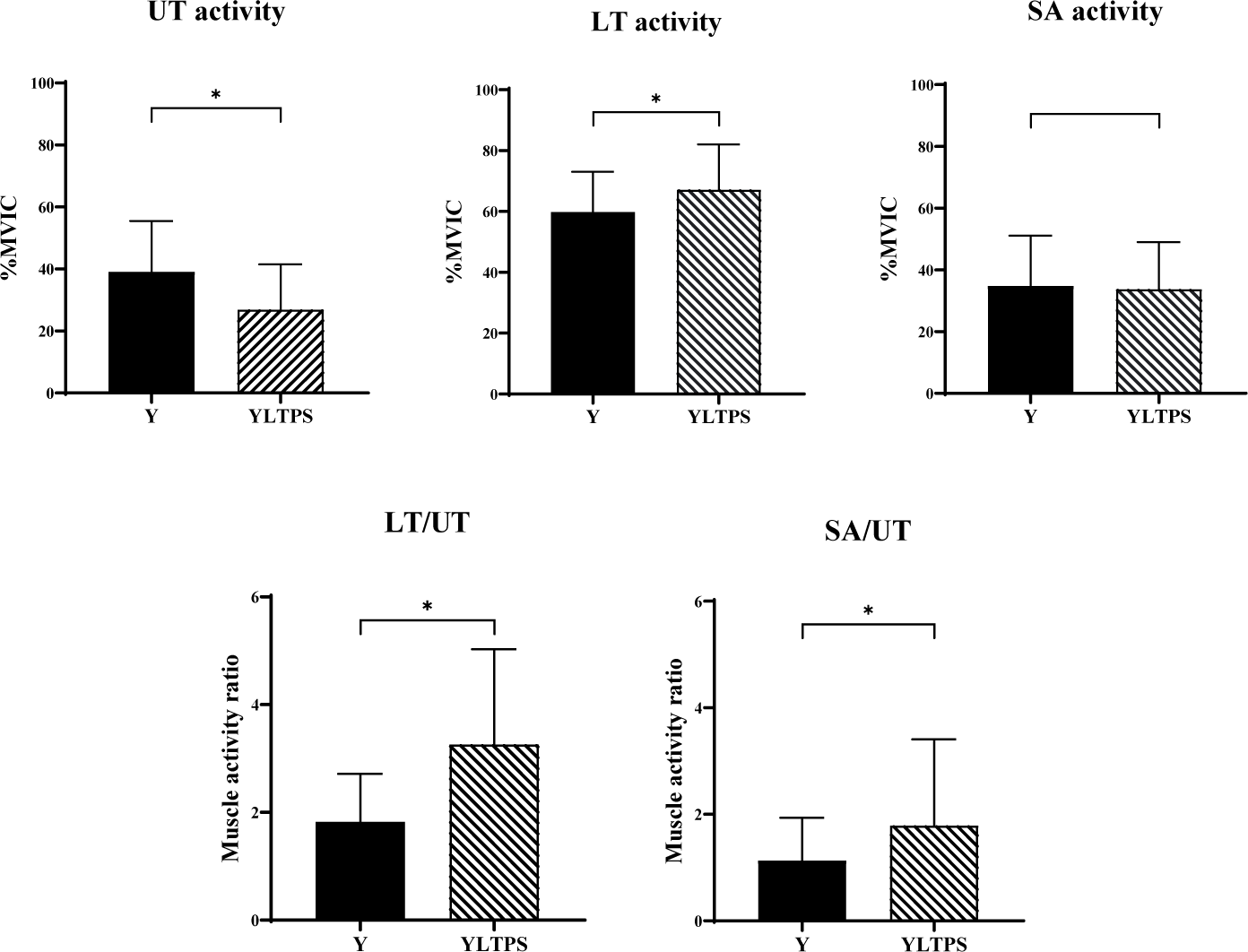
There was a statistically significant difference in the LT/UT muscle activation ratio and the SA/UT muscle activation ratio between the Y and the YLTPS (p<0.05). The LT/UT muscle activation ratio was calculated as 1.82 and 3.26, respectively, and it increased significantly for YLTPS compared to the Y (p<0.05). The SA/UT muscle activation ratio was calculated as 1.13 and 1.79, respectively, and it increased significantly for the YLTPS compared to the Y (p<0.05) (Table 3, Figure. 3).
| Mean ± SD | Difference value | t | p | ||
|---|---|---|---|---|---|
| Y | YLTPS | ||||
| LT/UT | 1.82±0.89 | 3.26±1.77 | 1.44 | 3.83 | .00* |
| SA/UT | 1.13±0.80 | 1.79±1.62 | 0.66 | 2.60 | .02* |
DISCUSSION
The purpose of this study was to compare the muscle activation and the muscle activation ratio of the UT and LT during the YLTPS. The research hypothesis was set that the YLTPS would show a significant increase in LT muscle activation and a significant decrease in UT muscle activation compared to the Y. Furthermore, it was hypothesized that there would be a significant difference in the LT/UT muscle activation ratio during the YLTPS. The muscle activation of the UT showed a significant difference, with a 31.29% decrease in the YLTPS compared to the Y (p<0.05). The muscle activation of the LT showed a significant difference, with a 12.31% increase in the YLTPS compared to the Y (p<0.05). The muscle activation of the SA decreased by 2.91% in the YLTPS compared to the Y, but this difference was not statistically significant (p>0.05). LT/UT muscle activation ratio increased significantly by 79.12% in the YLTPS compared to the Y, confirming a significant difference (p<0.05). The SA/UT muscle activation ratio increased significantly by 58.41% for the YLTPS compared to the Y, confirming a significant difference (p<0.05).
The research compared the results of the Y performed without intervention to the YLTPS. The reason for the increase in LT muscle activation and the decrease in UT muscle activation during the YLTPS is that the lengthening of the shortened Pm muscle improved the alignment of the scapula (shoulder blade). This is likely due to the viscoelastic response that occurs in the muscle-tendon unit by static stretching, which is thought to be the result of improved flexibility through a reduction in passive tension, regardless of the tissue's length.27 A previous study compared the effect of pec minor stretching on LT muscle activation when applying a scapular posterior tilting exercise in subjects with round shoulders. The study results showed that when the scapular posterior tilting exercise was performed after pec minor stretching, the LT muscle activation showed a statistically significant increase of 16.67% compared to performing the scapular posterior tilting exercise. This is because pec minor stretching improved the alignment of the scapula and contributed to the muscle activation of the LT, which is the primary mover for upward rotation of the scapula.13 Furthermore, a study that compared the muscle length of the UT by applying LT taping confirmed a significant reduction from 15.46 mm to 13.35 mm (13.65% decrease) after the intervention.27 Although direct comparison with prior studies is difficult, it is concluded that the reason for the increase in LT muscle activation and the decrease in UT muscle activation when performing the YLTPS in this study is that the intervention improved scapular alignment, optimizing the length-tension relationship, which inhibited the muscle activation of the UT and facilitated the muscle activation of the LT. While previous rehabilitation protocols often focused on either releasing tight soft tissues or strengthening weak muscles in isolation, this study proposes a comprehensive strategy that addresses both mechanical restrictions and neuromuscular control simultaneously.18 The application of Pm stretching creates an optimal scapular starting position by reducing anterior tilting, while the subsequent Dynamic Taping provides continuous tactile cues and mechanical support to the lower trapezius.18 This combined approach suggests a novel clinical pathway where 'structural preparation' (stretching) and 'functional facilitation' (taping) are integrated to maximize exercise efficiency.
Secondly, the reason the LT/UT muscle activation ratio increased when performing the YLTPS is that both the muscle activation of the LT increased, and the UT decreased. During the YLTPS, the LT is most recruited and stabilizes the movement of the scapula as the scapula undergoes upward rotation and posterior tilting. It is believed that this contributed to preventing the over-activation of the UT and increased the muscle activation of the LT. As mentioned earlier, it is believed that the muscle activation of the UT decreased due to the reduction in passive tension, regardless of the tissue's length.26 Previous study also showed that the LT/UT muscle activation ratio varied according to the shoulder abduction angle (140°, 150°, and 160°) applied during isometric adduction, measuring 2.06, 2.31, and 3.2, respectively, confirming that the 160° angle showed the highest ratio.29 In this study, the LT/UT muscle activation ratio was 1.82 when performing the Y and 3.26 when performing the YLTPS, showing a difference of 1.44 (in the ratio value, or a 79.12% increase as previously stated). This suggests that the LT was selectively activated while the UT decreased. Furthermore, this substantial improvement in the ratio observed in the YLTPS condition holds significant clinical value. An imbalance characterized by excessive activation of the upper trapezius and delayed or weak firing of the lower trapezius is a well-documented contributor to subacromial impingement and scapular dyskinesis.18 Achieving such a substantial improvement in this ratio suggests that the YLTPS intervention can effectively re-educate the scapular force couple. Therefore, this intervention offers a practical and time-efficient method for clinicians to selectively recruit the lower trapezius while inhibiting the upper trapezius, potentially reducing the risk of secondary shoulder pathologies during rehabilitation.
Third, the reason the muscle activation of the SA showed no statistically significant difference is that the Y posture does not selectively strengthen the serratus anterior. A previous study investigated the effect of scapular posterior tilting exercise on SA muscle activation in subjects with a rounded shoulder posture, depending on whether they performed pec minor stretching and/or wore a shoulder brace. That study reported that there was no significant difference in SA activation among the scapular posterior tilting exercise without intervention, the scapular posterior tilting exercise after pec minor stretching, and the scapular posterior tilting exercise after wearing a shoulder brace.24 Another preceding study also investigated the SA muscle activation during the Y in the prone position in athletes with rounded shoulder posture and forward head posture, with or without wearing a shoulder stabilization brace, and found no significant difference.30 The activation of the SA varies depending on the direction of scapular movement. According to a previous study, exercises performed in a posture that involves scapular protraction, such as the push-up plus, are recommended as exercises for SA strengthening.31 In this study, the scapula moves in the direction of depression, retraction, and upward rotation during the Y. It is believed that this is because the posture (Y) is relatively difficult for the SA to activate, resulting in its low muscle activation during the Y.31 However, no significant difference in SA activity between the Y and YLTPS conditions carries a clinical implication regarding safety. It indicates that the intervention did not inhibit the function of the SA, which serves as a stabilizer for the scapula.18 This finding suggests that the YLTPS allows for the selective enhancement of the LT/UT ratio without compromising the baseline stability provided by the SA. Therefore, this combined intervention can be considered a safe strategy that effectively targets LT while preserving the stabilizing muscle function of SA.
The limitations of this study are as follows: First, the study was conducted on healthy male subjects in their 20s. Future research intends to confirm the differences in various age groups. Second, randomization was not possible due to the inherent constraint of the experimental design, which required a comparison between a baseline condition and a subsequent, sequentially applied intervention (stretching and taping). Third, the changes in muscle strength per muscle following the application of the intervention could not be confirmed. Future research intends to measure both muscle strength and muscle activation together to confirm the effect of the exercise. Fourth, this study was conducted as a cross-sectional study. Future research intends to conduct the exercise over a long-term period to confirm the sustained effect of the exercise. Fifth, the study could not be randomized due to the exercise intervention. Future research intends to randomize to reduce bias and learning effects in the study.