


INTRODUCTION
Knee Osteoarthritis (KOA) is a chronic degenerative musculoskeletal disease that is considered a major problem causing functional impairment of the knee.1 The prevalence of KOA is mainly caused by aging and sports activities that cause strong impact on the knee, such as badminton.2 As a result, joint pain, functional decline, and decreased quality of life occur, requiring medical management. In particular, it has the problem that symptoms appear after a certain degree of irreversible damage to the joint occurs.3,4
The common diagnostic methods used to diagnose KOA mainly use X-rays or magnetic resonance imaging (MRI). However, these diagnostic methods have limitations in evaluating the structural and functional problems in the early stage of KOA.5-7 In response to these limitations, the Early Osteoarthritis Questionnaire (EOAQ) was developed as a self-reported screening tool to rapidly identify initial symptoms of KOA, such as pain, stiffness, and instability during daily activities. The EOAQ is often used in clinics and research studies to help identify people who might be at risk for early KOA.8,9 This questionnaire relies on patients’ subjective reports of symptoms in daily life, so it cannot objectively assess knee movement or functional status.7-9
Objective functional performance tests are needed to supplement subjective assessments. Among the various tests for the knee, the Y-balance test (YBT), which is widely used clinically, is a reliable and valid tool for measuring dynamic balance, neuromuscular control, and lower extremity functional symmetry. The YBT requires standing on one leg with maximum extension in three directions (anterior, posteromedial, and posterolateral), and evaluates lower extremity muscle strength, proprioception, and stability.10-12 Poor performance on the YBT is linked to a higher risk of injury and can effectively identify early declines in functional ability before structural damage becomes visible on imaging tests.11-13 In a recent study of subjects with early KOA, subjects who complained of subjective symptoms on the EOAQ showed more lateral knee displacement on the one-leg standing test compared to those who did not, confirming problems with dynamic balance.14 Previous EOAQ-based studies have also demonstrated that abnormal frontal plane knee movements, including increased horizontal displacement or frontal knee angulation (FKA), serve as early indicators of joint instability and altered load distribution, often preceding radiographic KOA changes and potentially accelerating cartilage degeneration.8,14 Assessing YBT which is representative dynamic movements can provide valuable information about early functional impairment that may be difficult to detect using KOA-based questionnaires alone. EOAQ is widely used tool for screening early symptoms of knee osteoarthritis, focusing on patient-reported pain and functional limitations.8 However, it has certain limitations, as it primarily relies on subjective measures and may not fully capture biomechanical or functional impairments that precede structural changes.15 The YBT is increasingly recognized as a functional assessment that evaluates dynamic balance and neuromuscular control, helping to identify subtle deficits not detected by questionnaires alone.10 However, while YBT reflects overall lower limb function, it does not directly quantify specific biomechanical parameters such as FKA, which is critically involved in the pathomechanics of early knee osteoarthritis.16 Therefore, incorporating quantitative measurements of FKA alongside functional tests like YBT and subjective tools such as EOAQ provides a more comprehensive assessment, enabling earlier detection of functional changes and more targeted interventions in individuals at risk of knee osteoarthritis.
The relationship between EOAQ-based risk classification and dynamic balance performance remains unclear, especially among recreational badminton players who frequently experience repetitive joint loading and multidirectional movements. Therefore, the present study aimed to (1) compare YBT performance between individuals identified as at risk for early KOA by the EOAQ and healthy controls, and (2) assess FKA during the YBT. We hypothesized that participants in the early KOA risk group would show significantly poorer YBT performance (shorter reach distances and lower composite YBT index) and greater FKA in all YBT directions compared to controls.
METHODS
This cross-sectional study included 20 recreational badminton players, aged 20–50 years, who had participated in badminton at least twice per week over the past year. The general characteristics of the subjects are shown in Table 1. Group classification was based on responses to the EOAQ, which assesses the frequency of knee pain, stiffness, and instability over the preceding six months. Participants reporting symptoms “frequently” or “rarely” in the clinical feature items were assigned to the EOAQ group (n=10), while those reporting “never” were assigned to the non-EOAQ group (n=10). Individuals were excluded if they had a lower extremity injury or surgery within the previous six months, a diagnosis of rheumatoid arthritis, neurological disorders, or established osteoarthritis, or any other condition affecting balance or lower limb function. All participants provided written informed consent, and the study protocol was approved by the Sangji University Institutional Review Board (No: 1040782-250217-HR-13-149).
All of the subjects performed the EOAQ and YBT after consent of this study. The EOAQ form was collected online using a tablet PC. The subjects wore short pants with bare feet to perform the YBT. When performing YBT, three directions were performed randomly. The mean values of the measurements obtained in the two trials were used for data analysis.
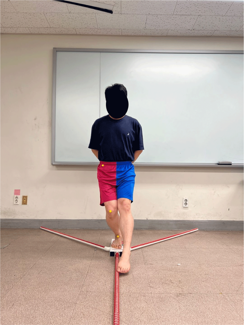
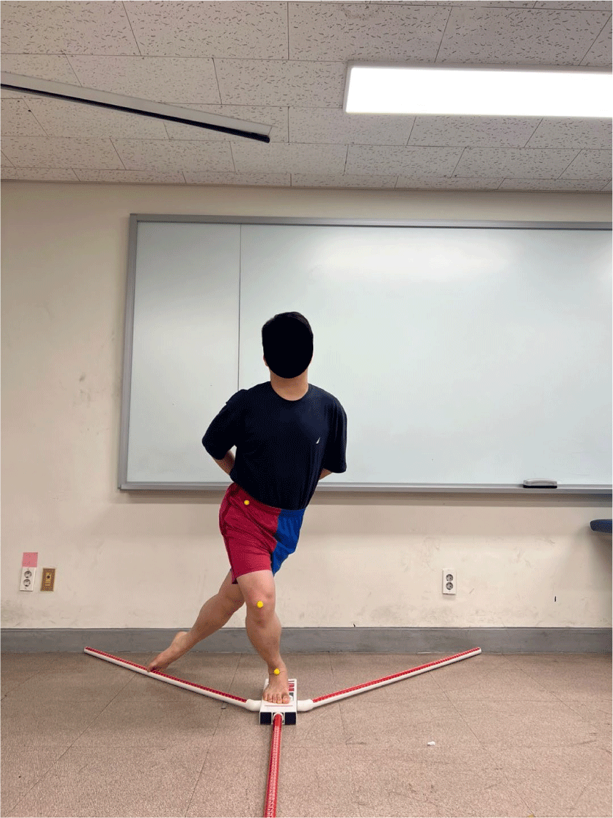
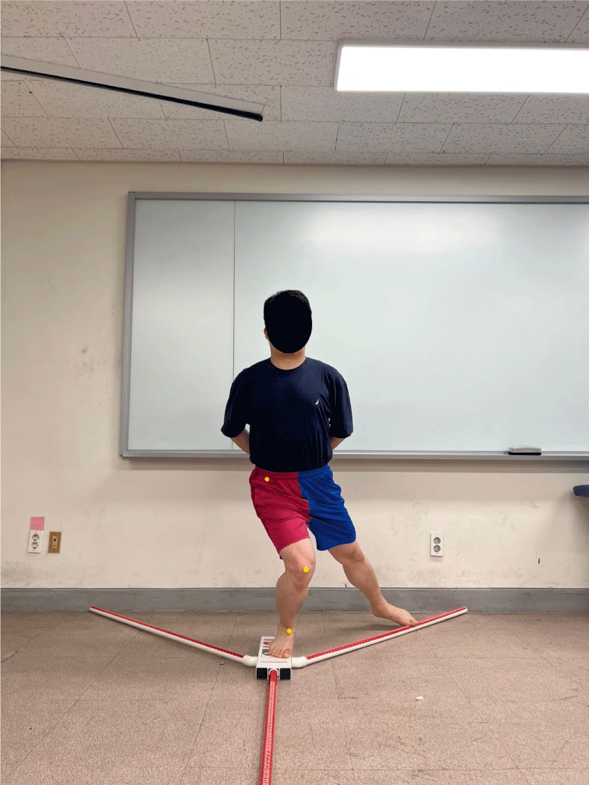
The YBT was performed using a tape measure attached to the floor in a Y shape, following the method used in previous studies.17 This tape-based method has been shown to provide reliable and valid measurements similar to those obtained using commercial Y-Balance kits. Three lines were marked on the floor with a tape measure. One was in the anterior (ANT) direction (Fig. 1), and two were in the posteromedial (PM) (Fig. 2) and posterolateral (PL) (Fig. 3) directions, and each line was separated by a 135° angle from the anterior line. Each line was marked in centimeters for accurate measurement. The subject stood barefoot at the center point, with the tested leg in the measurement position, and the other leg lifted off the ground to maintain balance. Both hands were crossed on the chest. The subject was instructed to extend the other leg along each line as far as possible. The supporting foot (the tested leg) was kept still and lightly touched the tape measure. The maximum reach in each direction was recorded in centimeters. Each participant performed six practice trials and then three tests in each direction. The average of the three test trials was used in the analysis. To normalize leg length differences, reach distances were normalized by dividing the reach distance by the subject’s leg length (measured from the anterior superior iliac spine to the medial malleolus) and multiplying by 100. Distances in each direction were normalized by dividing the leg length by each maximal reach distance and multiplying by 100. A composite YBT index was calculated by dividing the sum of the maximal reach distances in the three directions by three times the limb length and multiplying by 100. Normalized reach distances for each direction were calculated as: Normalized Reach Distance (%)=[Reach Distance (cm)/ Limb Length (cm)]×100. The composite YBT index was calculated as: YBI (%)=[(ANT+PM+PL)/(3×Limb Length)]×100.
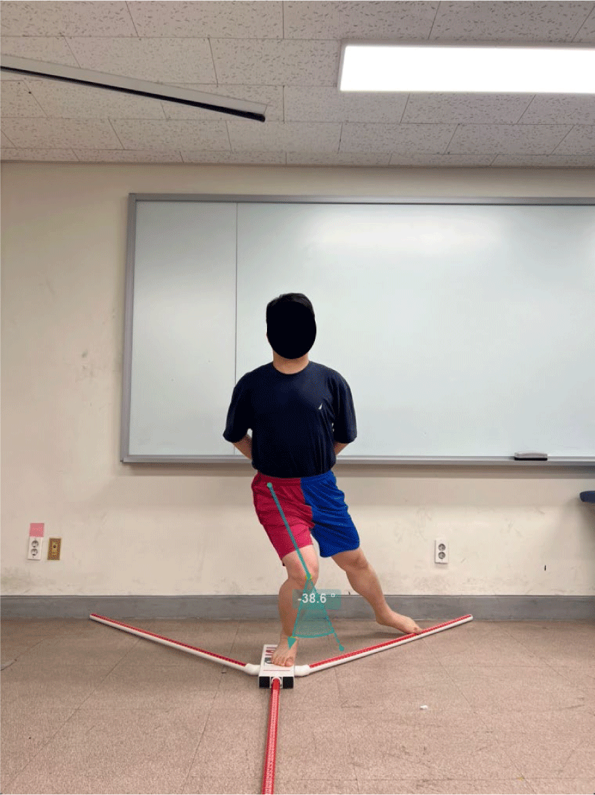
A regular smartphone (iPhone 13 pro; Apple Inc., USA) with video recording capabilities (4K, 2,556×1,179 pixels at 60 fps) was used to assess knee FKA during YBT (Fig. 4). Round yellow markers of 2 cm in diameter were attached on the anterior superior iliac spine (ASIS), mid-point of patellar, and mid-point of talus. The camera was positioned on a tripod that was 60cm in height and placed 250 cm in front of the subjects. The Knee FKA during YBT were analyzed using Kinovea software. All recorded videos were analyzed with the stable version of Kinovea (v. 0.8.16, Kinovea, Bordeaux, France). Kinovea is a free 2D motion analysis software that enables the establishment of kinematics parameters. Analysis of lower extremity movements in frontal plane using Kinovea software has been used in various studies with excellent intra-rater and inter-rater test reliability.18 In this study, the FKA was measured in Kinovea as the angle formed at the patella between a line from the ASIS to the patella and a line from the patella to the ankle, using video captured in the frontal plane. The measured values were analyzed without distinguishing between knee valgus and varus.
Statistical analyses were performed using the SPSS for Windows (ver. 29.0 software; IBM Co., Armonk, NY, USA). To confirm the normality of data distribution, Shapiro-Wilk test wad used. Independent t-tests were used to compare YBT performance and FKA of knee joint between groups. Statistical significance was set at p<0.05. The level of statistical significance was set at α of 0.05.
RESULTS
The Shapiro-Wilk test presented the normality of the data (p>0.05). Twenty subjects (EOAQ group n=10, non-EOAQ group n=10) were analyzed. There were no significant differences in age, height, or weight between groups (p>0.05). The results of the YBT performance for both groups are presented in Table 2. The EOAQ group showed slightly higher values across all reach directions and the composite index compared to the non-EOAQ group; however, there were no statistically significant differences in YBT performance between the EOAQ and non-EOAQ groups (Table 2). In contrast, FKA during the YBT was significantly greater in the EOAQ group across all directions. The EOAQ group demonstrated significantly higher FKA values for anterior (p<0.001), posteromedial (p<0.001), and posterolateral (p=0.006) reaches compared to the non-EOAQ group (Table 3).
| Characteristics | EOAQ group | Non-EOAQ group | t | p |
|---|---|---|---|---|
| ANT reach | 13.1 ± 5.1 | 4.0 ± 3.6 | –4.622 | <.001 |
| PM reach | 21.7 ± 8.9 | 7.2 ± 3.6 | –4.793 | <.001 |
| PL reach | 29.0 ± 16.5 | 12.2 ± 9.9 | –2.762 | 0.06 |
DISCUSSION
This study compared the YBT performance and FKA of the knee during the YBT between the EOAQ group and non-EOAQ group classified by the EOAQ in recreational badminton players. The results showed that there was no significant difference in YBT performance (reaching distance and composite index) between the two groups, but the FKA of the knee during the YBT was significantly greater in all directions (anterior, posteromedial, and posterolateral) in the EOAQ group.
These results suggest that the EOAQ group shows similar levels of functional performance ability to the non-EOAQ group, but there may be differences in joint stability or alignment during dynamic movements.19,20 Individuals in the KOA risk group report symptoms through the EOAQ, but general measures like reach distance in functional tests such as the YBT are not sensitive enough to detect early functional decline. However, quantitative motion analysis indices, such as KOA, are more sensitive to early changes like joint instability, alignment alterations, or abnormal load distribution during YBT. Previous studies have reported that increased horizontal displacement of the knee during functional movement, such as stair-up and walking, is associated with increased joint instability, malalignment, and increased risk of cartilage damage. 9,14,21 This is consistent with our results. These results demonstrate that early functional changes can be detected before structural damage appears on imaging modalities such as MRI or X-ray. This highlights the value of assessments that can identify the early stages of KOA progression. In our results, the EOAQ group showed significantly greater FKA in all directions compared to the non-EOAQ group. These findings imply that despite normal YBT performance, clinical impairments in knee joint stability or neuromuscular control may already exist. These changes may occur in the very early stages of osteoarthritis and may be missed by simple questionnaires or YBT performance (reach measurements) alone. Utilizing quantitative movement analysis measures, such as FKA during the YBT, may enhance the early detection and prevention of knee joint dysfunction. This study demonstrates that dynamic movement analysis is a useful method for detecting early signs of KOA, particularly in athletes exposed to repetitive and multidirectional knee stress, such as badminton players. Because self-report questionnaires like the EOAQ may not capture subtle functional changes, incorporating quantitative measures (such as FKA during the YBT) can improve early detection and help guide effective intervention strategies. The findings suggest that combining the YBT with the EOAQ provides a more comprehensive approach for identifying early functional decline, underscoring the clinical value of functional knee assessment in early-stage osteoarthritis.
Our study had some limitations. First, this study involved recreational badminton players who participated in the sport approximately two to three times per week. As the sample represents non-professional individuals rather than elite athletes, the findings should be interpreted with caution. The results may not be fully generalizable to populations with different levels of athletic performance, training intensity, or sporting backgrounds. Second, the sample size is small and it is difficult to generalize because it is a cross-sectional study. Second, there is a limitation of image-based two-dimensional analysis. Third, this study is limited as a cross-sectional study. Future research is needed to conduct follow-up studies using larger samples, various sports, three-dimensional motion analysis, and prospective study.
CONCLUSIONS
This study offers meaningful insights into the early diagnosis and screening of KOA. By combining quantitative movement analysis, such as FKA during the YBT, with subjective questionnaires, clinicians can more effectively detect early functional changes. This integrated approach not only enhances early detection but also supports the development of personalized intervention strategies for the KOA.