


INTRODUCTION
Pronated hindfoot deformity (PHD) is a common musculoskeletal dysfunction of the foot and ankle joints that affects approximately one-quarter of the world’s population.1-2 Hyper-pronation hindfeet can cause negative kinetic changes in the foot and ankle joints and segments with discomfort and pain problems during walking.3 PHD causes the medial aspect of the foot to roll inward during the stance phase of walking or running, increasing the medial weight load on the midfoot.4 This increased medial loading on the midfoot causes collapse of the medial longitudinal arch and increases lateral loading on the forefoot.5 Furthermore, PHD can contribute to lower extremity malalignment due to excessive internal rotation of the shank and thigh segments and pelvic drop during walking and running, which may increase the risk of musculoskeletal injuries.6,7
Previously reported evidence-based approaches to PHD include therapeutic stretching and strengthening exercises of the foot and ankle joints,8 various foot orthoses,9,10 conservative pain management,11 and surgical treatment.12 Therapeutic exercises, including conservative physical therapy, only shows temporary effects while no long-term studies have been conducted to evaluate their effectiveness and fundamental effects on skeletal alignment.8,11 Nonoperative interventions such as foot orthoses to correct the subtalar joint and support medial longitudinal foot arch are typically preferred in individuals with PHD.13 Therefore, orthotic interventions such as foot insole orthoses, which provide continuous assistance in supporting the alignment of PHDs while standing or walking, and that are also easy and simple to apply, would be the most idealistic and preferable.9 Although previous studies investigated the effects of foot orthoses to manage PHD and support medial longitudinal arch on three-dimensional (3D) ankle kinetics such as moments during gait, they did not considered using personalized 3D-printed foot orthoses based on 3D scanner device to obtain precise foot and sole silhouette.14,15 Furthermore, only few studies have investigated that the kinetic 3D ankle moments during gait based on a quantitative and objective motion analysis system with force platforms.
It is important to verify the kinetic characteristics of the ankle joints that occurred in stance phase of walking when applying foot ortho ses in individuals with PHD. A previous study investigated the kinetic effects of the 3D-printed foot orthosis fabricated through a 3D scanner and 3D printer system for eighteen participants but they did not use a 3D motion analysis system with force platforms and only verified the plantar pressure distribution.16 Additionally, most previous similar studies that verified the clinical effectiveness of foot orthotics for PHD applied prefabricated insole orthoses or did not performed kinetic verification using high-function gait analysis equipment. Therefore, advanced studies are needed to investigate the effects of 3D-printed foot orthoses fabricated using 3D scanner equipment to accurately measure the contours of PHD on the kinetic 3D ankle moments. The purpose of this study was to confirm the effects of personalized 3D-printed foot orthoses for PHD on the peak 3D ankle moments which are highly related to ankle and foot healthiness in individuals with both pronated feet during gait. The author hypothesized that participants who wear customized 3D-printed foot orthoses will show better the peak ankle moments compare with participants wearing prefabricated foot orthoses or general shoes.
METHODS
Twenty-one participants (13 males, 8 females) were participated in this study. The inclusion criteria for the study subjects were set as those with bilateral hyper pronated feet in a standing position, a foot posture index of 6 or more, and a plantar arch index of 0.9 or less indicating the PHD.17 G*Power was used to calculate a sample size based on the second peak ankle eversion moment when wearing the insole orthoses during gait in individuals with PHD. The sample size was calculated to be at least 14, considering an α level of 0.05, a statistical power of 0.08, and an expected effect size (d=0.4).18 This study was executed using a cross-sectional study design. All participants voluntarily participated in the study and provided written informed consent. The Institutional Review Board of Jeonju University approved the study design (jjIRB-230906-HR-2023-0713).
The participant’s 3D foot and plantar shape data were obtained to make a customized 3D-printed insole orthosis (3DPIO). The MediACE Scan (MS320F, RealDimension, Korea) device was used as a 3D scanner to acquire the subjects’ foot and sole shape (Figure 1A). The scanner configuration consisted of a seat, an acrylic footrest, a scan arm, and a scanner body. A USB 2.0 cable was used to connect the main computer for the operation of the scanner itself, and a USB 3.0 cable was used to transfer scan data obtained through a camera installed on the scanner arm to the main computer system. The foot and plantar sole shape contour models of the subjects collected through the scanner in a standing posture were finally processed using the computer-aided design (CAD) software (MediACE3D V1, RealDimension, Korea) for designing 3DPIO.
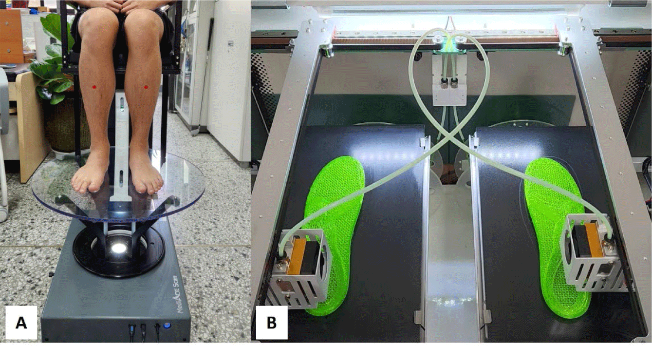
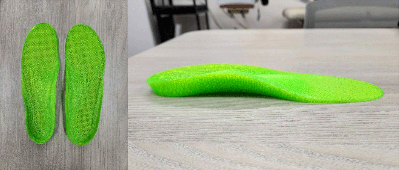
The design of the 3DPIO using a CAD program was produced and personalized by reflecting each participant’s individual foot shape model, the characteristics of the foot orthosis application, and the correction content. Mash smoothing process was executed on the orthosis sole contact surface, and the heel side based on the scanned foot and plantar contour. The boundary of the 3DPIO was extracted from the 3D scanned foot shape silhouette for the pattern layout. An insole bottom thickness was designed to be 3 mm, the usual thickness of general insole products.19 Moreover, we designed the insole by identifying the individual heel cup and arch support area, and added 10mm heel raise and 3mm arch support to printed insole orthoses. The completed insole models designed through CAD program were configurated to the 3D printing software (Simplify3D V4.0.1, Cincinnati, OH, USA).
According to the final insole CAD file completion, the 3DPIO was designed using a fused deposition modeling 3D printer (iSun3D Flx2, eSUN Industrial Co., Shenzhen, China) (Figure 1B). It has two printing flatform stations with a capability of printing two insole orthoses simultaneously, and is composed of a 3.5-inch HD touchscreen and a 0.8 mm diameter nozzle. The 3D printer device has a high-speed flexible technology with a maximum printing speed of 120 mm/s. The printing filament for making the insole orthosis was thermoplastic polyurethane (TPU) material (TPU-flexible 95A, Cubicon Co., Sungnam, Korea). The average filament injection temperature was 235°C, and the insole orthosis production time took an average of 45 minutes.
Two force platforms (BP400600, AMTI, USA) embedded in the middle walkway were used to obtain 3D GRF data during gait in different insole orthosis conditions: general shoe insole (GSI), prefabricated insole orthosis (PIO), and customized 3DPIO (Figure 2). The GSI was included in the general shoes (S260, Supercomet Co., Gimhae, Korea) provided according to each subject’s foot size. The PIO (TPS, BioMechanics Co., Goyang, Korea) was a foot insole orthosis to correct flatfoot deformity sold on the public market. A Vicon motion analysis device (Vicon Inc., Oxford, UK) with six cameras (model T10) was used to capture precise stance phase points of gait cycle such as heel contact or toe off. A Nexus 1.8.5 software (Vicon Inc., Oxford, UK) was used to process the ankle kinetic data and gait kinematic variables in 3D space. A 750 mm T-figure wand was used to calibrate the Vicon system and the force platforms, and a calibration reference object was used to identify the laboratory origin. The sampling rate of the motion capture system was set at 100 Hz, and the sampling rate of the force measuring plates was 500 Hz. The kinetic GRF data were processed using Nexus 1.8.5 software (Vicon Inc., Oxford, England) and exported as c3d files for final analysis. The c3d file data processed by Nexus software were sent in Visual3D motion analysis software (C-Motion, Rockville, MD, USA) for final processes and statistical analysis. The Visual3D program generated virtual lower joints and segments according to attached reflective markers to calculate the peak 3D ankle moment data during stance phase of free gait (Figure 3).
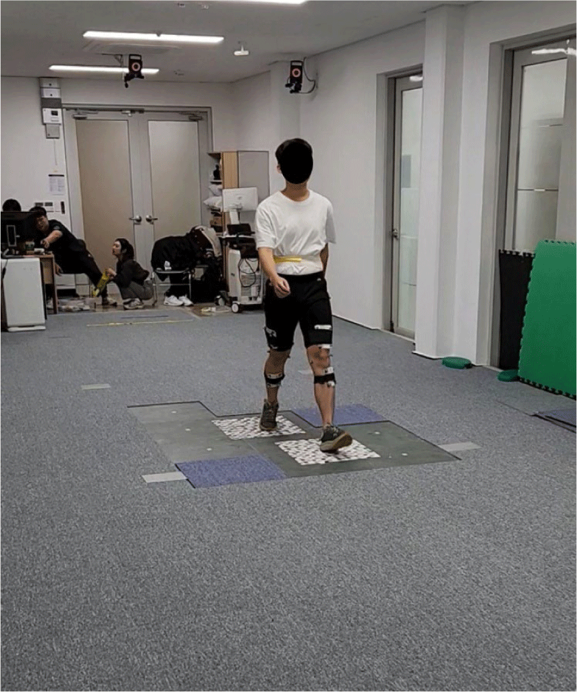
The participants wore 40 reflective markers (14 mm) with four-marker clusters attached to the participants’ both anatomical landmarks of the lower extremity joints and the segments according to the six-degrees-of-freedom (6DOF) model.20 To analyze the 3D peak ankle moment data during gait, three different insole orthosis conditions were applied to shoes, which are prepared according to the size of each participant’s feet. Under the one orthosis condition, walking experiments were performed without rest between each gait trial, except in unexpected situations such as not stepping on the force platforms. The participants were asked to walk a 10 m walkway at a free walking speed for a total of 8-10 repetitions. The experimental order of each insole orthosis condition was randomly assigned.
Descriptive analysis was executed to confirm general characteristics of the participants, as well as moment mean and standard deviation of the peak ankle moments. All ankle moments were normalized by participants’ weight. The Kolmogorov–Smirnov test was used to verify normal distribution and all analysis variables satisfied normal distribution. Two-way repeated-measures analysis of variance (ANOVA) with Bonferroni adjustment was used to confirm 3D kinetic ankle moment values for three different insole orthotic conditions and foot sides during free walking. If the main effect (orthosis intervention or foot side) was confirmed, post-hoc test was used to investigate the pairwise comparison according to the ANOVA results. Statistical significance was determined at an α level of 0.05. All analyses were executed using SPSS version 26.0 (IBM Corp., Armonk, NY, USA).
RESULTS
All peak ankle moment variables obtained for repeated measures ANOVA analysis satisfied the normal distribution and Mauchly’s assumption of sphericity. Table 1 showed the general characteristics of the participants and the means and standard deviations of typical gait parameters. There were no significant differences in walking speed, step length, and step width among the insole orthoses walking conditions (all p>0.05) (Table 1). The participants’ average FPI-6 score and plantar arch index were 7.33±1.19 and 0.68±0.20, respectively.
There were significant differences in the peak dorsiflexion moment (F=6.208, p=0.017), the plantar flexion moment (F=9.520, p=0.004), and the first peak eversion moment (F=12.662, p=0.001) that developed in the stance phase of gait according to the insole orthotic conditions (Table 2). There were no interaction effects between foot sides and insole orthotic conditions for any moment variables of the ankle joints (p>0.05) (Table 2). As a result of pairwise comparisons of peak ankle moments, the peak ankle dorsiflexion moment was significantly greater in the 3DPIO condition than in the GSI condition (p=0.017) (Table 3). Moreover, the peak ankle plantar flexion moments were significantly greater in the 3DPIO condition than in the GSI and PIO conditions (3DPIO compared to GSI, p=0.003; 3DPIO compared to PIO, p=0.009) (Table 3). The first peak eversion moments were significantly greater in GSI walking condition than in the PIO and 3DPIO conditions (GSI compared to PIO, p=0.002; GSI compared to 3DPIO, p<0.001) (Table 3). However, no significant differences were found in the second peak eversion moment and the peak internal/external rotation moment among the three different orthotic walking conditions (p>0.05) (Table 3).
DISCUSSION
Previous studies have reported that insole orthoses used to manage PHD are largely mass-produced in the medical market orthoses for PHD have some clinical inconveniences because they are not custom-made to the individual user’s feet, leading to discomfort and pain in the foot and ankle region.21 To overcome these discomforts, customized foot insole orthoses utilizing 3D scanners or 3D printing technologies have recently been developed to manage various foot deformities.22-24 They suggested that the 3D-printed foot insole orthoses offer superior fit and comfort of the foot and planar region, making them a promising clinical alternative to conventional insole orthotics. Therefore, clinical interventions using customized 3PIO based on 3D scanner acquisition of foot and plantar silhouette shape are necessary to address the disadvantages of previous prefabricated insole orthoses to support foot dysfunction, improve gait efficiency, and prevent various complications that may develop in the foot and ankle joints and segments.
The results of this study showed significant differences in some peak ankle moment variables during gait between the three insole orthosis conditions. This means that the 3D peak ankle moments during gait differs depending on the type of insole orthosis applied to individuals with PHD in this study. When the customized 3DPIO condition was applied compared to the other two insole conditions, a significant greater peak moment was confirmed in the ankle plantar flexion that occurred in frontal plane. A previous study was conducted to verify biomechanical variables during walking using personalized shoes and insole orthosis in 11 patients with pes planus and pronated feet.25 Contrary to the results of this study, they reported a significantly reduced plantar flexion moment in the walking with shoes and insole orthotic condition compared to the walking with barefoot condition. The reason why the results in this study were contradictory to the peak plantar flexion moment results can be considered as follows. First, the insole orthosis applied in this study was not a prefabricated orthosis manufactured by a conventional orthotic company, but a personalized insole orthosis using 3D scanner technology and a 3D printer device. Second, there was a difference in the walking experiment method because barefoot walking was not performed in the walking conditions of this study. Therefore, the results of previous studies are not considered to be sufficient to offset the plantar flexion moment results of this study. In a previous study supporting these claims, Lin et al.26 used 3D scanning and 3D printing to evaluate the mechanical properties of a 3D-printed foot orthosis and determine its biomechanical effects on individuals with flexible flat feet. They reported a significant increase 3% in the plantar flexion moment under 3D-printed foot orthoses walking condition compared to normal shoe walking condition. This was similar to the experimental conditions of this study in terms of research materials and walking comparison conditions, and the plantar flexion moment results were also the same. Compared to other conditions, the increase in ankle plantar flexion moment in the 3DPIO condition may provide a kinetic advantage to push off the ground in the final phase of stance during gait, and is expected to contribute to acceleration in the pre-swing phase and increased gait efficiency.
In the 3DPIO and PIO gait conditions, a significant decrease in the first peak ankle eversion moment was observed compared to the GSI condition. The first peak eversion moments occurred in the mid-stance phase, and the significant decreases observed in both orthosis conditions suggest that both orthoses positively impacted pronated foot correction and medial longitudinal arch support. Many previous studies support these results, a study reported that a significant 35% reduction in peak eversion moment when 3D printed foot orthoses were applied to patients with flexible flat feet compared to walking in regular shoes.26 Another study also reported a significant decrease in ankle eversion moment during walking in the arch support insole and arch support insole with cushion pads conditions compared to the general insole condition.27 The decrease in peak ankle eversion moment values is thought to have a positive effect on the reduction of the posture and alignment of the ankle and foot joints and segments in the frontal plane by providing sufficient arch support of the 3DPIO and PIO conditions. Therefore, it is thought that personalized insole orthoses such as 3DPIO based on high technology have a positive effect of an increased gait efficiency by affecting peak dorsiflexion, peak plantar flexion, and peak first eversion moments generated at the ankle joint compared to general insoles, and can limit the occurrence of secondary musculoskeletal pain and clinical symptoms of ankle osteoarthritis.
The strength of this study is that it analyzed the peak 3D kinetic moment values that occur in the stance phase during walking using two force platforms and a 3D motion analysis system, which is recognized as an objective and reliable evaluation technology in the field of biomechanics, to verify the clinical effectiveness of a personalized 3DPIO for hyper-pronated individuals fabricated using a 3D scanner and 3D printing equipment. To our knowledge, this study is the first experimental trial to investigate 3D peak ankle moments developed under the application of the 3DPIO, PIO, and GSI walking conditions in individuals with pronated flatfeet. The results showed that the application of the 3DPIO increased gait efficiency by improving ankle moments (dorsiflexion or plantar flexion) developed in the sagittal motion plane compared to other orthotic conditions. These results suggest that the clinical strengths of 3DPIO as follows. First, the technology for obtaining accurate foot models using 3D scanners enabled precise digitization of the physical characteristics of individual feet and soles. Obtaining such foot models using a 3D scanner reduces evaluation time and allows for easy and reliable acquisition of the patient’s foot shape. Second, using CAD software to create 3DPIOs enhances the quality of personalized orthoses before the final 3D-printed insole orthoses. CAD software allows for precise modifications and reinforcements to the insole orthosis, tailored to each patient’s foot deformity. The limitations of this study are as follows. The participants are generally healthy individuals with no musculoskeletal problems such as walking discomfort or pain in the foot and ankle joints and segments. Thus, it is difficult to apply and generalize the study results to all individuals with PHD. In addition, since the materials of the three different insole orthotic conditions used in the experiment were not identical, the influence of various physical factors such as the weight and elasticity of the orthoses could not be confirmed. Therefore, further studies are needed under a research design that can overcome our study limitations.
CONCLUSIONS
The clinical intervention of the customized 3DPIO to individuals with PHD has significant effects on the peak ankle plantar flexion moment occurred in the stance phase of gait compared to the prefabricated foot insole and general shoe walking conditions. The 3DPIO, designed and fabricated based on 3D scanning and 3D printing technologies for the customization of individual foot conditions, can overcome the previous prefabricated foot orthoses. Abnormal increased or decreased ankle moments occurred during gait may cause to osteoarthritis of the ankle joints and foot segment deformities such as pronated foot, pes planus, or hallux valgus. Therefore, insole orthotic interventions, such as the 3DPIO introduced in this study, can be considered as an effective method to manage and prevent musculoskeletal problems in individuals with PHD.