INTRODUCTION
More than 85% of individuals experience hemiplegia after a stroke, and 55-75% of stroke survivors experience permanent disabilities such as motor function impairments.1 Hemiplegic symptoms lead to difficulties in postural control and balance.2,3 Independent gait after a stroke becomes a crucial element for performing activities of daily living. Previous studies indicate that sensory-motor dysfunction leads to restrictions in muscle and joint range of motion, causing difficulties in functional activities such as sitting and standing, and gait due to lower limb weakness.4,5,6 Stroke patients experience motor function impairments such as instability due to decreased sensation and loss of muscle, as well as reduced gait ability.7 These experiences induce asymmetric postures and result in the loss of the ability to shift the center of gravity within the base of support, leading to problems with balance maintenance and gait performance, further negatively influencing the performance of daily living activities.8,9 One of the commonly observed functional movement abilities in daily life is sitting down and standing up.10 It has been reported that repetitive standing exercises can enhance the function of stroke patients.11
Restrictions in the ankle joint of stroke patients ne-gatively impact functional activity performance.12 When joint mobilization was applied to the ankles of elderly adults, there was an increase in ankle range of motion. This improvement in ankle range of motion can resolve functional difficulties caused by sensory loss. Furthermore, it has been suggested that an adequate range of motion in the ankle joint is necessary for the performance of functional movements.13 Comparing muscle activity and mechanical load based on ankle joint angles during the sit-to-stand, proper movement of the ankle joint and muscle contractions are essential for improving gait stability and efficiency.14 Functional gait requires interventions to improve both active and passive joint range of motion. Mobilization with Movement (MWM) has been proposed by Mulligan.15 There are studies reporting that improving the dorsiflexion range of motion in stroke patients is effective for enhancing gait speed. This suggests that the reduction in muscle tone associated with a rigid ankle positively affects gait speed and stride length.16,17
So far, numerous prior studies have presented improve-ments in static and dynamic balance due to enhanced dorsiflexion range of motion of the ankle joint using MWM and stretching in stroke patients. 18,19 Changes in the performance of the Five Time Sit to Stand (FTSTS) test showed statistically significant differences in the Timed Up and Go (TUG) test and the 10 Meter Walking Test (10MWT). This indicates that sit-to-stand ability affects dynamic balance and gait and further confirms that interventions focusing on sit-to-stand can have a positive impact on dynamic balance, gait speed, and quality of life.20 It has also been reported that significant differences exist in muscle activity of the rectus femoris and gastrocnemius during sit-to-stand training associated with improved dorsiflexion range of motion of the ankle joint.21 However, prior research is considered insufficient in investigating the impact of sit-to-stand training based on the dorsiflexion range of motion of the paralyzed side ankle on functional movements, such as gait ability due to imbalance between the non-paralyzed and paralyzed legs in patients with hemiplegia due to stroke. This study hypothesized that sit-to-stand training using MWM would be more effective than self-stretching-based training in improving lower extremity function, balance, and gait in patients with chronic stroke. Therefore, this study aims to examine the effects on lower limb function, balance, and gait performance when applying MWM and passive stretching to improve the dorsiflexion range of motion of the ankle joint during functional movements such as sit-to-stand.
METHODS
This study selected 36 patients diagnosed with stroke and hospitalized at Hospital, located in Daegu city, from August 2024 to September 2024, who met the selection criteria. All participants were informed about the purpose and content of the study, and voluntary consent was obtained. The study was conducted after obtaining approval from the Daegu University Institutional Review Board (Approval Number: 1040621-202407-HR-058).
After explaining the purpose and content of the study in advance, general characteristics such as gender, age, height, and weight were investigated in the patients who provided voluntary consent. Participants recruited for the study were randomly assigned to two groups using a lottery method. However, due to the discharge of 2 participants during the study, a total of 34 participants were divided into an experimental group (Sit-to-stand using MWM group, SUG) of 17 members and a control group (Passive stretching group, PSG) of 17 members to complete the study.
All groups underwent twice-daily gait training, twice-daily functional electrical stimulation therapy, twice-daily occupational therapy, and twice-daily Neuro-Developmental Treatment (NDT). Changes in lower limb function, balance and gait performance were measured for all participants before the experiment and after 4 weeks of intervention.
Before conducting the experiment, the study details were accurately communicated, and consent was obtained through signatures. The general characteristics of the participants, including gender, age, weight, height, scores from the Korean Version of the Mini-Mental State Examination, time since onset, type of stroke, and affected side were measured (Table 1).
The participants were randomly assigned to either the experimental group (SUG) or the control group (PSG). All participants underwent assessments of lower limb function, balance ability, and changes in gait ability before and after the intervention, which included range of motion, the five-times sit-to-stand test, and the 10-meter walk test.
Before starting the treatment, participants were briefly informed about the purpose, content, and methods of the treatment. The treatments were performed by skilled physical therapists with over four years of clinical experience who had completed training in NDT. The therapist demonstrated the treatment methods and procedures to ensure proper implementation. To ensure patient safety and prevent falls, treatments were conducted with the presence of the therapist, and both the experimental and control groups received treatment in the same environment under the therapist's assistance and supervision. If participants reported fatigue during treatment, they were provided with a break. In cases of dizziness, shortness of breath, pain, complaints, or refusal of treatment, the exercise was immediately discontinued.
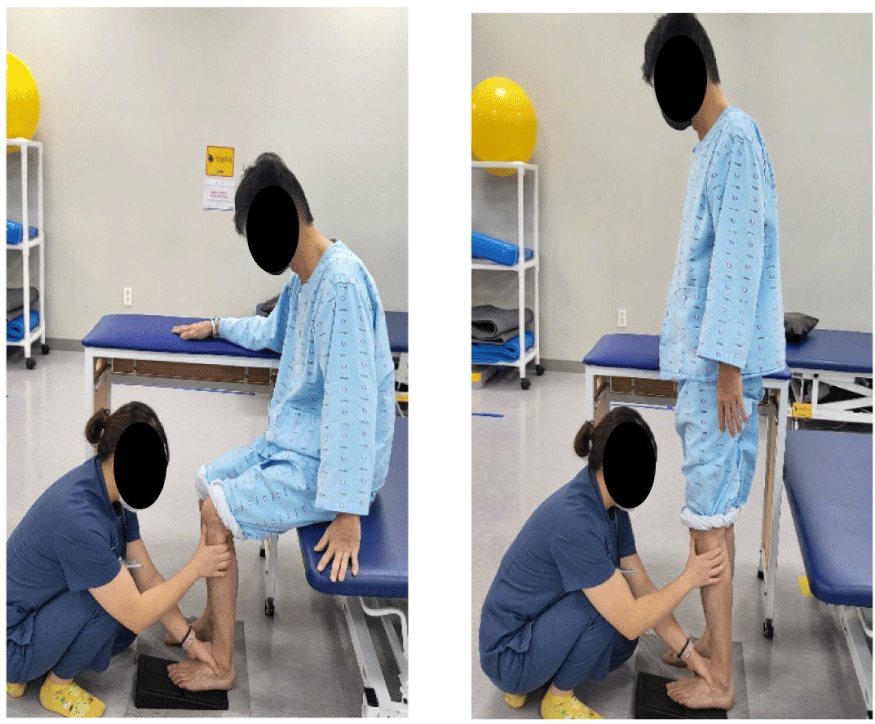
During the performance of the sit-to-stand movement, training will be conducted sit-to-stand exercises combined with MWM for the ankle joint's dorsiflexion. The starting position was set with the patient's affected foot positioned behind the non-affected foot on an ankle ramp at a chair height where the knees are flexed at 90 degrees. While performing the sit-to-stand movement with the verbal instruction from the therapist to "stand up comfortably," the therapist conducts mobilization with movement for the dorsiflexion of the ankle joint by sliding the talus while keeping the heel bone fixed. The sliding direction of the talus was performed in a posterior-inferior direction.
To prevent risks such as falls, a non-slip pad was placed under the ankle ramp, as shown in Figure 1, and a support table was provided on the unaffected side during treatment. After practicing dorsiflexion through MWM for about 5 minutes in a standing position, the sit-to-stand combined with MWM training was carried out for 4 weeks, three times a week, with 15 repetitions in 3 sets (Figure 2).

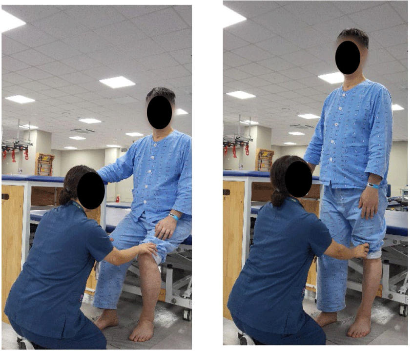
After performing passive stretching for dorsiflexion of the ankle joint using an ankle ramp, sit-to-stand training was conducted (Figure 1). The starting position was set with the affected foot positioned behind the non-affected foot at a chair height where the knees are flexed at 90 degrees. The sit-to-stand movement was performed with the verbal instruction from the therapist to "stand up comfortably." To prevent risks such as falls, a non-slip pad was placed under the ankle ramp, as shown in Figure 2, and a support table was provided on the unaffected side during treatment (Figure 2).
The participants placed the ankle ramp under the affected foot and performed passive stretching for dorsiflexion of the ankle in a standing position. After approximately 5 minutes of passive stretching for dorsiflexion using the ankle ramp in a standing position, sit-to-stand training was conducted for 4 weeks, three times a week, with 15 repetitions in 3 sets (Figure 3).

All participants were guided to participate in the experi-ment according to their respective exercise programs thro-ughout the study period. Dorsiflexion range of motion of the ankle joint, the FTSTS and 10MWT were measured on the first day before starting the exercise and again on the last day, four weeks after the exercise began, using the same examiner and the same testing methods.
An electronic goniometer was used to measure the dorsiflexion range of motion. The FTSTS measures the time taken for the participant to stand up and sit down five times without using their arms, while seated in a chair with a backrest and no armrests, with their arms crossed over their chest. The timing began when the participant's back moved forward from the backrest and ended when the back made contact with the backrest again.22 The upright posture was defined as having the torso straight and the knee and hip joints fully extended. Before the test, the examiner demonstrated the FTSTS test once to the participant to teach them the correct procedure. The 10MWT was conducted to assess walking speed. During the total distance of 14 meters, the time taken to traverse the 10-meter section, excluding the predefined 2-meter acceleration and deceleration zones at the start and finish points, was measured using a stopwatch. The measurement of walking speed over the 10-meter distance has been reported to have high intra-tester and inter-tester reliability (r = .89 to 1.00).23
All statistical analyses were conducted using SPSS version 23.0 for Windows. In this study, the data processing of the participants' general characteristics utilized the chi-square test for categorical variables and the independent T-test for continuous variables. Normality was assessed using the Shapiro-Wilk test to confirm the normal distribution. A two-way ANOVA was used to examine the interactions between the two groups. A paired t-test was conducted for within-group com-parisons. The statistical significance level (α) was set at 0.05.
RESULTS
There were statistically significant differences in the dorsiflexion AROM, the FTSTS and 10MWT of the SUG before and after the intervention (p < 0.05) (Table 2). In the dorsiflexion AROM of the PSG, there were no statistically significant differences before and after the intervention (p > 0.05) (Table 2). However, the FTSTS and 10MWT of the PSG showed statistically significant differences before and after the intervention (p<0.05) (Table 2). However, the interaction values for dorsiflexion AROM, the FTSTS and 10MWT between the groups showed no statistically significant differences (p>0.05) (Tabel 3, 4 and 5).
| Dorsi flexion AROM (unit: angle) | Pre (M±SD) | Post (M±SD) | Group F(p) | Time F(p) | Group×Time F(p) |
|---|---|---|---|---|---|
| SUG | 6.0±7.21 | 11.58±6.69 | 4.014(0.054) | 7.393(0.010*) | 0.256(0.616) |
| PSG | 6.12±5.33 | 10.24±5.87 |
| FTSTS (unit: number) | Pre (M±SD) | Post (M±SD) | Group F(p) | Time F(p) | Group×time F(p) |
|---|---|---|---|---|---|
| SUG | 22.93±5.32 | 15.75±5.36 | 7.525(0.010*) | 20,173(0.000*) | 1.528(0.225) |
| PSG | 22.84±4.76 | 18.46±4.91 |
| 10MWT (unit: sec) | Pre (M±SD) | Post (M±SD) | Group F(p) | Time F(p) | Group×time F(p) |
|---|---|---|---|---|---|
| SUG | 31.22±24.58 | 25.64±19.64 | 6.440(0.016*) | 0.468(0.001*) | 0.540(0.468) |
| PSG | 31.05±19.64 | 27.09±17.17 |
DISCUSSION
This study aimed to demonstrate that improving the range of motion of the ankle joint is effective for performing functional movements such as sit-to-stand and gait, and to present an accessible approach for patients experiencing difficulties with balance and gait. A total of 34 participants with chronic stroke lasting more than six months were divided into two groups: the SUG (17 participants) who underwent sit-to-stand exercises combined with MWM, and the PSG (17 participants) who underwent passive ankle stretching followed by sit-to-stand exercises. Both groups received treatment three times a week for four weeks, performing three sets of 15 repetitions, and the changes in lower limb function, balance, and gait performance were assessed. The range of motion of the ankle joint was evaluated, and the FTSTS and the 10MWT were conducted.
In this study, the active range of motion was measured to evaluate dorsiflexion of the ankle joint. Significant effects were observed in the active range of motion of the SUG before and after the intervention (p<0.05). Alamer et al.24 stated that utilizing mobilization with movement improves the range of motion of the joint by enhancing the accessory movement of the talus through posterior gliding. In a study by Cho and Park,18 it was reported that joint mobilization was more effective than stretching in improving the range of motion of the ankle joint in stroke patients. A study by Ahn found that applying MWM at the talocrural joint, while the weight was shifted onto the affected limb, stretched the plantar flexors limiting active dorsiflexion, and that anterior gliding of the tibia by the therapist improved passive range of motion. 25 Furthermore, it was reported that the improvement in the strength of the ankle joint increased the active range of motion. These findings are consistent with the results of this study. However, there were no significant effects between the groups (p > 0.05). This may be attributed to the short intervention duration of 4 weeks, which was insufficient to mitigate the limitations in range of motion resulting from sensory-motor dysfunction following a stroke.4
To assess balance ability, the FTSTS was conducted, showing significant effects before and after the intervention in both the SUG and PSG (p < 0.05). Limitations in the range of motion of the ankle joint can lead to balance and gait disabilities.26 There is a relationship between balance and ankle joint range of motion, and improvements in the range of motion can enhance balance ability.27,28 Additionally, with increased spasticity, the movement of the foot decreases, leading to difficulties in maintaining balance due to reduced adaptability of the foot to the ground.29 In a study by Kwag et al.,30 mobilization with movement of the ankle joint was shown to relax plantar fascia stiffness and increase the range of motion of the ankle, thereby enhancing the foot's adaptability to the ground and improving balance ability. This supports the findings of the current study. No significant differences between the groups were observed (p > 0.05).
To assess gait performance, 10MWT was conducted. Significant effects were observed before and after the intervention in both the experimental and control groups (p < 0.05). There were no significant effects between the groups (p > 0.05). In a study by Cho,18 it was reported that improvements in ankle joint range of motion were more effective in enhancing spatiotemporal gait variables, such as walking speed and steps per minute. This report supports the findings of the current study. However, there were no significant differences between the experimental and control groups regarding interactions in the three evaluations (p > 0.05). This may be due to the fact that, unlike the study by An and Won31 which applied MWM using a non-elastic treatment belt, the intervention in this study involved the manual application of MWM by the therapist, which did not clearly define the intensity and direction of the intervention. As a result, this may not have been sufficient to derive insights into the interaction.
It is thought that the 4-week intervention period was in-sufficient to confirm interactions. Future research will re-quire longer-term interventions and follow-up tests to assess long-term effects and retention capabilities. During the experiment, it was not possible to control the participants' daily activities, and since they received conventional inpatient treatment, such as NDT, it could not be completely ruled out that daily life and other treatments influenced the range of motion of the ankle joint, lower limb function, balance, and gait performance. Future studies should address these limitations to enhance rehabilitation treatment for stroke patients and to facilitate diverse and ongoing research aimed at improving quality of life and recovery in daily activities.
CONCLUSIONS
This study applied the intervention of sit-to-stand exer-cises combined with MWM without clearly defining the intensity and direction, which hindered the ability to determine the interactions between groups. If the limitations of this study are addressed, it can be suggested that sit-to-stand exercises combined with MWM may have a positive impact on lower limb function, balance, and gait in stroke patients, as significant differences within the groups were observed. In the future, the application of MWM during functional movements such as sit-to-stand can be proposed as part of rehabilitation treatment programs for lower limb function, balance, and gait performance in stroke patients.


