


INTRODUCTION
Non-specific low back pain (NSLBP) is a musculoskeletal disorder that affects a large number of individuals worldwide.1-4 This condition extends from the lowest rib to the gluteal fold and may also affect both thighs and the area above the knees.2,3 For individuals with NSLBP, exercise therapy is easier, safer, and more cost-effective than surgical treatment. Exercise interventions for low back pain include core stabilization training, bridge exercises, sling exercises, and stretching.5 In particular, the bridge exercise not only strengthens the gluteus maximus (GM), which contributes to lumbar and pelvic stability, but also trains the coordinated activation of global and local trunk muscles.? This exercise improves the function of the lumbo-pelvic-hip complex and enhances coordination among the muscles surrounding the spine and hip joints, thereby reducing the risk of spinal injury.7 As a closed kinetic chain exercise, it also helps improve joint proprioception and stability.8 A previous study comparing nine exercises, including bridge, plank, side plank, and lunge, reported that the bridge exercise could improve endurance, stability, and activation of the GM and multifidus (MF) without external load.9 In particular, the bridge exercise has been shown to increase the activation of the GM, MF, and biceps femoris (BF) muscles.10,11 In addition, it enhances pelvic mobility and promotes smooth interaction among the spinal stabilizers, thereby preventing spinal injury,10 while also increasing joint stability and the sensitivity of proprioceptive feedback.8
The GM contributes to the stability of the sacroiliac joint and plays an important role in hip stabilization and extension movements.12 The MF functions to stabilize the trunk when it is in a neutral position.13 In addition, the BF is the only muscle that prevents anterior pelvic tilt during closed kinetic chain exercises.14 According to previous studies, weakness of the GM can reduce lumbar and pelvic stability, potentially leading to low back pain.15,16 Such pain occurs in individuals with low back pain due to insufficient contraction of the GM. As a result, the BF contracts more rapidly and becomes excessively activated compared to the GM.17,18 This phenomenon can impair intermuscular coordination and lead to functional deficits.19 In such cases, activation of the muscles surrounding the spine and hip joints may contribute not only to single-leg stance performance but also to functional abilities such as balance control.20
Another important factor to consider when performing the bridge exercise is joint position and muscle fiber orientation. Muscle activation increases when the direction of movement aligns with the orientation of the muscle fibers.21 A previous study examined the effects of varying hip abduction angles 0°, 15°, and 30° during the bridge exercise and found that GM activation was significantly higher at 30° compared to 0° and 15°.11 In addition, the activation ratio between the GM and hamstring muscles also increased significantly.11 Therefore, when the goal is selective strengthening of the GM, setting the hip abduction angle to 30° is recommended.11 Another study investigated the effect of knee angle on muscle activation during the bridge exercise.22 The results showed that performing the bridge exercise with a 90° knee angle led to greater activation of the GM compared to 40° and 60°, and the activation ratio between the GM and BF was also increased. 22 A separate study compared GM activation during the bridge exercise according to different pelvic tilt angles.23 Performing the bridge exercise with a 5° posterior pelvic tilt resulted in significantly greater activation of the GM than with a neutral or 5° anterior pelvic tilt.23
Heart rate variability (HRV) is a non-invasive indicator used to assess the autonomic regulation of the heart.24 It refers to the physiological responses in the intervals between heartbeats.25 HRV can be positively influenced by high-intensity intermittent resistance exercise, particularly in balancing sympathetic and parasympathetic nervous system activity.26 The overall autonomic nervous system regulation can be evaluated through the standard deviation of normal-to-normal intervals.27 A high resting heart rate is associated with an increased risk of mortality from cardiovascular disease and sudden cardiac death due to myocardial infarction, even in healthy individuals.28 Conversely, failure to reach a low maximal heart rate or a certain percentage of the predicted maximal heart rate is also linked to a higher risk of cardiovascular-related mortality.28 Therefore, adjusting resistance training sessions based on cardiac autonomic regulation may enhance training adaptation and performance, and also help prevent injury and overtraining.29 Resistance training can reduce cardiac vagal modulation for 12 to 48 hours after exercise, and the magnitude and duration of this acute response may vary depending on training variables such as load intensity. Therefore, assessing cardiac autonomic regulation using HRV during resistance exercises such as squats and shoulder presses may provide valuable insights into autonomic function and exercise-induced stress.30
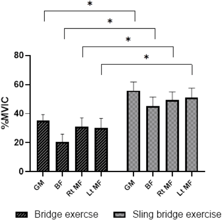
Sling exercises are a type of closed kinetic chain training commonly used in clinical settings and are effective in activating stabilizing muscles such as the transversus abdominis, rectus abdominis, and pelvic floor muscles.31 In addition, performing exercises targeting specific muscles under unstable conditions contributes to the activation of core muscles and the improvement of intermuscular balance.? One study investigated the effects of sling bridge exercises by positioning the sling at the knees and adjusting the angle between the strap and the ground to 10°, 20°, and 30°. The results showed that muscle activation of the GM, erector spinae, and MF was significantly higher at 30° compared to 10° and 20° (Figure 1).31
Compared to traditional bridge exercises, sling bridge exercises reduce the base of support, requiring greater balance ability to maintain postural stability and thereby increasing the level of difficulty. In such an unstable support environment, the appropriate use of body weight to sustain posture further enhances the activation of the muscles involved in the exercise.32 Therefore, performing bridge exercises using a sling is recommended to provide a higher level of difficulty and to maximize muscle activation.
An individual’s perception of physical activity intensity during exercise is important. Therefore, applying the same exercise intensity to everyone is not recommended, as it may lead to compensatory movements and muscular imbalances. To prevent such issues, the rating of perceived exertion (RPE) is widely used as a tool to assess an individual’s subjective perception of exercise intensity based on their physical condition.33 A previous study compared the effects of three different types of prone hip extension exercises on hip and lumbar muscle activation and pelvic compensation.34 The exercises included: basic prone table hip extension (PTHE), PTHE with abdominal drawing-in (PTHEA), and PTHEA performed on a chair while flexing the contralateral knee (PTHEAC).34 The results showed that the PTHEAC was the most effective in activating the GM, without causing excessive activation of the BF or semitendinosus, or inducing lumbopelvic compensation. In addition, the PTHEAC recorded the lowest RPE among the three exercises.34 Therefore, exercises that effectively activate the target muscles without significantly increasing perceived exertion may reduce the burden of exercise performance in individuals with NSLBP and contribute to minimizing compensatory movements, thereby enhancing the effectiveness of the exercise.
To date, no studies have compared traditional bridge exercises and sling bridge exercises in individuals with NSLBP. Therefore, the purpose of this study was to investigate the effects of performing traditional bridge and sling bridge exercises based on previous studies on bridge exercises by setting the hip abduction angle to 30°, knee flexion angle to 90°, and posterior pelvic tilt to 5°, on the activation of the GM, BF, and MF, as well as on the activation ratios of GM to BF and MF to BF.11,22,33 In addition, this study aimed to measure changes in RPE and HRV while performing the two exercises. It was hypothesized that sling bridge exercise would result in significantly greater activation of the GM, BF, and MF compared to traditional bridge exercise. However, it was also hypothesized that the activation ratios of GM to BF and MF to BF would be significantly higher during traditional bridge exercise than during sling bridge exercise. Furthermore, it was assumed that both heart rate and perceived exertion would be significantly higher during sling bridge exercise than during traditional bridge exercise.
METHODS
The sample size was calculated based on the results of a pilot study involving five participants, using G*Power software (version 3.1.2; Franz Faul, University of Kiel, Kiel, Germany). The power analysis was performed with a power of 0.95, an alpha level of 0.05, and an effect size of 3.44. The analysis indicated that a minimum of four participants was required. To compensate for potential dropouts, the final sample size was set at 20 participants.11-23,31 Accordingly, twenty male participants with non-specific low back pain (NSLBP) were voluntarily recruited for this study. The participants had a mean age of 24.2 ± 1.9 years, a mean height of 175.7 ± 3.8 cm, a mean weight of 67.9 ± 4 kg, and a mean visual analog scale (VAS) score of 2.5 ± 0.5 (Table 1). Participants were selected based on the following criteria: 1) no specific disorders of the hip or knee joints; 2) a history of visiting a hospital for low back pain within the past 12 months,35 3) experience of low back pain symptoms for more than one week before the start of the study,35 and 4) intermittent mild NSLBP (VAS ≤ 3). Exclusion criteria were as follows: 1) a history of disease in the lower back, pelvis, or legs,35 2) pain in specific areas during leg movement,36 3) neurological symptoms such as numbness during exercise, 4) musculoskeletal problems that cause pain during exercise, 5) diagnosis of femoroacetabular impingement syndrome; 6) restricted joint range of motion in the prone position (external rotation < 45°, internal rotation < 45°),37,38 and 7) VAS score greater than 3 for low back pain. The study procedures were thoroughly explained to all participants, and written informed consent was obtained before the experiment. This study was approved by the Institutional Review Board of Hoseo University [1041231-240708-HR-180].
| Parameter | Group (N = 20) |
|---|---|
| Age (years) | 23.9 ± 1.80 |
| Height (cm) | 176.45 ± 3.84 |
| Weight (kg) | 68.65 ± 4.89 |
| VAS | 2.45 ± 0.51 |
To measure the muscle activity of the GM, BF, and MF, an electromyography (EMG) system (Ultium EMG system, Noraxon, USA) and its dedicated software were used. The device settings were configured with a band-pass filter of 20–450 Hz, a notch filter at 60 Hz, and a sampling rate of 1,024 Hz. To improve signal accuracy, the skin was shaved and cleaned with an alcohol pad before electrode placement to minimize skin resistance. The surface electrodes were placed horizontally in alignment with the direction of the muscle fibers, with an inter-electrode distance of 2 cm. Electrode placement sites were determined based on a previous study.39 The electrode for the GM was placed at the midpoint of the diagonal line connecting the sacrum and the greater trochanter of the femur. The electrode for the MF was positioned 2 cm lateral to the line connecting the posterior superior iliac spine and the spinous process of L5. The electrode for the BF was attached 2 cm lateral to the point located at two-thirds of the line between the greater trochanter and the posterior aspect of the knee.39 The GM and BF were measured on the dominant side (all participants: right side), while the MF was measured bilaterally. Before starting the experiment, maximum voluntary isometric contraction (MVIC) was measured to standardize muscle activation data. Subsequently, the muscle activity of the GM, BF, and MF was normalized using %MVIC values. The postures for MVIC measurements of each muscle were determined according to the guidelines by Kendall et al.40 For the GM, the participant lay prone on a table with the hip abducted at 30° and the knee flexed at 90°, and then performed maximal hip extension on the dominant side. During the measurement, the examiner stabilized the participant’s PSIS and applied maximal resistance in the direction of hip flexion at the posterior aspect of the knee. For the MF, the participant lay prone on a height-adjustable table with the trunk fully extended. For the BF, the participant lay prone with the knee flexed at 90° and performed maximal knee flexion on the dominant side. The examiner stabilized the PSIS and applied maximal resistance in the direction of knee extension at the posterior aspect of the ankle.
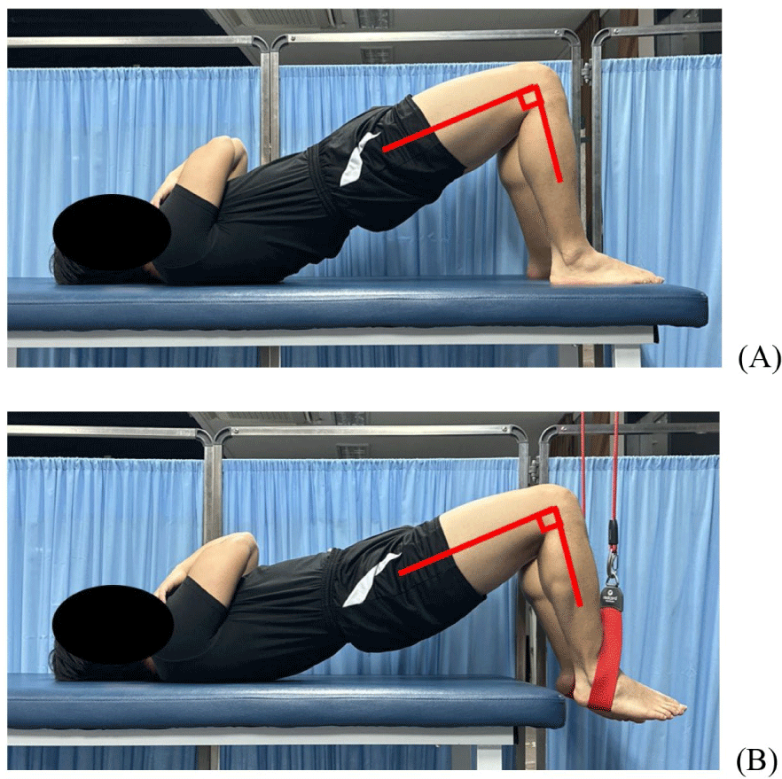
Participants were given a 10-minute practice session to become familiar with the bridge exercise. The bridge exercises included a traditional bridge exercise and a sling bridge exercise, and the order of the exercises was randomized using Microsoft Excel (Microsoft, Redmond, WA, USA). To maintain consistent exercise timing, a metronome set at 60 beats per minute was used. A 10-minute rest period was provided between each exercise session to reduce muscle fatigue, and additional rest was allowed if the participant reported muscle fatigue.23 According to the exercise protocol, heart rate was systematically measured. First, participants were instructed to rest, and resting heart rate was measured before each exercise. Immediately after performing three repetitions of the exercise, heart rate was measured again. A sufficient rest period of approximately 10 minutes was provided until the heart rate returned to resting levels, after which the next exercise was performed.41 This procedure was applied equally to both exercises. Heart rate measurements were conducted using the Apple Watch Series 7 (Apple Inc., Cupertino, CA, USA), which is known to have high accuracy (ICC = 0.96) and validity (r = 0.97) in measuring heart rate during exercise.42,43 During the experimental procedure, participants wore the Apple Watch Series 7 on their left wrist, and heart rate was measured at rest and immediately after each exercise. This protocol was designed to accurately observe changes in heart rate before and after exercise and to minimize inter-exercise effects in order to obtain reliable data. RPE was assessed using the Modified Borg Scale, where 0 indicates ‘no exertion at all’ and 10 indicates ‘maximal exertion.’ Participants were asked to report the score that best represented their perceived level of exertion. The RPE was measured immediately after the completion of each exercise to quantitatively assess the subjective level of difficulty experienced during the exercise. The muscle activity was measured for 5 seconds, and the data obtained from the 2nd to the 4th second, excluding the first and last seconds of the measurement period, were used for analysis.
Participants were instructed to lie on their backs on the floor with their arms crossed and feet placed flat on the ground, and to position their hips according to a pre-marked 30° hip abduction angle on the floor.11 To maintain consistency in exercise conditions aside from the independent variables, a target bar was placed next to the participant’s hip joint to standardize the end position of the bridge exercise at 5° of hip extension.23 To control pelvic tilt, the starting position was set such that the anterior superior iliac spine contacted an adjustable-height target bar. At the final phase of the exercise, the examiner used a goniometer to confirm a 90° knee flexion angle and a 5° posterior pelvic tilt.23 Finally, participants were instructed to hold the final posture for 5 seconds.
Data analysis was performed using IBM SPSS Statistics 20.0 (IBM Co., Armonk, NY, USA). The Shapiro–Wilk test was conducted to assess the normality of muscle activity and RPE data. A paired t-test was used to compare the muscle activity of the GM, BF, and MF, the activation ratios of GM/BF and MF/BF, RPE between the traditional bridge exercise and the sling bridge exercise. Since HRV data were not normally distributed, the non-parametric Wilcoxon Signed-Rank Test was used. The level of statistical significance was set at p<0.05.
RESULTS
The muscle activity of the GM, BF, and bilateral MF showed significant differences between the traditional bridge exercise and the sling bridge exercise (p < 0.05; Table 2, Figure 2A). During the sling bridge exercise, the muscle activity of the GM, BF, and bilateral MF was significantly higher than during the traditional bridge exercise (p < 0.05; Table 2, Figure 2A).
The muscle activation ratios of GM to BF and MF to BF showed significant differences between the traditional bridge exercise and the sling bridge exercise (p < 0.05; Table 3, Figure 3). During the traditional bridge exercise, the GM/BF activation ratio was significantly higher than during the sling bridge exercise (p < 0.05; Table 3, Figure 3). Similarly, the MF/BF activation ratio also increased during the traditional bridge exercise compared to the sling bridge exercise (p < 0.05; Table 3, Figure 3). However, there was no significant difference in the GM/MF activation ratio between the traditional and sling bridge exercises (Table 3, Figure 3).
DISCUSSION
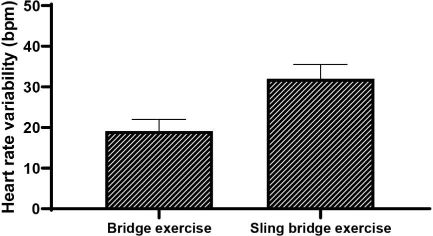
The purpose of this study was to compare the changes in the muscle activity of the GM, BF, and MF during the performance of traditional bridge and sling bridge exercises. According to the results, the GM activity during the sling bridge exercise increased significantly by 59.58% compared to the traditional bridge exercise. The muscle activity of the BF increased significantly by 121.66% during the sling bridge exercise compared to the traditional bridge exercise. The right and left MF also showed significant increases of 59.25% and 68.56%, respectively, during the sling bridge exercise. During the sling bridge exercise, the activation ratio of the GM to the BF was significantly lower by 32.43% compared to the traditional bridge exercise. Similarly, the ratio of the MF to the BF was significantly lower by 29.81% during the sling bridge exercise compared to the traditional bridge exercise. HRV was significantly higher by 67.54% during the sling bridge exercise compared to the traditional bridge exercise.
Since the sling bridge exercise is performed on an unstable support surface, it requires a higher level of muscle coordination and neuromuscular control compared to the traditional bridge exercise. As a result, the muscle activity of the GM, BF, and MF increases significantly, which may also lead to an increase in heart rate. In the present study, the average heart rate during the sling bridge exercise was significantly higher than that during the traditional bridge exercise, indicating that the sling bridge exercise demands a higher level of intensity. Because the sling bridge exercise is performed on an unstable surface, it requires greater muscle coordination and neuromuscular control than the traditional bridge exercise.44,45 In particular, sling-based exercises necessitate additional recruitment of lower extremity muscles to maintain postural stability and balance, which is believed to further stimulate cardiovascular responses.45 These findings also indicate that the sling bridge exercise is an effective exercise modality for stimulating the cardiovascular system and increasing muscle activation. Therefore, the sling bridge exercise may be usefully applied in rehabilitation or exercise programs aimed at improving heart rate or promoting cardiovascular stimulation. However, when prescribing exercise, it is important to consider the discrepancy between subjective perception and objective physiological responses, suggesting the need for an individualized approach to exercise programming(Figure 4).
The significant increase in the muscle activity of the GM, BF, and MF during the sling bridge exercise can be attributed to the difference in the stability of the support surface at the heels compared to the traditional bridge exercise. In the sling bridge exercise, the heels are supported by unstable slings, which likely led to increased activation of the GM compared to when the exercise was performed on a stable surface. Moreover, the unstable surface likely required greater muscle coordination, particularly contributing to the facilitation of BF activity through its role in stabilizing the lower extremities. In the case of the MF, the statistically significant increase in activity observed during the sling bridge exercise may be explained by the need to generate counterbalance forces in response to increased activation of the GM and BF, while maintaining a posterior pelvic tilt of 5° and 90° knee flexion. Therefore, when performing the sling bridge exercise, simultaneous contraction likely occurred due to the muscular balance formed by the connection between the GM at the hip joint, the MF above, and the BF below. This simultaneous co-contraction of muscles is considered a key factor in the ability to successfully perform the exercise.
In contrast, the activation ratio of the GM to BF was significantly lower during the sling bridge exercise compared to the traditional bridge exercise. Because the traditional bridge exercise was performed on a stable surface, the difficulty of maintaining 90° knee flexion was reduced compared to the sling bridge exercise. This likely led to a lower RPE, allowing for a more comfortable exercise experience and minimizing compensatory movements from the lower back.18 A previous study evaluated the effects of three exercises, including prone hip extension, on GM activation, pelvic muscle engagement, compensatory actions, and perceived exertion.34 The results showed that the perceived exertion scores were 13.7 for PTHE, 17.2 for PTHEA, and 10.3 for PTHEAC.34 The higher RPE for PTHE was attributed to the need to perform hip extension in a posterior pelvic tilt position, with one leg in contact with the floor and abdominal drawing-in, which made the exercise more challenging. 34 Although a direct comparison with previous studies is difficult, it is presumed that the sling bridge exercise, which required maintaining 90° knee flexion while performing 5° of hip extension, demanded greater balance in the legs and more core stability. This may have contributed to the overall increase in muscle activation of the GM, MF, and BF. In addition, this suggests that the sling bridge exercise imposed relatively greater load on the lower back compared to the traditional bridge exercise, increasing exercise difficulty as indicated by descriptive statistics. During the traditional bridge exercise, the phenomenon of active insufficiency may have led to inhibition of the BF, which could explain the higher GM/BF activation ratio observed in the traditional bridge exercise compared to the sling bridge exercise, where maintaining a 90° knee angle on an unstable surface was required.46-48
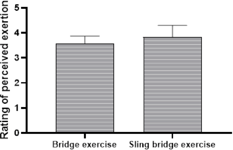
Although descriptive statistics indicated a difference in exercise difficulty based on RPE between the traditional bridge exercise and the sling bridge exercise, no statistically significant difference was found. This result may be attributed to the fact that participants with mild symptoms of NSLBP did not perceive the sling bridge exercise as being substantially more difficult. Given the subjective nature of the RPE scale, it is possible that participants did not perceive a significant difference in exercise intensity, despite objective differences in lower extremity muscle activity and heart rate. Since RPE is based on the participant’s subjective perception, the results may not have shown consistent enough values to produce statistically significant differences.49 This highlights the discrepancy between objective physiological responses and subjective perception, emphasizing the need to consider multiple indicators when prescribing exercise intensity. On the other hand, in individuals with more severe low back pain or those who experienced greater difficulty performing bridge exercises on an unstable surface, RPE scores may have been higher.
This study found that HRV and the activation of the GM, BF, and MF were significantly higher during the sling bridge exercise compared to the traditional bridge exercise. However, the GM to BF activation ratio was significantly higher during the traditional bridge exercise than during the sling bridge exercise. Therefore, for individuals with NSLBP, the traditional bridge exercise is recommended in the early stages of rehabilitation to selectively activate the GM. In the later stages of rehabilitation, the sling bridge exercise is clinically recommended to promote co-contraction of the overall lower extremity muscles, including the GM, BF, and MF, as well as to facilitate heart rate stimulation.
There are several limitations in this study. First, because all participants were young men with NSLBP, the results may not be generalizable to older individuals or women. Therefore, future studies should include participants from diverse genders, age groups, and patient populations. Second, the activation of hip and lumbar muscles, excluding the GM, BF, and MF, as well as the onset time of muscle activation, was not measured during the bridge exercise. Third, this study was a cross-sectional study, and thus, it was not possible to assess the long-term effects of the exercises over an extended period. Future research should investigate the long-term effects of sling-based exercises through comparisons between experimental and control groups.
CONCLUSIONS
In the early stages of rehabilitation, it is recommended to prioritize the traditional bridge exercise to selectively activate the GM and restore its function in individuals with NSLBP. In addition, in the later stages of rehabilitation, it is recommended to apply the sling bridge exercise to facilitate overall lower extremity muscle activation and heart rate, while facilitating coordination among various muscles. This approach can be used as a rehabilitation strategy to achieve a balanced selective strengthening and functional recovery of muscles.