


INTRODUCTION
The muscle activity of the upper trapezius (UT), lower trapezius (LT), and serratus anterior (SA) is influenced by the shoulder abduction angle.1 The UT is recruited earliest during the initial phase of shoulder abduction and indicates approximately 40% of muscle activity at 60° of abduction.1 The SA shows relatively high muscle recruitment between 60° and 100° of shoulder abduction, with a muscle activity of approximately 46%.1 The LT exhibits low recruitment during the early and middle phases of shoulder abduction, but its muscle activity increases steeply at angles exceeding 140°.1 A previous study examined the muscle activity of the UT and LT during scapular retraction exercises performed at several shoulder abduction angles (0°, 45°, 90°, and 120°).2 The muscle activity(%MVIC) of the UT gradually increased at shoulder abduction angles of 0°, 45°, and 90°, to 15.8%, 33.3%, and 54.7%, respectively, but decreased to 40.0% at 120°.2 In contrast, the LT activity was not significantly changed despite increases in the shoulder abduction angle from 0° to 120°.2 Therefore, it is necessary to perform exercises by adjusting the shoulder abduction angle for the specific activation goals of the UT, LT, and SA.
Reciprocal inhibition is a neural mechanism in which afferent impulses activate inhibitory interneurons within the spinal cord. These interneurons in turn inhibit the alpha motor neurons of the antagonist muscles.3,4 A previous study conducted that individuals with scapular winging performed a scapular protraction exercise with isometric horizontal abduction resistance using an elastic band. As a result, the muscle activity of the antagonist pectoralis major decreased and the selective activation of the SA increased.5 Another study confirmed that individuals with rounded shoulders who performed a one-arm lifting exercise with isometric adduction resistance using an elastic band. The activity of the LT was selectively increased while the muscle activity of the antagonist UT decreased.6 Therefore, an elastic band can be used to induce reciprocal inhibition and facilitate the selective activation of the target muscle.55,6
The arm-lifting in prone position can be used as an exercise to activate the UT, LT, and SA.7 The Y-raise minimizes compensatory trunk rotation and activates the UT, LT, and SA by lifting both arms simultaneously.8 The Y-raise is performed at shoulder abduction angles greater than 140°. According to a previous study, a universal Y-raise indicated moderate and similar levels of muscle activity in the UT, LT, and SA. The Y-raise with isometric adduction is recommended in early-stage rehabilitation to selectively activate the LT, as it reduces UT and SA activity through reciprocal inhibition.8 However, excessive resistance during reciprocal inhibition may cause co-contraction of adjacent muscles instead of effectively inhibiting the antagonist.9 Therefore, appropriate elastic resistance should be applied to induce effective reciprocal inhibition during the Y-raise with isometric adduction.
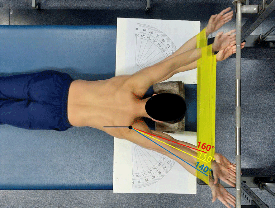
A previous study confirmed the muscle activity of the UT, LT, and SA at three different shoulder abduction angles (160°, 150°, and 140°) during the Y-raise with an elastic band.9 The muscle activity of the UT was significantly lower at 160° compared to 140° and 150° of shoulder abduction. Additionally, the SA showed a significant increase in muscle activity at 160° compared to 140° and 150°. Although there was no statistically significant difference in LT activity between the angles, descriptive statistics indicated the highest activation at 160° of shoulder abduction. Furthermore, the LT/UT activity ratio was significantly higher at 160° of shoulder abduction.9 To date, studies have compared the muscle activity of the UT, LT, and SA at different shoulder abduction angles using % MVIC. However, the patterns of muscle activity changes have not been investigated through normalization of individual muscle activities using Z-scores. This study aimed to investigate the correlation between normalized UT, LT, and SA activity and shoulder abduction angles (160°, 150°, and 140°) during the Y-raise with an elastic band. This study hypothesized that increasing the shoulder abduction angle from 140° to 160° would decrease UT activity (negative correlation) and increase SA activity (positive correlation).
METHODS
15 healthy males (23.3 ± 1.9 years; 175 ± 5.9 cm; 78 ± 9.5 kg; 25.48 ± 2.9 kg/m2) participated in this study. The inclusion criteria required participants to test negative on the following scapular instability assessments: (1) internal rotation test, (2) eccentric lowering test, and (3) shoulder flexion test. Each test was considered negative when participants performed shoulder internal rotation to 70°, eccentric arm lowering with a 2 kg load, and full-range shoulder flexion without compensatory scapular movements.10 The exclusion criteria were as follows: (1) inability to perform full range of motion in shoulder flexion, (2) reporting pain during exercise, and (3) history of shoulder pain within the past six months. This study was approved by the Institutional Review Board of Hoseo University [1041231-230706-HR-163] and was conducted in accordance with the Declaration of Helsinki. Written informed consent was obtained from all participants prior to the study.
Surface electromyography (Ultium EMG system, Noraxon, USA) was used to measure the muscle activity of the UT, LT, and SA during Y-raise exercises. The EMG signal was preprocessed using a band-pass filter (10–450 Hz), a 1024 Hz sampling rate, a 60 Hz notch filter, and a 50 ms moving window. The data were processed using the root mean square (RMS) method.11 Muscle activity was measured on the participant’s dominant arm. Before electrode placement, the skin surface was shaved and cleaned with alcohol to minimize impedance-related errors. The UT electrode was placed parallel to the muscle fibers middle point between the spine and the lateral acromion.12 The LT electrode was attached diagonally at the inferomedial border of the scapula, about 5 cm below the scapular spine.12 The SA electrode was positioned on the medial side of the latissimus dorsi, below the scapula and axillary region.12 Electrode placement for the UT, LT, and SA followed the methods of previous studies.8,13 Muscle activity was normalized for each muscle using the maximum voluntary isometric contraction (MVIC), and the MVIC measurement procedures followed guidelines.13 2-minute rest was provided between MVIC measurements, and a 10-minute rest was given between different muscles to minimize learning effects and muscle fatigue. Additional rest was provided if the participant reported experiencing muscle fatigue.
Participants practiced each exercise posture for 5 minutes to ensure correct performance. To control the shoulder abduction angle, an angle plate marked at 10° intervals from 90° to 180° of shoulder abduction was used. Participants were asked to perform a prone position with 180° of shoulder abduction as the baseline. A height-adjustable table was used to set the target bar at a height corresponding to 180° of shoulder flexion, allowing participants to reach it during the Y-raise. To induce reciprocal inhibition of the muscles, an elastic band was applied to the participant’s wrists during the Y-raise. No resistance was applied by the elastic band at the initial position of 180° of shoulder abduction and flexion. However, resistance increased as the shoulder abduction angle decreased. A decrease in shoulder abduction angle means that both arms move from an overhead position to the sides of the trunk. Participants performed the Y-raise with an elastic band at three different shoulder abduction angles (160°, 150°, and 140°) in a randomized order, and the shoulder abduction angles were controlled by the examiner (Figure 1). In each trial, participants maintained the position with hands on the target bar for 5 seconds and repeated each condition three times. For muscle activity analysis, the initial and final second of each trial were excluded to obtain stable values.14 The mean value from the three repetitions was calculated.
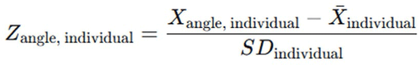
Muscle activity was initially collected as %MVIC and standardized to a 0–100% range. However, Z-score normalization was performed using each individual’s mean and standard deviation to account for inter-individual variability and examine relative muscle activity changes at three different shoulder abduction angles for correlation analysis.15 The formula for this normalization was presented in Figure 2. The Z-score was calculated by subtracting the individual’s mean muscle activity from the value at a specific shoulder abduction angle, divided by the individual’s standard deviation.15 Since the mean of the normalized values was calculated as zero, positive and negative Z-scores represent relative increases and decreases in muscle activity compared to the individual’s average, respectively.
The collected data were analyzed using SPSS Version 20.0 software (SPSS Inc., USA). The Shapiro–Wilk test was conducted to assess the normality of the data (p > 0.05). A non-parametric Spearman correlation was performed. The statistical value range of the correlation coefficient was r = –1 to 1. If the value was closer to −1 or 1, it could be considered to represent a perfect linear relationship. The ranges of r values were interpreted as follows: |0.90|–|1.00| = very strong, |0.70|–|0.89| = strong, |0.40|–|0.69| = moderate, |0.10|–|0.39| = weak, and |0.00|–|0.10| = negligible correlation.16
RESULTS
The normalized muscle activity values of the UT, LT, and SA were presented in Table 1.
The correlations between shoulder abduction angles (160°, 150°, and 140°) and the Z-scores of UT, LT, and SA were as follows: UT_Z-score, r = –0.85, p = 0.001 (negative correlation); LT_Z-score, r = 0.22, p = 0.16; and SA_Z-score, r = 0.93, p = 0.001 (positive correlation) (Table 2). Accordingly, the correlation coefficients were interpreted as strong (UT_Z-score), weak (LT_Z-score), and very strong (SA_Z-score).
DISCUSSION
The correlations between shoulder abduction angles (160°, 150°, and 140°) and the normalized muscle activity-of the UT, LT, and SA were r = –0.85, p = 0.001; r = 0.22, p = 0.16; and r = 0.93, p = 0.001, respectively. Among the 15 participants, 11 (78%) showed a gradual decrease in UT_Z-score as the shoulder abduction angle increased from 140° to 160°. 12 participants (80%) showed a gradual increase in SA_Z-score as the shoulder abduction angle increased from 140° to 160°. This study aimed to examine the correlation between shoulder abduction angle and the normalized muscle activity of the UT, LT, and SA during the Y-raise with an elastic band. We hypothesized that increasing the shoulder abduction angle would indicate a negative correlation for the UT and a positive correlation for the SA.
The negative correlation between shoulder abduction angle and UT activity may result from compensations caused by excessive elastic resistance. Reciprocal inhibition can be used to inhibit antagonist muscles by facilitating the activation of agonist muscles.3 Therefore, an elastic band was used in this study to provide shoulder adduction resistance and inhibit the antagonist UT. Muscles such as the UT can be frequently overactivated during external resistance and shoulder movements in daily activities.17,18 In particular the Y-raise at 140° likely produced greater elastic resistance than other angles. Despite maintaining an isometric end position the variable elastic resistance may have increased UT activation.9 Although reciprocal inhibition was intended by using an elastic band, the Y-raise at 140° shoulder abduction may have caused compensatory scapular upward rotation and elevation. These compensations likely occurred to maintain posture under excessive resistance and resulted in UT overactivation.
The length of the UT at each shoulder abduction angle was considered to influence its muscle activity. Muscles generate optimal tension at an appropriate length. Excessive shortening reduces their tension-generating capacity.1 Therefore, the UT may have become excessively shortened at 160° of shoulder abduction, resulting in active insufficiency and thus reduced muscle activity. A previous study reported that hamstring activity was inhibited due to active insufficiency during prone hip extension with knee flexion. This facilitated selective activation of the gluteus maximus as the target muscle.19 Although direct comparison was limited due to differences in limbs, those studies observed a similar reduction in muscle activity from active insufficiency caused by shortened muscle length. In contrast, the Y-raise performed at 140° shoulder abduction may have increased UT activation despite applying reciprocal inhibition with the elastic band. This was likely due to decreased active insufficiency associated with the length–tension relationship. Therefore, increased muscle activity may be due to the length of the UT and excessive elastic resistance leading to UT overactivation.
The positive correlation between shoulder abduction angle and SA activity was likely influenced by reciprocal inhibition of the UT. As shoulder abduction increased from 140° to 160°, the angle of scapular upward rotation also increased. Reciprocal inhibition resulted in the inhibition of the UT as a primary muscle responsible for scapular upward rotation during this process. Therefore inhibition of the UT likely facilitated increased SA activation to enhance scapular upward rotation.20 Additionally, the differing activity patterns between the SA and UT may be explained by the posterior tilt of the scapula occurring at the end range of the Y-raise.8 Posterior tilting of the scapula occurs to maintain contact with the target bar at the end of the exercise. Posterior tilting of an upward rotated scapula moves the inferior angle anterolaterally along the thorax, resulting in strong scapular protraction and upward rotation.1 Particularly, increasing the shoulder abduction angle requires the maintenance of posture more in the sagittal plane rather than the frontal plane during the Y-raise performed with 180° of shoulder flexion.1 Therefore, performing the Y-raise at 160° of shoulder abduction with 180° of shoulder flexion likely facilitated muscle activity necessary for scapular protraction and upward rotation. As a result, the SA demonstrated a different activation pattern compared to the UT.
This study has several limitations. First, the Y-raise was performed by healthy adult males in their 20s, limiting the generalizability of the results to different age groups and females. Future studies should include participants of various ages and analyze muscle activity data in female populations. Second, the small sample size may limit the generalizability of the results, and future studies with a larger sample size are needed. Third, kinematic movements of the scapula during the exercise were not measured. Further studies should consider using kinematic sensors or dynamic MRI to analyze real-time scapular movements occurring under isometric adduction resistance. Fourth, reciprocal inhibition should be examined not only during the Y-raise exercise used in this study but also during functional movements commonly performed in daily-living, such as overhead reaching and arm elevation. Lastly, the changes in resistance of the elastic band at each shoulder abduction angle were not quantified. Future research should quantify elastic resistance at each angle to determine the optimal tension generated during the exercise.
CONCLUSIONS
A strong negative correlation and a very strong positive correlation were identified between shoulder abduction angle and UT_Z-score and SA_Z-score, respectively. Therefore, a shoulder abduction angle of 160° can be recommended for inducing selective inhibition of the UT and efficient muscle activation of LT during the Y-raise with an elastic band.