INTRODUCTION
The Morton foot syndrome (MFS), known as a hereditary syndrome, is radiologically characterized by a short first metatarsal bone and a distally longer the second metatarsal head compared to the head of the first metatarsal bone.1-3 Due to posterior displacement of the first metatarsal sesamoid bones and excessive mobility of the medial cuneiform and first metatarsal bones, relatively the second metatarsal head is exposed over weight load during standing or walking.4,5 The cause of MFS is still unclear, but previous studies have reported heredity, inflammation of the first sesamoid bone, vascular replacement, and traumatic injury of the medial feet and toes as the main etiology.6-8
MFS is accompanied by several clinical symptoms when standing or walking while wearing shoes with narrow balls of the metatarsophalangeal joint area and causes dysesthesia or metatarsophalangeal neuroma in the intermetatarsal nerve area.9,10 However, most individuals with Morton foot deformity rarely have clinical symptoms such as metatarsal pain or neuroma, and do not have limitation in their ability to perform daily activities such as standing or walking.2 In a previous study of over 3,600 people with MFS, approximately 1,150 (32.1%) of the participants had a longer second metatarsal compared to the first, but they had no clinical symptoms in their feet or toes.11 Although previous studies have reported a few or no clinical symptoms of MFS, most study participants were young males, making it difficult to generalize the results to all individuals with MFS. In addition, previous studies on the effects of MFS on biomechanical variables during walking have shown negative effects on ground reaction force and foot pressure distribution in the first and second metatarsophalangeal areas.1,2 These negative effects can cause severe pain in the metatarsal and toe bones, paresthesia, and spread of symptoms to adjacent foot-toe joints and segments.6
Among the various clinical treatments for alleviating symptoms and improving gait function of MFS, foot-toe orthosis intervention should be considered as a non-invasive treatment with relatively few side effects.2,12 Foot-toe orthoses for MFS should reduce medial forefoot pain during walking as well as abnormal body weight pressure concentrated on the lower extremity joints and foot segments, contributing to improvement of gait efficiency. In the case of hallux valgus (HV) deformation, which exhibits a similar musculoskeletal deformity of the forefoot as MFS, the abnormal alignment due to the lateral deviation of the first metatarsophalangeal joint is associated with a decrease in walking efficiency.13-15 The biomechanical function of the medial forefoot during walking or jogging is to provide body weight support and sufficient GRF in the terminal stance phase, thereby generating propulsive force in the early swing phase and maintaining the efficiency of walking. From this perspective, individuals with MFS or HV deformities have a decreased gait efficiency and more clinical symptoms compared to the general population.2,15
Previously, there have been reports on the clinical and biomechanical effects of a pad-type foot-toe orthosis on the lower extremity joints during gait in subjects with MFS.1,2 However, few studies have investigated the influences between pelvic kinematics and structural changes in the forefoot musculoskeletal system of MFS according to the application of a pad-type foot-toe orthosis. In addition, the pelvis is a segment that plays an important role in the efficiency of walking, and it does not only affect the biomechanics of the lumbar and hip joints but also contributes to postural stability.16 A smaller value of pelvic movement is considered indicative of better pelvic stability during gait. Therefore, the purpose of the study was to investigate biomechanical influences of a pad type foot-toe orthosis on 3D kinematic pelvic motion in individuals with MFS using a reliable and objective 3D motion analysis equipment. The study hypothesis established that when applying the foot-toe orthosis condition, the subjects would have increased pelvic stability during walking compared to those without orthotic condition.
METHODS
The subjects were twenty-five individuals (15 males and 10 females) with MFS. All participants had both feet MFS and shorter length of the hallux end than the second toe. They had no other feet deformities such as HV and reported no foot or toe pain during gait. Inclusion criteria were at least 8 mm longer length of the second metatarsal bone compared to the first metatarsal head in both feet and no surgical history of the feet and toes.2 Individuals with musculoskeletal or neurological problems of the foot and toe joints and foot-toe osteoarthritis were excluded due to the low efficacy of foot-toe orthosis walking condition. In addition, if participants had systemic rheumatoid arthritis, painful ankle, or pathologic conditions affecting gait, they were excluded from the study. MFS assessment was performed by a licensed clinical professional to determine the difference in length between the first and second metatarsal heads.11 Participants had an average age of 34.4 years, a height of 166.9 cm, and a weight of 63.0 kg. A research expert explained all the experimental procedures to the participants, and all participants participated in the experiment voluntarily. The study design and experimental protocol were approved by the Institutional Review Board of Jeonju University (jjIRB-190611-HR-2019-0602).
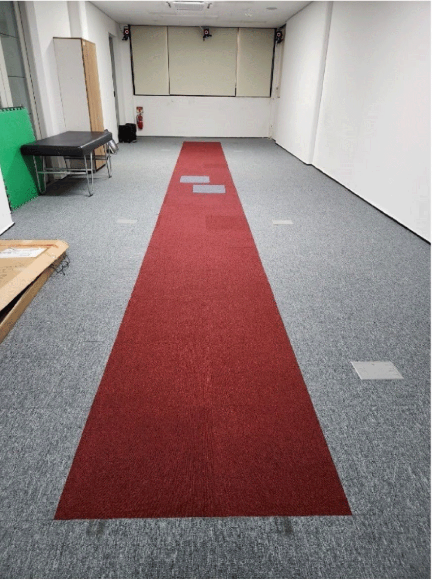
This study design was a cross-sectional and repeated measures design with and without foot-toe orthosis. The motion analysis laboratory room had a 3D motion capture system (Vicon Inc., Oxford, England) composed of eight infrared cameras, two force platforms (AMTI, Watertown, MA, USA), and a 10 m walking pathway (Figure 1). This study utilized the force platforms installed in the center of the walkway to measure more accurate gait cycles. The sampling rate of the force platform was set to 500 Hz. The sampling rate of the motion capture camera operation was set at 100 Hz. A 7.5 cm T-frame wand was used as a calibration reference object for identifying the lab origin and to calibrate the motion capture system. 3D pelvic kinematic data during gait were acquired in the motion analysis laboratory of Jeonju University.

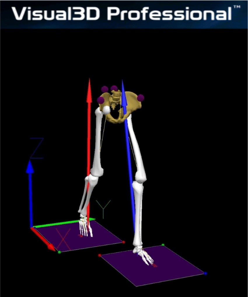
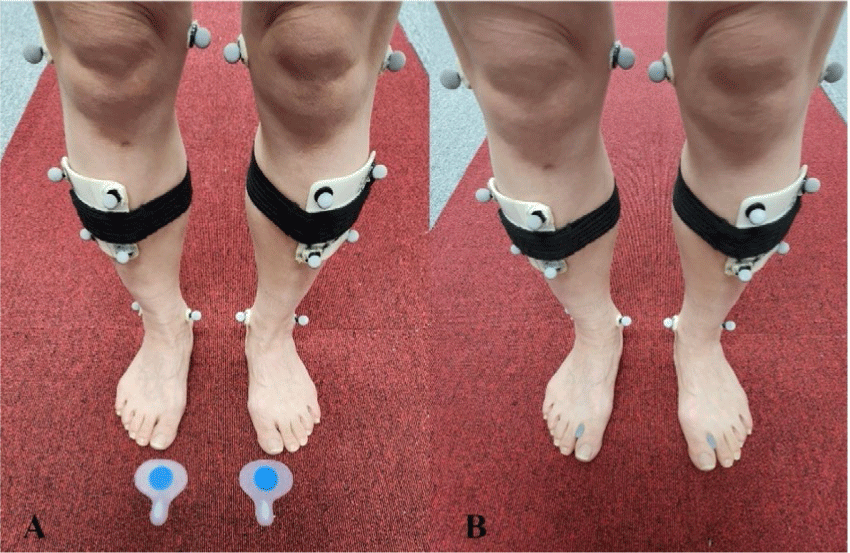
A Nexus software program (version 1.8.5, Vicon Inc., Oxford, England) was used to analyze camera capture and force platform data. Additionally, it was used to create the basic musculoskeletal model of each participant through the attached reflective markers on the anatomical landmarks of the joints and segments.2 Kinetic force platform data were low-pass filtered using a 4th order Butterworth filter with a cut-off frequency of 15 Hz and Kinematic camera capture data were low-pass filtered with a 4th order Butterworth filter with a cut-off frequency of 6 Hz.17 The c3d files finally obtained through the Nexus program were transferred to the Visual3D v6 professional program (C-Motion Inc., MD, USA) which was used to analyze all final graph reports and 3D kinematic pelvic data based on the Calibrated Anatomical System Technique according to foot-toe orthosis walking conditions (Figure 2).17 The foot-toe orthosis (ZESPA Co., Seoul, Korea) for MFS used as the orthotic condition in this study was made of soft silicone. It was inserted between the hallux and the second toe to support the sole area of the medial forefoot (Figure 3).


The experimental procedures included with and without foot-toe orthosis gait trials. To maintain a constant walking condition, tape guidelines were attached to the walking path to maintain a walking velocity and step width so that each participant could be aware of the free gait achieved during walking practice. To obtain kinematic pelvic range of motion (ROM) data through gait analysis, 40 reflective markers (1.4 cm) were attached to the pelvic segment and bilateral lower limbs to feet, malleoli, femur condyles, grater trochanters, anterior superior iliac spines, and posterior superior iliac spines. Each four-marker cluster was fixed by a Velcro on the calf and thigh segments according to six degrees of freedom (6DOF) model.17 After setting the reflective markers, an initial static camera capture of the standing posture was performed to generate a template model that was used to analyze the dynamic pelvic ROM values under with and without foot-toe orthosis gait conditions. For foot-toe orthotic condition, the participants were asked to walk along a 10 m walking pathway with a free gait speed. A total of 8 successful walking trials were performed with and without orthosis condition, and the kinematic data of the pelvic segment obtained through these trials were used for final analysis. If the walking trial was not completed successfully due to marker loss or foot position errors in an orthotic condition, an additional gait trial was performed. The experimental order of each orthosis condition was randomly assigned by dice throwing.
To compare the pelvic ROM occurred with and without orthosis gait trial, peak ROM variables developed in each 3D motion plane during gait were used. Six maximal pelvic ROM variables (anterior tilting and external rotation developed in 30%–60% of the gait cycle; posterior tilting and elevation developed in 50%–80% of the gait cycle; and depression and internal rotation developed in 0%–30% of the gait cycle) were used in final statistical analysis. Repeated measures analysis of variance (ANOVA) with Bonferroni’s adjustment was used to compare kinematic data of the pelvic segment according to orthotic conditions and lower limb sides. If the interaction effect (foot-toe orthotic condition and limb side) was significant, post-hoc testing was used to verify pairwise comparison based on ANOVA results. The Kolmogorov-Smirnov test was used to investigate that the pelvic ROM data were distributed normally. All analyses were conducted using SPSS version 29.0 (IBM Corp., Armonk, NY, USA). Differences were considered significant at α=0.05 level.
RESULTS
Normal distribution and Mauchly’s assumption of sphericity were satisfied for all maximal pelvic ROM variables required for repeated measures ANOVA analysis. There were significant differences in some maximal pelvic ROM variables between the foot-toe orthotic conditions during gait (Table 1). The pelvic ROM values of the maximal depression (F=6.447, p=0.008), total ROM in coronal plane (F=4.665, p=0.021), and total ROM in the transverse plane (F=3.990, p=0.038) were significant differences according to orthosis conditions (Table 1). However, there were no significant differences in any maximal pelvic ROM values between the lower limb sides (p>0.05) (Table 1). In addition, there were also no interactive effects between the orthosis conditions and the foot sides in any of the pelvic maximal ROM variables (p>0.05) (Table 1).
Table 2 presented the mean values of pelvic maximal ROM between with and without foot-toe orthosis conditions during gait. Under the orthosis walking condition, the participants walked with less maximal depression, less external rotation, and less total coronal pelvic motion than in the without orthotic condition in right foot side (Table 2). In left foot side, the participants walked with less maximal depression and less total transverse pelvic motion in the applying orthotic condition (Table 2). There were no significant differences in anterior and posterior tilting variables between with and without foot-toe orthotic conditions (p>0.05) (Table 2).
DISCUSSION
Reliable and quantitative biomechanical examination is an important process in acquiring clinical results for patients with musculoskeletal foot and toe deformation such as MFS.18 In this respect, the strength of this study is that it analyzed the kinematic data of the pelvic segment during gait using a motion analysis system, which is widely recognized as the most objective and quantitative technology in the field of biomechanical analysis. In addition, this study investigated the 3D kinematic maximal ROM values of the pelvic segment according to foot-toe orthotic condition using a motion analysis system while walking in individuals with MFS.
Study results partially supported our hypothesis, as the foot-toe orthosis did affect some maximal pelvic ROM variables during free walking. The results showed that the pad-type foot-toe orthosis affected maximal pelvic depression while gait compared to the without orthotic condition. Additionally, total pelvic ROM developed in coronal and transverse motion planes showed less pelvic movement during gait compared to the without orthotic condition. It was difficult to review the results of this study because there were only few previous studies that performed biomechanical analysis of the lower extremities using gait analysis individuals with MFS. Hypertrophy of the second metatarsal causes disability and pain (metatarsalgia) in the forefoot in patients with MFS.18 Inflammation and pain of the medial metatarsal joint is common when walking or after exercise and prolonged standing.2,18 These musculoskeletal disorders are due to excessive pressure on the second metatarsal head.
The results of this study showed that the application of the foot-toe orthosis for MFS contributed to pelvic stability during walking by reducing pelvic movements, especially in the coronal and transverse planes. The reason for these results is thought to be those musculoskeletal disorders of the foot and ankle joint, such as ankle sprain, ankle instability, and HV, which commonly occur in patients with MFS, also contribute to the instability of the pelvic segment during gait.3,4,19,20 Therefore, the silicone based foot-toe orthosis applied to the first and second metatarsal head areas on the medial side of the forefoot may contribute to the prevention and improvement of various foot deformities and pain and improve walking efficiency in individuals with MFS.
Similar to the pelvic elevation of the coronal motion plane, total range in transverse motion of the pelvic segment mainly decreased applying the foot-toe orthosis compared to non-orthotic condition. We could not find any comparative studies that examined the effect of foot-toe orthosis on 3D pelvic kinematics for MFS. In a previous study that verified kinetic ground reaction force by applying foot-toe orthosis for MFS in individuals with Morton toes, the application of the foot-toe orthosis had a clinically positive effects on peak ground reaction force developed in frontal plane compared to without orthotic condition.2 The application of pad-type toe orthotics for MFS is thought to have contributed to pelvic stability and walking efficiency by improving the push-off force of the medial forefoot in the terminal stance during walking. When the second metatarsal bone is longer, its head area is in touch with the floor first, resulting in a transfer of body weight to the front of the medial forefoot.2,18,19 This musculoskeletal imbalance, associated with an abnormal gait and an incorrect postural alignment causes biomechanical problems during gait. Additionally, these problems can cause pain in the ankles, hips, back, neck, and even headaches.18 The physical stress caused by this abnormal posture causes small but painful contractions called myofascial triggers in the horizontal muscles of the ankle and foot segments.2,19
The result of this study showed no significant difference in the pelvic anterior and posterior tilting movements in the sagittal plane between two foot-toe orthosis conditions among the 3D pelvic movement variables. These results are presumed to be because the toe orthosis applied in this study improved the insufficient walking support of the first and second metatarsal heads arranged in the frontal plane, thereby contributing to the stability of the movement of the lower extremity joints and pelvic segments. As shown in the results in Table 2, the same results were not observed in some variables of maximal pelvic motion during gait on the right and left sides depending on whether the foot-toe orthosis was applied or not. For example, there was a significant difference in maximal external rotation of the right pelvic segment before and after application of the foot-toe orthosis, but there was no significant difference in the left pelvic segment. This is a factor based on the general characteristics of the subjects, such as dominant and non-dominant feet, and most participants were right-foot dominant. In addition, the gait analysis was conducted only in one direction on the 10 m walk pathway, not in both directions.
The limitations of the study were that the experimental procedure was performed on relatively healthy MFS individuals without painful feet and toes that would interfere with walking. Therefore, it is difficult to consistently apply these results to all MFS individuals who already have MFS signs and symptoms in the foot and toe joints. In addition, due to the difficulty in recruiting subjects with MFS, a small number of subjects participated in the study. This study also has limitations in that it verified the temporary biomechanical effectiveness of the cross-sectional experimental application of the foot-toe orthosis for MFS. Therefore, future studies will need to overcome these limitations and verify the mid- to long-term effects of clinical interventions such as the application of the foot-toe orthoses through many MFS subjects with various clinical signs and symptoms.
CONCLUSIONS
This study was executed to confirm the influences of the silicone pad-type foot-toe orthosis on the 3D kinematic pelvic movements using a quantitative and reliable 3D motion analysis system. The results supported that applying the foot-toe orthosis for MFS increased the pelvic stability as it reduced maximal pelvic movement during gait compared to baseline barefoot walking condition. Therefore, clinical interventions using relatively easy-to-apply foot-toe orthoses, as introduced in this study, are needed to manage and prevent future development of various musculoskeletal deformities and pain in individuals with MFS who have no signs or symptoms.


