


INTRODUCTION
Various types of short leg braces (SLB) and insole orthoses are used for the purpose to correct musculoskeletal deformities of the foot, ankle, and knee joints or pain relief of the lower extremities.1-4 A general-purpose SLB to correct deformities of lower extremity joints and segments is a method of applying lateral traction force through pressure pads or straps on a metal frame support directly to the joint area.5 In addition, the general SLB is bulky and heavy. It tends to slip down and cause discomfort when worn because it is not personalized.6 For this reason, it is not widely used to correct deformities of common musculoskeletal deformities.5,6 Existing insoles and SLBs provided for the purpose of correcting or protecting various deformities of lower extremity joints are mostly produced as mass-produced products rather than customized for each individual.7,8 Personalized orthoses also have the disadvantage of requiring a lot of cost and time to obtain the appearance of each patient’s foot and evaluating lower extremity function.9-11 Therefore, research and development of SLB and insole orthoses that can compensate for shortcomings of previous manufacturing processes for orthoses are very important for clinical application and improvement of treatment effectiveness.
Recently, three-dimensional (3D) printing output of the foot and insole orthoses based on computing 3D scanners has been rapidly spreading and disseminated for the production of personalized plastic SLBs and insole orthoses.12 3D printing equipment is a device that divides the 3D human body shape into 2D cross-sectional data and then stacks various materials layer by layer to print the desired shape.13,14 For this process, modeling of the human body through a 3D scanner is essential.15 3D lower leg and foot modeling using a 3D scanner can save time and cost by complementing shortcomings of the traditional plaster model production method. It enables acquisition of more accurate and convenient lower leg and foot segment imaging data.16-18 Reliable and quantitative data are needed to understand the accuracy of 3D measurements around the lower leg and foot joints and segments. Telfer et al.19 have verified the reliability of length, width at forefoot, width at rearfoot, and peak medial arch height of foot segments. Their study showed that only some 3D scanner measurements met the criteria for good reproducibility because it only evaluated a portion of anatomical landmarks related to SLB fitting.19 As the production of medical orthoses using 3D printing becomes more accessible, interest in personalized 3D lower extremity scanning is growing.20,21 Personalized SLB and insole orthoses manufactured using 3D scanning and printing technologies have reported similar functional fit to conventional hand-fabricated orthoses.8,21 Although existing studies have reported measurement accuracy within 1 mm for SLBs produced through 3D scanning and printing technology, the reliability and validity of the lower leg and foot shapes captured during 3D scanning are not well known.
High-quality and consistent production of SLB and insole orthosis through 3D printing depends on the ability to obtain accurate and reliable human geometry through 3D lower extremity scanning systems. The purpose of this study was to verify test-retest reliability and criterion validity of various lower limb anatomical measurement data related to the SLB fitting process obtained through 3D lower limb scanning equipment.
METHODS
To recruit research subjects, a recruitment notice was posted on online social networking service. This study was conducted on 40 adults who voluntarily expressed their intention to participate. Inclusion criteria were: those who had no pain in the lower extremities, those who had no history of surgery on the lower extremities, those who were able to communicate, and those who could understand instructions. Exclusion criteria were: those who had uncomfortable sitting posture and those who had various musculoskeletal pain or dysfunction in the joints and segments of the lower extremities. All subjects received a sufficient explanation of the purpose and research method of this study before experimental participation and agreed to participate in this study. The Institutional Review Board (IRB) of Jeonju University approved study methods and study design (IRB approval number: jjIRB-220728-HR-2022-0713). Table 1 shows general characteristics such as mean age, height, and weight of participants.
| Right measures (n=22) | Left measures (n=18) | p value | |
|---|---|---|---|
| Age (year) | 22.6±1.6 | 22.1±1.5 | 0.609 |
| Height (cm) | 168.4±8.2 | 167.8±7.8 | 0.107 |
| Weight (kg) | 62.2±11.8 | 66.1±13.0 | 0.326 |
| Sex (male/female) | 10/12 | 10/8 |
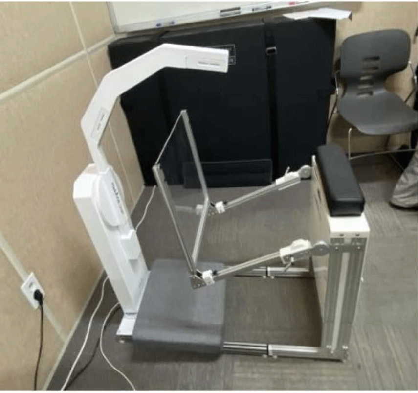
MediACE Scan (MS320A, RealDimension, Korea) was used as a 3D scanner to verify inter-rater reliability and criterion validity (Figure 1). Components of the scanner included a lower extremity holder, an acrylic footplate, a scan arm, and a scanner body. A USB 3.0 cable was used to connect between the scan arm and the main computer system and a USB 2.0 cable was used to connect between the controller and the main computer. The lower leg and foot models of subjects obtained from the scanner were processed through personalized 3D printing orthosis design CAD software (MediACE3D V1, RealDimension, Korea) installed on the main computer to obtain anthropometric lower leg and foot data necessary for inter-rater reliability and criterion validity analysis.
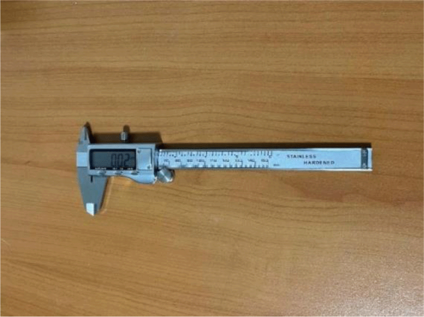
A digital vernier caliper (PSTTL833-US, Proster, USA) was used as a physical measurement tool to verify the criterion validity of the 3D scanner equipment (Figure 2). The caliper consisted of a reinforced stainless-steel shaft with a depth-measuring blade, precision internal and external measuring jaws, and a digital display with precise lapping finish. Measurement range, resolution, and accuracy of the digital vernier caliper were 0–150 mm, 0.01 mm, and 0.02 mm, respectively.22 The digital caliper has an advantage to measure inside, outside, depth, and step with two sets of jaws and the probe.22
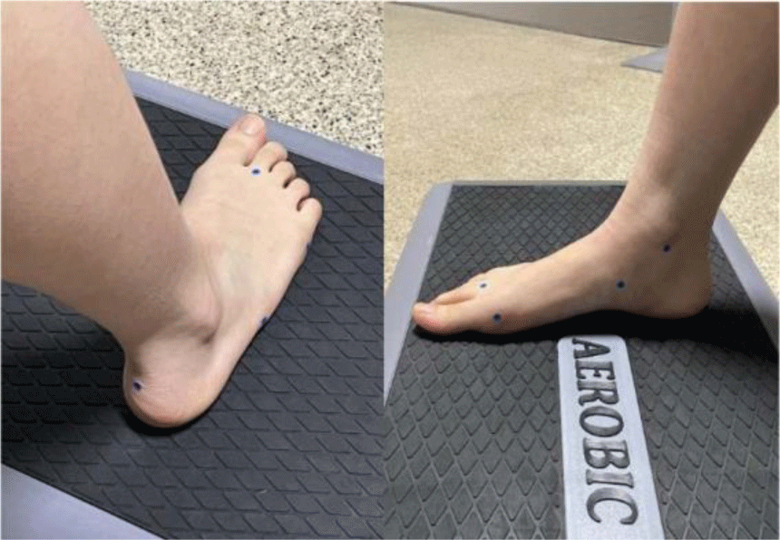
The research process to determine the reliability and validity of 3D scanner equipment is shown as follows. A sticker was attached to anatomical landmarks needed to measure the length, width, and height of one randomly selected lower limb (Figure 3). Anatomical landmark points of the lower leg and foot for anthropometric measurements were the lateral malleolus center, the styloid process of the fifth metatarsal base, the fifth metatarsophalangeal joint (MTPJ) lateral border, the second metatarsal head, the medial malleolus center, the navicular tuberosity, the first MTPJ medial border, the plantar medial forefoot center, the plantar lateral forefoot center, the plantar heel center, and calcaneal tuberosity center (Figure 3). The attached sticker was used as a reference point for direct comparison between physical measurements and 3D scan-based measurements. Anthropometric measurement variables based on the two anatomical landmark points were the navicular tuberosity height, medial malleolus height, lateral malleolus height, first MTPJ frontal length, fifth MTPJ frontal length, foot length, first MTPJ length, fifth MTPJ length, metatarsal length, and foot breadth length (Table 2).
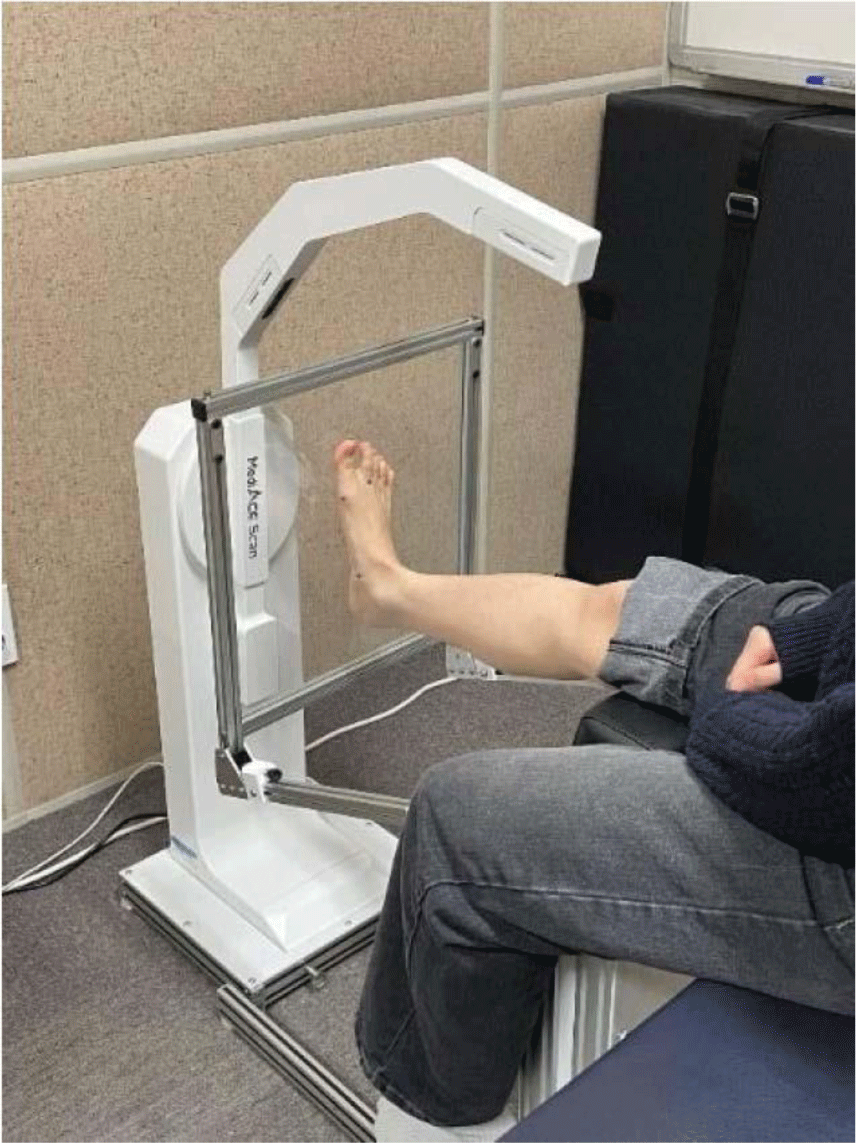
Digital vernier caliper measurements were performed three times for each measurement landmark in sitting position of the subjects. A one-minute break was taken after measuring one anatomical point. For 3D scanner measurements, the subject sat comfortably on a chair and placed the foot to be measured on the acrylic footplate of the 3D scanner (Figure 4). To verify the reliability and validity of the 3D scanner, the average value of measurements scanned three times was used.
To ensure inter-rater reliability of the 3D scanner, two testers performed measurements within three days of each other. Additionally, the criterion validity of the 3D scanner was verified through comparison with physical measurement values. The Kolmogorov-Smirnov test was used to confirm normal distribution of all measurement variables. Intraclass correlation coefficients (ICCs) were used to investigate inter-rater reliability and criterion validity for measurement variables of the 3D scanner equipment using the ICC (2,k) method. ICC correlation coefficients less than 0.5, between 0.5 and 0.75, between 0.75 and 0.9, and exceeding 0.9 indicated a low reliability, a medium reliability, an excellent reliability, and a very good reliability, respectively.23 The Pearson correlation coefficient was used to verify the relationship between the 3D scanner and physical anthropometric variables assessed with the caliper. SPSS IBM version 22.0 (IBM Corp, Armonk, NY, USA) was used to verify the inter-rater reliability and criterion validity of the 3D scanner. The statistical significance level was set at 0.05.
RESULTS
Mauchly’s assumption of sphericity was satisfied for all physical measurement and 3D scanner variables required for ICCs and Pearson correlation analysis. Table 3 presents Pearson’s correlation coefficient and mean values of the caliper and 3D scanner measurements. All caliper and 3D scanner variables between measurement correlations were greater than 0.815, presenting a high correlation. In detail, the mean correlation for foot and sole measurements used in SLB fabrication (medial malleolus height, lateral malleolus height, first MTPJ frontal length, fifth MTPJ Frontal length, foot length, and metatarsal length measures variables) was 0.894 (p<0.01).
Inter-rater reliability for the 3D scanner showed excellent correlation with ICCs of 0.90 or higher for most measurement variables (Table 4). As a result of verifying the validity of the 3D scanner, ICC values of measurer A ranged from 0.84 to 0.96 (p<0.01) and those of measurer B ranged from 0.91 to 0.99 (p<0.01) (Table 4). Most ICC values except some variables (navicular height, medial malleolus height, and lateral malleolus height) showed very good reliability (Table 4).
DISCUSSION
This study verified the inter-rater reliability and validity of human anatomical measurement landmarks of a 3D scanner that designed and operated SLB and insole orthosis fabricated through 3D printing in comparison with physical measurement. As a result of examining the relationship between physical measurements through a caliper device and scanner measurements in this study, significantly high correlations were found for most measurement variables. Although the correlation between most measurement variables through the two measurement devices was significant at r=0.81 or higher, measurement variables of first MTPJ frontal length, foot length, metatarsal length, and foot breadth length showed very high correlations at r=0.91 or higher. The difference in correlation between the two measure devices depending on the measurement variable was thought to be due to measurement characteristics of the caliper tool. Specifically, there is a method of measuring the length by aligning ends of the two internal and external measuring jaws of the caliper with the measurement point. There is also a method of measuring the length by contacting measuring jaw bars with the surface of the object to be measured, like measuring foot width. Therefore, the method of measuring length by matching point with measuring jaws of the caliper showed a relatively lower correlation than the method of measuring length by contacting the caliper with the surface of the object to be measured in this study.
Results of this study verified that meaningful data on 3D lower leg and foot shapes could be obtained using a measurement system that could be easy to use and save time and cost. As a result of this study, 3D scanning-based measurements showed excellent reliability compared to anthropometric data using caliper measurement tools. Results of this study were similar to results of a previous study that verified the reliability and validity using anthropometric 3D scanner equipment and physical measurement tools.23 Powers et al.23 have verified the reliability and validity between ankle and foot measurement data obtained through a physical measurement device and a 3D limb scanner for 30 participants. Their results showed that the average correlation between foot and ankle measurements was 0.88 and that the average correlation between circumference and length measurements was 0.96. Although the scanner system used in this study was very easy to fabricate SLBs and insole orthoses, the reliability and validity of anthropometric values that indicate the accuracy of the 3D scan are not known. Most existing studies that verified the reliability and validity of 3D scan scans verified the accuracy through data that measured limb volume.24-26 Therefore, the strength of this study is the provision of accurate anthropometric data from a 3D scanner, which is essential for orthotic manufacturing.
Few studies investigating the validity and reliability of 3D leg and foot scan have reported ICCs for measurement related to SLB and insole fabrication. In this study, the inter-rater reliability of the 3D scanner showed ICC values of 0.90 or higher for most measurement variables. In addition, excellent ICC values were shown between anthropometric values measured with calipers by two measurers and 3D scanner measurements. ICC values provide an objective basis for how reliable a measurement is across different raters and measurement environments.23 Although previous studies that directly compare ICC reliability results of this study are rare, previous studies that evaluate limb volume through 3D scanners have generally reported excellent ICC values.27,28 As a result of the validity verification of this study, differences were noticed in ICC values of several measurement variables (navicular height, medial malleolus height, and lateral malleolus height) between rater A and rater B using the caliper tool. This reason might be due to differences in clinical experience and measurement proficiency between raters.
This study has some limitations. During physical measurement and 3D scanning, knee and ankle angles of subjects were not fixed, which might have had a minor effect on measured values. Although the experiment was conducted according to a strict manual, the same time period and the same anatomical marker location were not confirmed in the test-retest performed within 3 days of interval. Additionally, most of the study subjects were in their 20s, which limited the generalization of study results. Therefore, in future studies, it is recommended to fix the knee and ankle angle during scanning experiment and confirm marker attachment sites. Although data on calf circumference were not collected in this study, the reliability of calf circumference measurements is necessary for clinical use of data obtained with a 3D scanner when manufacturing SLB and insole orthoses.
CONCLUSIONS
This study investigated the clinical applicability of personalized orthosis production by verifying the inter-rater reliability and criterion validity of a new 3D scanner for the production of SLB and insole orthoses. The inter-measurement reliability of each measurement variable through test-retest of the 3D scanner equipment showed a high level of correlation. Most measurement variables showed a high level of correlation between caliper measurements directly performed by the rater and 3D scanner measurements. Therefore, the production and clinical application of 3D printing-based customized SLB and insole orthoses can correct, manage, and treat various musculoskeletal diseases of the lower extremities through clinical use of 3D scanner equipment whose reliability and validity are verified in this study.