INTRODUCTION
Knee osteoarthritis (OA), known as a degenerative joint disease, is a common disease that occurs in approximately one-third of the population over the age of 60. It is rapidly increasing due to recent increase in average human lifespan and aging.1,2 Joint moments, such as knee adduction moment (KAM), are often used to evaluate clinical manifestations of knee OA.3 The moment that occurs during the stance phase when the body weight is supported during gait is a biomechanical variable calculated by multiplying the size of the three-dimensional (3D) ground reaction force and the distance of the lever arm from the joint center.4–6 In biomechanics, KAM typically has two butterfly figure peaks in the stance phase during gait. The first peak develops around the initial 25% stance and the second peak occurs around the 75% stance during walking.7 The peak KAM is influenced by various factors such as pain avoidance strategy, walking characteristics, and foot progression angle (FPA). Excessive increase in KAM is closely related to the exacerbation of knee OA.8,9
Previous studies verifying kinesiologic effects through changes in FPA, such as foot internal or external rotation during gait in patients with knee osteoarthritis, have shown a significant decrease in the second peak KAM10 with increased FPA and a significant decrease in the first peak KAM11 with decreased FPA walking modification. Alteration of gait characteristics such as FPA during walking may affect the kinematic and kinetic value of the lower extremities because changes in biomechanical variables that affect one joint and segment of the lower extremity can also affect other joints and segments connected to the musculoskeletal system.12,13 Many previous studies have reported that walking with foot internal or external rotation status can reduce two peak KAM in patients with OA in the medial compartment of the knee joint. However, peak KAM during walking varies depending on the target FPA level, individual anatomical characteristics, and kinesiologic specificity of the musculoskeletal link.8,10,11 Thus, verifying the effect of FPA modification gait training through consistent experimental settings on biomechanical variables of lower extremity joints using objectively reliable, high-performance equipment such as a 3D motion analysis system is important for patient evaluation and prognosis.
Previous studies applying the FPA modification walking intervention to patients with knee osteoarthritis have mainly verified its effect on KAM of the knee joint.14–16 However, studies on the effect of FPA alteration walking on the 3D moment of the hip joint in patients with knee osteoarthritis are insufficient. According to previous studies, individuals with knee OA commonly develop hip OA because they share risk factors for OA in other joints of the lower extremities.17,18 Thus, the aim of this study was to determine effects of three FPA modification walking conditions on 3D biomechanical moments elicited to hip joints in individuals with knee OA using an objective and quantitative 3D motion analysis system.
METHODS
Study subjects were 27 individuals (9 males and 18 females) with medial compartment knee OA. Inclusion criteria were: 1) no pain in both knee joints that would interfere with free walking; 2) no previous surgery knee or hip joints; 3) Kellgren-Lawrence grade 3 or less; and 4) able to walk free without any walking aids. If subjects had clinical healthy conditions affecting gait or systemic disease such as rheumatoid arthritis, they were excluded from this study. An average Kellgren-Lawrence grade of all participants was 1.5±0.42 (Grade I: n=10, Grade II: n=17). The Kellgren-Lawrence classification is a common method of classifying the severity of knee OA using five grades.3 Most participants in this study were of grade I (doubtful joint space narrowing and possible osteophytic lipping) or grade II (definite osteophytes and possible joint space narrowing). They were evaluated by an orthopedic specialist. The Institutional Review Board of Jeonju University approved our study methods and study design (jjIRB-200714-HR-2020-0708). All subjects voluntarily provided written informed consent. Mean age, height, and weight of all participants were 60.6±3.8 years, 159.7±11.6 cm, and 60.9±9.6 kg, respectively.
This study was executed with a cross-sectional study design. The experimental protocol included three different FPA walking conditions: freely normal foot position (FNFP), maximal possible internal rotation foot position (MIFP), and maximal possible external foot position (MEFP). To maintain a consistent foot rotation angle in each FPA modification condition, the foot rotation when subjects walked freely was set as the baseline FNFP. MIFP and MEFP conditions were then set by adding or subtracting about 20° to this angle.19 To assure consistent walking speed and step width, a tape line was emplaced on the gait way of experiment room.
Two force platforms (AMTI, Watertown, MA, USA) were set for sampling at 500 Hz to obtain kinetic moment data. A 3D motion capture system (Vicon Inc., Oxford, England) composed of six cameras and 8 m walking pathway was used to obtain moments of hip and knee joints during gait. The sampling rate of the infrared camera operation was set at 100 Hz. All biomechanical moment values obtained from the motion capture system and force platforms were processed with Nexus software version 1.8.5 (Vicon Inc., Oxford, England). The Nexus software created a lower limb segment model based on anatomical labeling of reflective markers.7 The final c3d file was created through the Nexus program which was used for processing all kinetic moment data.
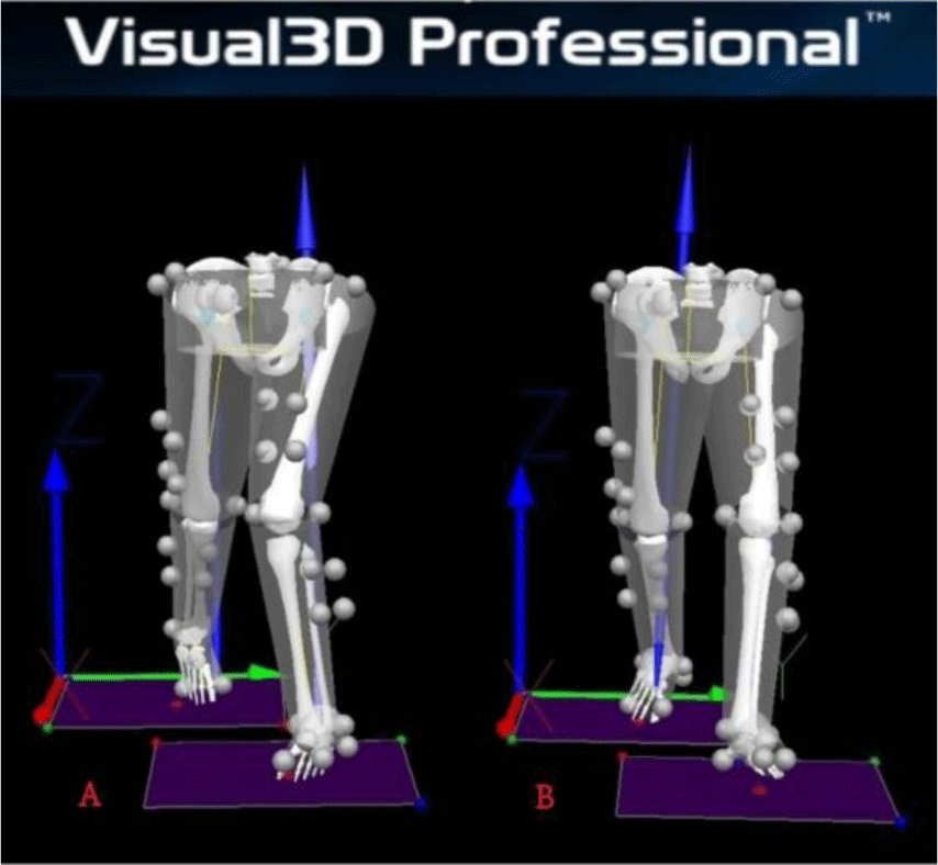
Visual3D v6 professional program (C-Motion Inc., MD, USA) was used to create a virtual skeletal model based on previous c3d files of the Nexus software to analyze all biomechanical moment data through the Calibrated Anatomical System Technique (Figure 1).20 Kinematic motion data were low-pass filtered with a 4th order Butterworth filter with a cut-off frequency of 6 Hz. Kinetic moment data were low-pass filtered using a 4th order Butterworth filter with a cut-off frequency of 15 Hz.20 All kinetic moment data of the hip and knee joints were normalized by each subject’s body weight and analysis time of moment was normalized to 100% of the stance phase during gait.

To conduct the walking experimental procedure, four cluster markers were attached bilaterally on the lower leg and thigh segments according to a six-degree-of-freedom (6DOF) model. In addition, twenty-four reflective markers (1.4 cm) were attached bilaterally on the first and fifth metatarsophalangeal joints, the dorsal center of the midfoot, medial and lateral hind foot, medial and lateral malleoli, medial and lateral femoral epicondyles, greater trochanters, anteriosuperior iliac spine, and posteriorsuperior iliac spine (Figure 2).7 After setting markers, static calibration was conducted to obtain hip and knee moments. First, the static motion capture process was conducted to analyze dynamic FPA gait for each subject. After acquiring static capture data, subjects were asked to walk freely according to FPA walking conditions. A total of 8–10 gait trials for each FPA condition that achieved the target walking line were performed. Hip and knee moment data were calculated for each FPA modification trial and averaged over each gait condition. The FPA walking order was randomly assigned.

A sample size was obtained with G*Power based on variables for two peak KAMs during internal or external rotation foot position walking in individuals with medial knee OA.14 The sample size obtained using study data was 23 people, with an effect size of 1.0, an alpha level of 0.05, and a power of 90%. For comparing hip moments developed with each FPA application, maximal peak moment variables elicited in each 3D motion plane were used. Peak 3D hip moment values for comparison according to FPA conditions were flexion, extension, abduction, adduction, internal rotation, and external rotation moments that occurred in stance phase during each FPA walking condition. KMA values (first peak KAM during 0%–50% of stance phase, peak KAM at mid stance, and 2nd peak KAM during 50%–100%) of stance phase were compared between main FPA walking trials. Statistical analyses were executed using SPSS version 26 (IBM Corp, Armonk, NY, USA). The Kolmogorov-Smirnov test was used to investigate normal distribution. Two-way repeated-measures analysis of variance (ANOVA) with Bonferroni adjustment was used to verify moment values of FPA conditions. If ANOVA results showed significant differences of main effects, a post hoc test was conducted to confirm pairwise comparisons.
RESULTS
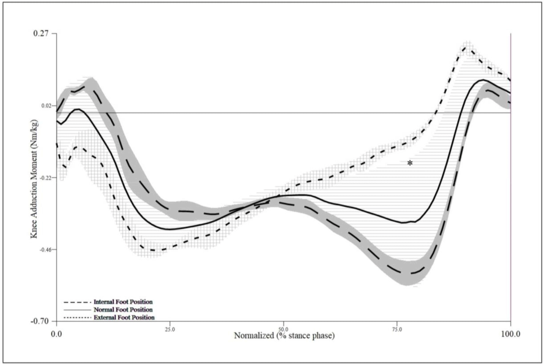
Mauchly’s assumption of sphericity was satisfied for all moment variables required for repeated measures ANOVA analysis. There were significant differences in the peak KAM during walking among FPA modification conditions according to ANOVA results (p<0.05) (Table 1). The second peak KAM (F=5.624, p=0.014) occurring at 75%–100% stance phase during gait was significantly different among FPA gait conditions (Table 1). The second peak KAM of the MEFP walking trials showed a significant decrease compared to that of the FNFP condition through post-hoc test (p<0.05) (Figure 2). On the other hand, all peak KAM moment variables except the second peak KAM showed no significant differences among FPA modification gait trials (p>0.05) (Table 1). There were no interactive effects between FPA condition and knee sides on any KAM values (p>0.05) (Table 1).
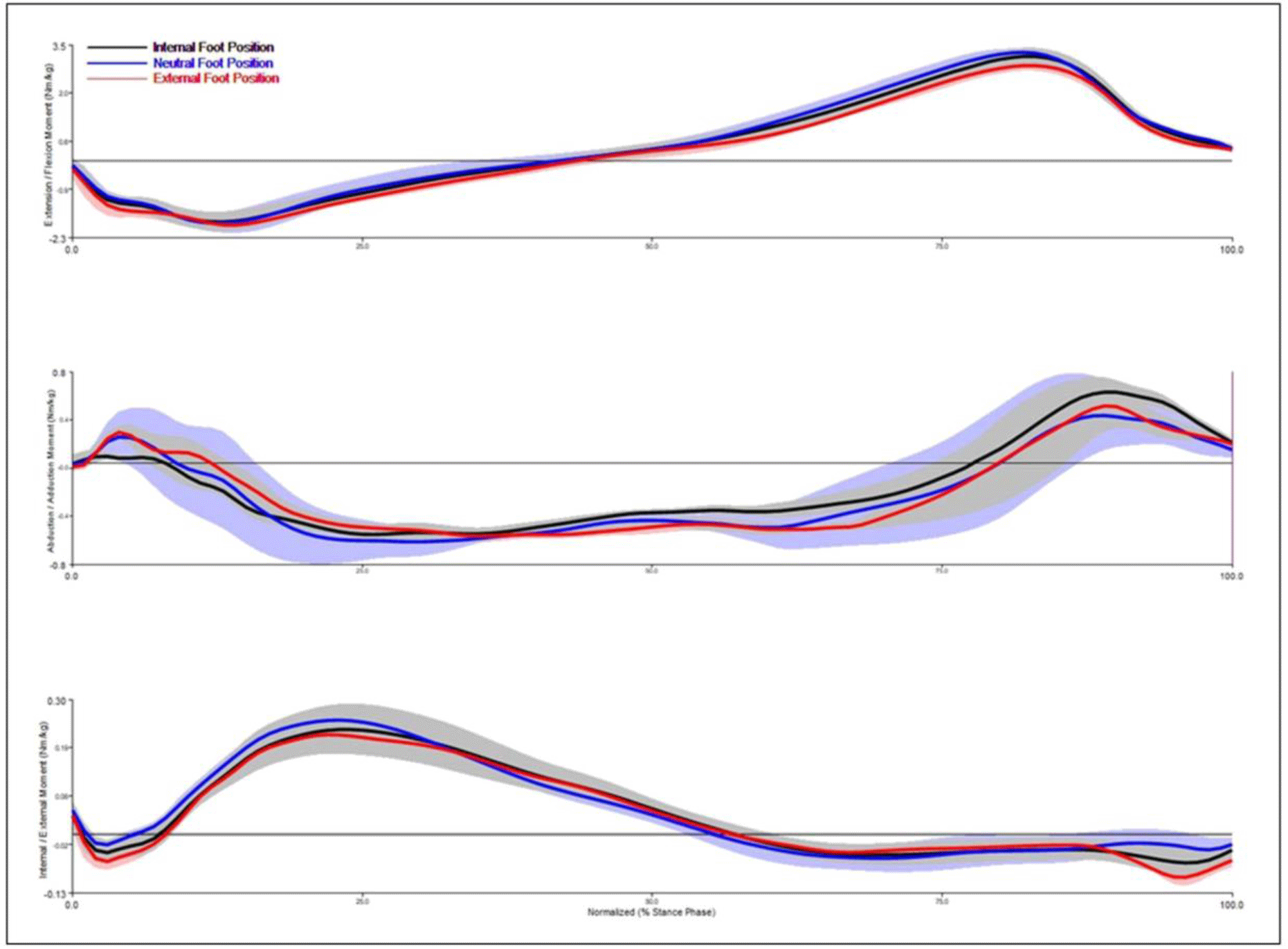
There were no significant differences in any peak moments generated at the hip joint in the stance phase during walking among FPA modification walking trials (p>0.05) (Table 2). There were no interactive effects between FPA walking conditions and hip sides on any hip moment variables (p>0.05) (Figure 3).

DISCUSSION
This study was executed to investigate influences of gait retraining such as internal or external foot rotation gait on KAM and peak hip moments developed in the stance phase during gait in 27 individuals with knee OA. Results of this study showed that the first peak value of KAM in the FNFP walking condition was not significantly different from that in MIFP or MEFP walking condition. Many previous studies have reported that the first peak KAM is significantly reduced when walking in the MIFP modification condition compared to the baseline walking condition, the FNFP.3,8,21 The reason for such conflicting results might be due to differences in the number of study subjects, clinical characteristics of subjects, and inter-individual variability in biomechanical variables such as moments.
In contrast to the MIFP modification walking that showed no significant difference compared to FNFP or MEFP in the first peak KAM, the MEFP gait condition showed a significant decrease in the second peak KAM compared to the FNFP modification walking. These results were similar to previous studies reporting a decrease of the second peak KAM during foot external rotation walking in individuals with knee OA.6,10,19,22 The reason why the MEFP modification condition significantly reduced the second peak of the KAM compared to other FPA conditions might be because the loading center moved to the medial side as the knee joint axis moved laterally.4,6
Many studies have determined effects of toe-in and toe-out walking conditions on biomechanical variables of peak KAM of the knee joint in individuals with medial compartment knee OA.2,6,10,14,23,24 However, very few studies have examined effects of these walking conditions on the moment of the hip joint in patients with knee OA. The present study analyzed effects of different walking conditions on 3D hip moments along with their effects on peak KAM according to FPA during walking in individuals with knee OA. Results showed that there was no significant increase in hip joint moment value according to FPA walking conditions. In a previous study conducted on 50 patients with medial compartment knee OA, on average, there were no significant differences in the total peak hip moment in stance phase during gait.3 In addition, there was no difference in the change in total hip moment from baseline for 10° internal foot rotation walking condition (–1.9%±0.8% [range: −17.3%, 9.2%], p>0.05) or 10° external foot rotation walking condition (–1.4%±0.8%, [range: −16.5%, 12.4%], p>0.05) compared to baseline condition.3 However, they reported that 37 participants (74% of participants) showed a decrease in their total hip moment while 13 participants (26% of participants) showed an increase in their total hip moment when walking with internal or external foot rotation conditions that maximally reduced KAM.3 Such conflicting results compared to the current study might be due to differences in the number of study subjects, clinical characteristics of subjects, and inter-individual variability in biomechanical variables such as moments. Similar to this study, a previous study conducted on 12 normal subjects to determine the effect of walking under three FPA modification walking conditions on hip moment25 also reported no significant differences in peak flexion, extension, abduction, or adduction moments of the hip joint among three FPA modification walking conditions.25 Although experimental design and characteristics of study subjects of that study were different from those of the present study, these results were similar to those of this study.
Our results showed no significant increase in the hip joint moment value in the stance phase during walking under maximal toe-in or toe-out FPA walking condition. Therefore, the clinical implication of this study is that FPA modification gait training to manage KAM in individuals with knee OA can generally be performed without considering kinetic moments normally generated at the hip joint. However, in some participants, the IFP gait modification increased the peak hip adduction moment when ankle joint contact forces peaked. This indicates that for patients with musculoskeletal deficits of the hip joint such as hip OA, the effectiveness of toe-in or toe-out walking trials at the hip joint should be verified case by case before adopting FPA gait to manage the KAM. Although maximal internal or external rotation FPA gait trials showed a significant reduction of the first or second peak KAM, clinical application of the toe-in or toe-out gait trial for patients with knee OA needs caution. Since the first peak KAM is more important for deciding the current clinical status, disease stages, and prognosis of knee OA compared to the second peak KAM, reduction of the first peak KAM is considered more important.6 However, considering the negative impact of a 1% increase in KAM on knee OA symptoms,4 although not statistically significant, clinicians may notice a trend to increase the first maximum KAM during the MEFP walking condition and a trend to increase the second maximum KAM during MIFP walking.
This study had some limitations. First, since most participants had mild knee OA, results of this study could not be generalized to all knee OA patients. Second, because of difficulties in recruiting participants with knee OA, this research could not be performed with many subjects. Third, musculoskeletal characteristics such as ankle pronation or supination angle, hip anteversion or retroversion, and so on could not be assessed. Therefore, further studies are needed to investigate the influences of FPA modification walking interventions on kinematic and kinetic variables of lower joints and segments in a large number of individuals with knee OA.
CONCLUSIONS
This study was conducted to investigate influences of artificial FPA modification walking trails on the first and second peak KAM and peak hip moments using a 3D motion analysis system and force platforms. Results showed that MEFP modification walking reduced the second peak KAM compared to baseline walking and MIFP walking conditions. In addition, all hip moments were unaffected by applying the three FPA modification conditions during gait. Results of this study suggest that FPA walking trial, which affects the peak KAM of the knee joint, does not have a significant effect on the peak hip moment. However, influences of the FPA retraining on various types of knee musculoskeletal disorders such as lateral compartment knee OA cannot be overlooked. Therefore, interventions of FPA walking retraining to patients with knee OA should be applied to each patient based on accurate biomechanical evaluation.


