


INTRODUCTION
The hip joint plays an important role in connecting the trunk and legs and has inherent joint stability.1 Therefore, impaired range of motion of the hip joint can affect an individual’s ability to perform functional activities and athletic activities. Additionally, impaired range of motion of the hip joint can affect biomechanical properties and cause damage to joints adjacent to the hip joint.1 Previous researchers have suggested an association between hip range-of-motion deficits and disorders including chronic low back pain,2-4 hip osteoarthritis,5-6 chronic athletic groin injuries,7 and sports hernias.8 In particular, clinical practice guidelines for hip osteoarthritis suggest that limitations in hip flexion and or internal rotation (IR) are criteria that can be used to identify patients with osteoarthritis of the hip.6 Groin pain is a concern for many athletes, especially those playing rugby, football, soccer, ice hockey or any other sport that requires repetitive and strenuous use of hip adductors. Previous researchers have shown reduced strength of hip adductors and range of motion in hip IR are the outcome measures that best differentiate athletes with hip/groin pain from those without this pain.9
In clinical practice, mobilization with movement (MWM) is a frequently used technique to reduce pain and improve an individual’s available joint range of motion. Three studies have reported that MWM of hip joint increased the IR of the hip joint. The first study reported that caudal MWM of the hip joint in a quadruped posture increased the IR range of motion of hip joint in healthy young adults with limited IR of hip joint.10 The second study showed that lateral MWM with hip flexion in supine position in patients with hip osteoarthritis showed an increase in hip flexion of 12.2° and IR of 4.4°.11 The third study reported that inferior-lateral MWM with hip flexion in supine position increased in hip IR of 3.7° in subjects with reduced IR of the hip.1
However, there has been no study yet on comparison of MWM in non-WBP (weight-bearing position) and WBP and for increasing hip IR. The purpose of this study was to compare the immediate effect of lateral MWM in non-WBP and WBP on the hip IR in subjects with limited hip IR. Thus, it was hypothesized that a lateral MWM in non-WBP and WBP would significantly increase hip IR range of motion and there would be a significant difference of hip IR range of motion between lateral MWM in non-WBP and WBP. Through this study, we aim to introduce evidence-based exercise methods that can improve the IR range of motion of the hip joint in various musculoskeletal patients related to limitations of the hip IR.
METHODS
The participants in this study were 15 volunteers (15 male; mean age: 22.6±1.8 years old; height: 173.9±4.9 cm; weight: 69.7±8.2 kg). The subjects were required to have an IR range of motion of both hip joints of less than 30° in the prone position. Subjects are excluded if they have experienced a lower extremity injury in the past 6 months or have previously been diagnosed with hip surgery, rheumatoid arthritis, osteoarthritis, or neurological conditions. Additionally, subjects who had a positive sign in anterior impinge test are also excluded. The anterior impinge test was performed as follows: The examiner flexes the patient’s hip to 90° and then places the hip full of adduction and then medially rotates the hip to end range. The test is considered positive if anterior hip pain is produced.12 All subjects were told about the procedures of this study and offered informed consent form Before the experiment. This study was approved by the Joongbu University Institutional Review Board (JIRB-2023053001-01).
This study relied on a One-group Pre-Post design to compare the immediate effects of MWM in non-WBP and WBP. All participants were subjected to MWM in non-WBP and WBP for bilateral hip joint. The order of the MWM type and tested leg was assigned randomly.
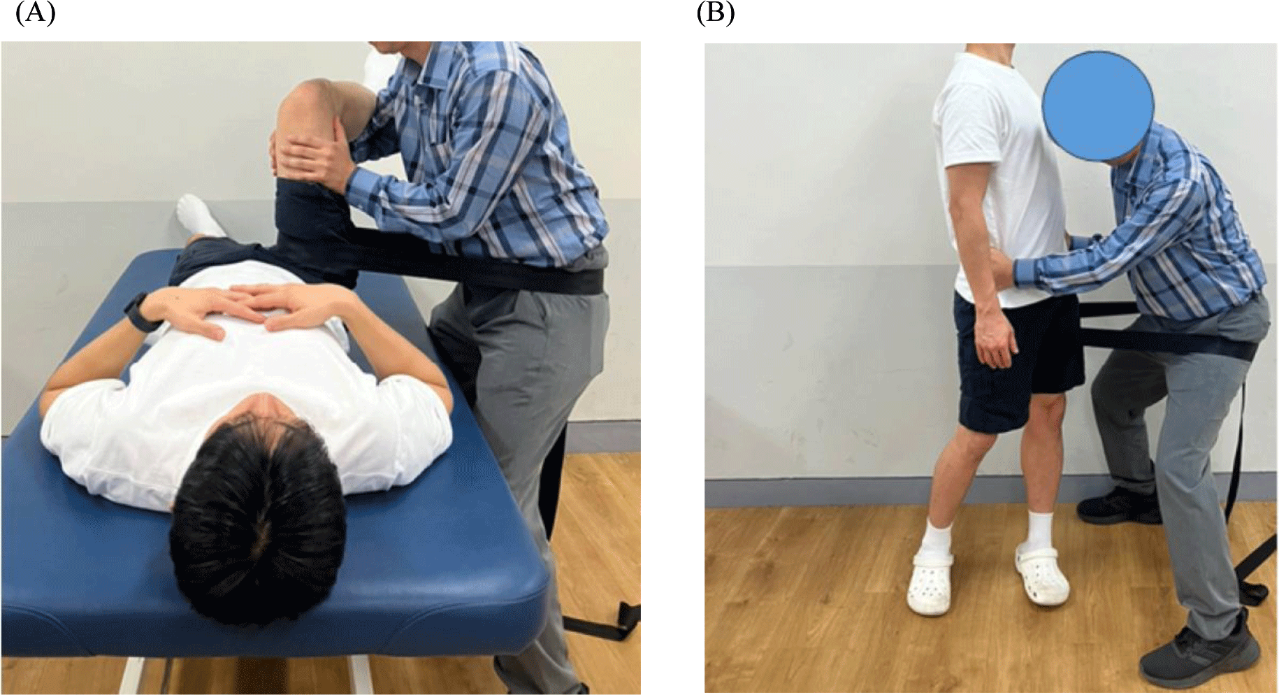
The MWM in non-WBP was performed in supine with the physical therapist standing next to the subject. A belt was looped around the pelvis and the subject’s thigh contacting medial side of the subject’s upper thigh closest to the joint. The belt was positioned such that it was always perpendicular to the subject’s thigh. The therapist supported the subject’s leg with hip flexion 90° and the subject’s hip was moved passively into hip IR to maximum pain-free range (Figure 1A).11 The MWM in WBP was performed in standing and the belt place around subject’s thigh and around physical therapist’s thigh. The belt lies horizontally. The physical therapist’s hands are placed on the subject’s ilium and a lateral distraction force is applied using the therapist’s thigh to stabilize it. Sustaining this, the subject rotated on the tested leg with subject’s other leg held just off the floor. The distraction force need only be applied just short of the limited range of rotation (Figure 1B).13 Three sets of 10 repetitions were applied with a one-minute rest interval between each set.
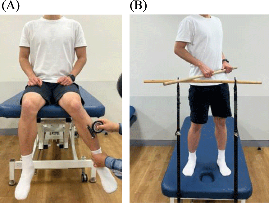
For the seated internal rotation test (SIRT), subjects were secured to a treatment bed with a belt in a seated position. The leg to be tested was passively internally rotated to the full range of motion determined by the first firm resistance keeping the pelvis neutral. The angle was measured with an inclinometer (Biomechanics Inc., Koyang, New York, Korea) positioned 5 cm distal from the tibial tuberosity (Figure 2A).14 The functional internal rotation test (FIRT) was performed in standing. Subjects were positioned in front of a horizontal bar equidistant from the floor as the subjects’ posterior superior iliac spines. The subject’s body weight is distributed over the tested leg and the other leg is used only for support. Another bar is placed across the subject’s anterior superior iliac spine so that it is parallel to the horizontal bar. The subject actively internally rotates the hip joint of the tested leg. The subjects were instructed to ensure the bar stayed in contact with both anterior superior iliac spines and physical therapist monitored there was no additional movements of other joints, thus potentially increasing the IR without intention (Figure 2B).10 The outcome measure was the angle of intersection between two bars using a plastic goniometer.
The mean, standard deviation, and 95% confidence interval (CI). All data were tested for normal distribution by using the Kolmogorov-Smirnov test (p>0.05). Statistical differences between right and left hip IR range of motion were analyzed with an independent t-test. The two factors were time (before and after interventions) and in MWM type (non-WBP vs. WBP). A two-way repeated analysis of variance (ANOVA) was used to determine the effects of intervention on SIRT and FIRT. The level of significance was set at p<0.05.
RESULTS
No significant difference were noted between right and left hip IR range of motion (p>.05). For SIRT measurement, the two-way ANOVA revealed no significant MWM type × time interaction (p>.05) and no significant main effect of MWM and time (p>.05). For FIRT measurement, the two-way ANOVA revealed no significant MWM type × time interaction (p>.05) and no significant main effect of MWM type (p>.05). However, there was significant main effect of time (p<.05). Both MWM techniques improved significantly hip IR range of motion for FIRT measurement (p< .05) (Table 1).
DISCUSSION
The purpose of this study was to evaluate differences in hip ROM when MWM in non-WBP and WBP was applied to a population with limited hip IR. There was not a significant difference between MWM in non-WBP and WBP in both SIRT and FIRT. However, both MWM techniques improved significantly hip IR range of motion for FIRT measurement.
There are probably only three studies that have evaluated hip MWM to determine its impact on hip range of motion, pain and physical performance. Beselga et al.11 reported that pain level decreased by 2 points in the numeric rating scale, hip flexion and IR increased by 12.2° and 4.4° respectively in a group of patients with hip osteoarthritis, who received lateral MWM. Walsh et al.10 suggested significant differences in hip ROM with caudal self-MWM, but caudal MWM hip flexion MWM applied by therapist significantly increased immediately functional IR in standing position (mean difference in pre- and post-intervention=5.8°). Torres et al.1 reported that inferior-lateral MWM in non-WBP increased hip IR by 3.7° in the sitting position in subjects with reduced hip IR. In similar to Walsh et al’s study, in this study, both MWM in non-WBP (mean difference=5.5°) and WBP (mean difference=5.1°) improved significantly hip IR range of motion for FIRT measurement.
The theory explaining the effects of MWM is still unknown or debated in the literature. The concept of positional fault proposed by Brian Mullian, is difficult to explain changes in hip range of motion due to lack of substantive evidence recording changes in bony position before and after MWM intervention. In this study, it is intended to determine the hip capsular-ligament stretching positions for IR through knowledge of anatomy and kinesiology. Knowledge of anatomy and kinesiology can be used to explain hip capsular-ligament stretching positions for improving hip IR. The soft tissues around the hip joint, including the fascia, ligaments, muscles, skin, tendons, affect mobility of hip joint.15 In particular, there are many researches on stretching positions for ligaments around hip joint.16-18 Hidaka et al.18 reported that superior iliofemoral, inferior iliofemoral, pubofemoral, and ischiofemoral was anatomically presumed to be stretched during external rotation, extension, abduction, IR, respectively. In this study, active hip IR during lateral traction using belt caused to stretching the ischiofemoral ligament. Therefore, both MWM in non-WBP and WBP improved significantly hip IR range of motion for FIRT measurement.
In this study, hip IR was measured by the SIRT and FIRT. The SIRT is the most frequently used method to measure passive ROM. Charlton et al.19 reported that intra-class reliability was high (ICC=.82–.84), and measurement errors for hip IR using bubble inclinometers and smartphone was 3.3–3.4°. Although, hip range of motion is normally generally in a non-WBP, most activities and sports are performed in WBP.20 Therefore, measuring the range of motion of the hip joint in WBP is functionally meaningful. Walsh and Kinsella10 reported that hip IR were measured by 47.1°, 57.2° and 49.0° in control, MWM, and self-MWM group, respectively. In this study, the hip IR was measured at 43.8° and 44.1°, which is smaller than in previous studies. The reason for these results is believed to be that thorough control was taken to prevent as much movement as possible in the trunk, knees, and subtalar joints adjacent to the tested hip joint.
There are several limitations in this study. First, it may not be generalized to hip joint in older and female populations or patients with hip pain because this study investigated restricted hip IR range of motion in young healthy male. Second, only immediate effects were evaluated, although one group pre- and post-design may help develop future research protocols. Further study is needed to determine the long-term effects of MWM in non-WBP and WBP in randomized controlled trial. Third,
CONCLUSIONS
In the present study, both MWM in non-WBP and WBP improved immediately hip IR range of motion for FIRT measurement in healthy subjects with limited hip IR. Further study is needed to compare the long-term effects of MWM in non-WBP and WBP in female or elderly with hip pain in randomized controlled trial.