Research Report

Validity and Reliability of Digital Navicular Drop Equipment in Young People with and without Pes Planus

Sang-Yeol Lee1, Du-Jin Park2,*
Author Information & Copyright
1Department of Physical Therapy, Kyungsung University, Busan, South Korea
2Department of Physical Therapy, College of Health Sciences, Catholic University of Pusan, Busan, South Korea
*djpark35@cup.ac.kr, Du-Jin Park, Department of Physical Therapy, College of Health Sciences, Catholic University of Pusan, Busan, South Korea

© Copyright 2023, Academy of KEMA. This is an Open-Access article distributed under the terms of the Creative Commons Attribution Non-Commercial License (http://creativecommons. org/licenses/by-nc/4.0) which permits unrestricted non-commercial use, distribution, and reproduction in any medium, provided the original work is properly cited.

Received: Oct 17, 2023 ; Revised: Nov 06, 2023 ; Accepted: Nov 07, 2023

Published Online: Dec 31, 2023

ABSTRACT

Background

The navicular drop test (NDT) is clinically useful, but its reliability varies depending on the examiner’s skill. As an alternative, digital navicular drop test equipment (D-NDT) has been developed, but it needs to be validated.

Purpose

This study investigated the diagnostic validity and inter-rater reliability of D-NDT in adults with and without pes planus.

Study design

A case–control study

Methods

We enrolled 52 participants: 26 with normal feet and 26 with pes planus. To assess the diagnostic validity of the D-NDT, all participants randomly underwent the NDT and used the D-NDT at least three times. Three raters assessed the reliability of the D-NDT based on the data for 10 participants randomly selected from each group.

Results

The correlation between the NDT and D-NDT grades was good in the normal (p<0.001, r=0.650) and pes planus (p<0.001, r=0.740) groups. The inter-rater reliability of the total D-NDT grade was good (ICC2,1=0.702) in the normal group and excellent (ICC2,1=0.773) in the pes planus group.

Conclusions

D-NDT showed high diagnostic validity, with excellent inter-rater reliability for participants with pes planus.

Keywords: Digital navicular drop test equipment; Navicular drop test; Pes planus; Reliability; Validity

Key Points

Question Are the diagnostic validity and reliability of digital navicular drop equipment (D-NDT) sufficient for clinical application?

Findings The D-NDT shows good validity and reliability in adults with and without pes planus.

Meaning The D-NDT could be used to measure pes planus in clinical practice.


INTRODUCTION

Developing children have soft bones, lax ligaments, increased adipose tissue, and immature neuromuscular control.1,2 Consequently, “flat” arches are normal for children up to 8 years of age.3 Although various ligaments prevent a decrease in the medial longitudinal arch (MLA) height, such as the plantar fascia and spring ligament,4 weakness of the foot intrinsic muscles is also essential.5 Adults with a flexible pes planus experience more back and lower extremity pain6 and reduced quality of life.7 In America, around 5 million people are diagnosed with pes planus8 and >40% of young adults are at risk of the condition.9 In Germany, 8% of pes planus patients are prescribed foot orthotics, and the related costs reached 466.6 million Euros in 2019.8,10 A flexible pes planus cannot maintain normal inversion in the terminal stance phase, resulting in potential foot and lower extremity fatigue.11 Evaluation of pes planus is important for avoiding flexible pes planus.

Lateral weight-bearing X-rays are commonly used to assess pes planus, as well as to measure Meary’s talo-first metatarsal angle, the angle of plantar flexion of the talus, also called the talo-horizontal angle, and the talocalcaneal angle.11 These examinations have some restrictions due to radiation exposure, low accessibility, and high costs.12 Clinical tests such as the navicular drop test (NDT) and arch height index are commonly used in clinical practice because they are convenient, cost-effective, feasible, readily available, and non-invasive, and do not require specific devices.12

For this reason, NDT is continuously used to identify the intervention effects related to pes planus, from past study24 to recent study.25 Although the NDT is a widely used clinical test, its intra- and inter-rater reliability is controversial. The intra-rater reliability reportedly ranged from 0.33 to 0.76 and was slightly higher than the inter-rater reliability.13 Evans reported that while the intra-rater reliability of the NDT ranged from 0.51 to 0.77, the inter-rater reliability was low, at only 0.46.14 A manual clinical test may lead to errors due to a lack of training.15

In addition, placing the body weight unequally on each foot can cause measurement error. Participants should widen their stance more than usual. The criterion for the neutral position of the subtalar joint differs among examiners, and there is a difficulty in accurately positioning it. To solve these problems, Park and Park developed digital navicular drop test equipment (D-NDT), which was shown to have diagnostic validity and reliability for participants with normal feet.16 However, their data were insufficient for generalization to all people. Therefore, this study investigated the diagnostic validity and inter-rater reliability of D-NDT in people with and without pes planus.

METHODS

Participants

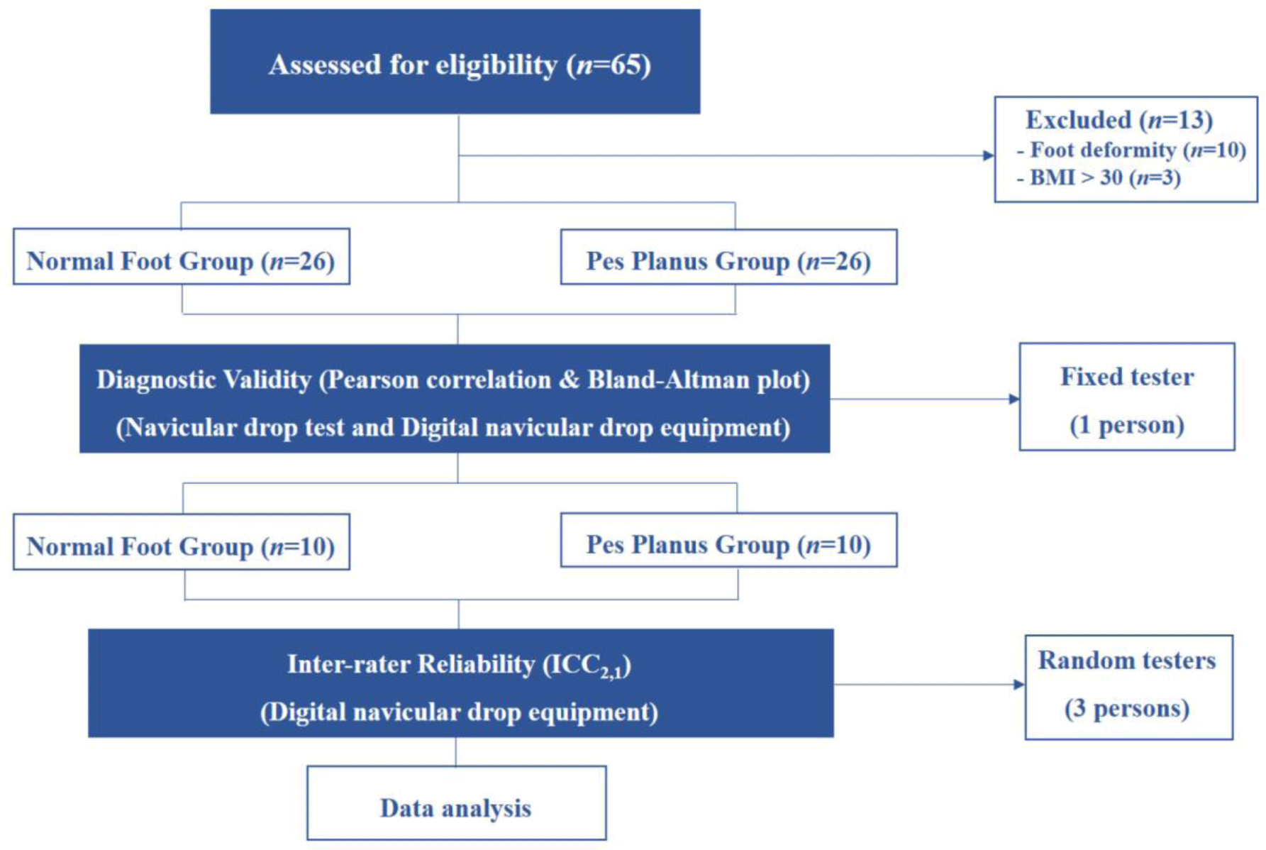
The sample size was determined using the G-Power program (3.1.9.6, Düsseldorf University, Germany) based on the correlation between NDT and D- NDT grades. In a pilot study, the correlation coefficient of the total NDT and D- NDT grades was ≥0.5. Therefore, we needed to enroll 46 people assuming a correlation of 0.50, with a power of 0.95 and significance level of 0.05. Considering the potential for dropouts, 3 participants were added to each group, such that there were 26 people with normal feet and 26 with pes planus (Figure 1). The participants had no foot deformities or diseases, and no foot pain or neurological disorders. Pes planus was defined as ≥10 mm in the NDT,17,18 the MLA angle of >131 degrees,19 and the body mass index (BMI) <30.18 The dominant foot, as determined by kicking a ball, was recorded in all participants.18 The entire study procedure was approved by the University Institutional Review Board. Table 1 summarizes the general characteristics of the two groups.

jkema-7-2-80-g1
Figure 1. Diagram of the study design. Abbreviations: BMI, body mass index; ICC, intraclass correlation coefficient.
Download Original Figure
Table 1. Descriptive statistics for participants (n=52)
Variable Normal group (n=26) Pes planus group (n=26) t p
Age 24.38±4.28 25.88±4.74 –1.198 0.237
Height (cm) 167.27±8.20 166.42±7.28 0.394 0.696
Weight (kg) 66.23±18.63 71.12±9.22 –1.198 0.239
BMI (kg/m2) 23.37±5.01 25.61±2.26 –2.081 0.045*
Gender Female 15 (57.7%), Female 11 (42.3%) - -
Male 11 (42.3%) Male 15 (57.7%)
Dominant foot Right 26 (100%) Right 20 (76.9%) - -
Left 6 (23.1%)

BMI, body mass index.

* p<0.05.

Download Excel Table
Navicular drop test

The NDT was performed to assess the diagnostic validity of the NDT and D- NDT.16 Participants removed their shoes and were barefoot. The navicular tuberosity was marked with a pen. Then, the participants maintained the subtalar joint in a neutral position while seated in a non-weight-bearing posture, and the distance between the navicular tuberosity and the ground was measured using ruler (Stainless hardened 300 mm, Minemura, Janpan). All assessments were repeated three times in each posture.

Digital navicular drop test equipment

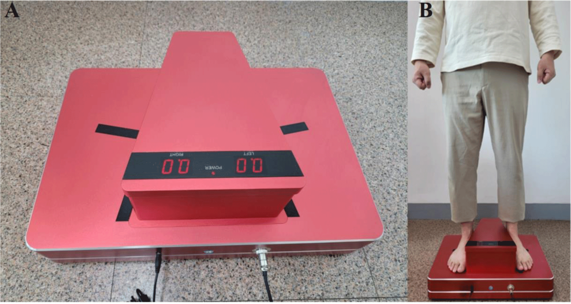
After learning how to use the D-NDT,16 reflective markers were attached to the navicular tuberosities of all participants. Subsequently, they were instructed to place their bare foot on a reflector sensor along a guide line and relax the foot. In a sitting position, the examiner relaxes the subject’s foot and then places it comfortably in the sensor area. Next, the distance between the navicular tuberosity and the ground was measured while both seated and standing (Figure 2). The examiners were experts with sufficient knowledge of the NDT and D-NDT. All assessments were repeated three times in each position.

jkema-7-2-80-g2
Figure 2. Digital navicular drop test equipment (A) and operating the equipment in standing position (B).
Download Original Figure
Medial longitudinal arch angle

The medial longitudinal arch angle between the line from the medial malleolus to the navicular tuberosity and the line connecting the head of the first metatarsal bone and the navicular tuberosity was measured in degrees.19

Inter-rater reliability

The inter-rater reliability of the D-NDT was assessed based on the grades of three raters: a physical therapy senior student, a physical therapist with <10 years of clinical experience, and a physical therapist with >10 years of clinical experience. All raters participated in the experiment after receiving >30 minutes of training on use of the D-NDT.

Statistical Analysis

Independent t-tests were used to compare general characteristics between two groups. The diagnostic validity of the NDT and D-NDT was determined using Pearson’s correlation coefficient and Bland–Altman plots. The inter-rater reliability of the D-NDT was assessed using an intraclass correlation coefficient (ICC). For the statistical analyses, SPSS for Windows (ver. 18.0; SPSS Inc., Chicago, IL, USA) was used, and the level of statistical significance was set to 0.05.

RESULTS

Diagnostic validity

Regarding the diagnostic validity of the two tests (Table 2), in the normal foot group, the correlation between the NDT and D-NDT was excellent in the sitting (r=0.938, p<0.001) and standing (r=0.926, p<0.001) positions. The correlation between the results of the two tests was significant in the normal foot group (r=0.650, p<0.01). In the pes planus group, the correlation between the NDT and D-NDT grades was excellent in the sitting (r=0.980, p<0.01) and standing (r=0.977, p<0.01) positions. The correlation between the results of the two tests was significant in the pes planus group r=0.740, p<0.01).

Table 2. Diagnostic validity of digital navicular drop test equipment (n=52)
NDT D-NDT r p
Normal group (n=26) Sitting (mm) 42.83±7.22 42.40±8.01 0.938 <0.001
Standing (mm) 39.68±7.55 39.89±7.99 0.926 <0.001
Difference (mm) 3.15±1.84 3.65±2.17 0.650 <0.001
Pes planus group (n=26) Sitting (mm) 42.87±6.69 42.81±6.45 0.980 <0.001
Standing (mm) 31.83±6.60 35.77±6.48 0.977 <0.001
Difference (mm) 11.04±0.99 10.46±0.72 0.740 <0.001

NDT, navicular drop test; D-NDT, digital navicular drop test equipment.

Download Excel Table
Bland-Altman plots

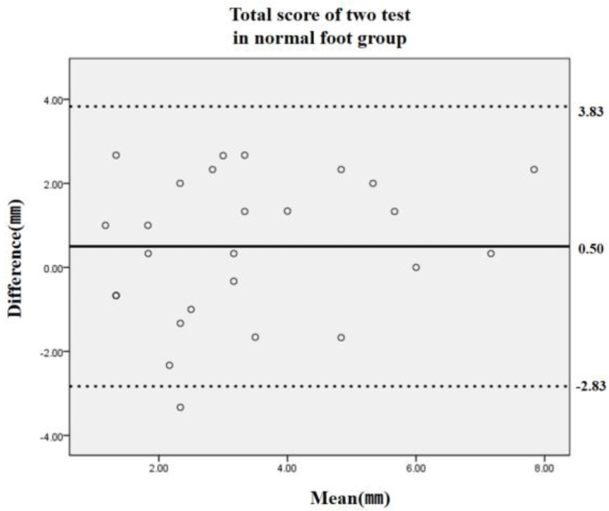
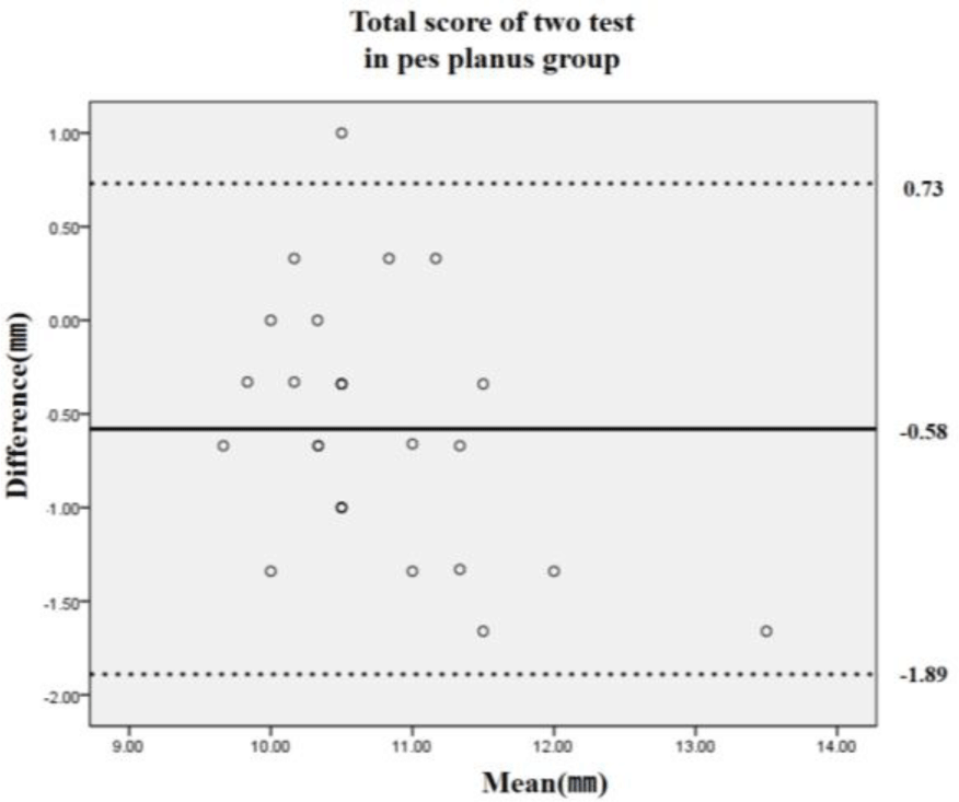
The mean difference between the two tests was 0.50 (95% limits of agreement: –2.83 to 3.83) mm in the normal foot group (Figure 3) and –0.58 (95% limits of agreement: –1.89 to 0.73) mm in the pes planus group (Figure 4).

jkema-7-2-80-g3
Figure 3. Bland-Altman plot of result of navicular drop test in normal group.
Download Original Figure
jkema-7-2-80-g4
Figure 4. Bland-Altman plot of result of navicular drop test in pes planus group.
Download Original Figure
Inter-rater reliability of the D-NDT

Examining the inter-rater reliability of the D-NDT (Table 3), the ICC of the normal and pes planus groups was 0.94 and 0.958, respectively, in the sitting position, and 0.978 and 0.957, respectively, in the standing position, with all values indicating excellent reliability. The total grade had an ICC of 0.702 in the normal foot group, demonstrating good reliability, and 0.773 in the pes planus group, reflecting excellent reliability.

Table 3. Inter-rater reliability of digital navicular drop test equipment (n=20)
ICC2,1 95% Confidence interval
Lower bound Upper bound
Sitting Normal group 0.944 0.851 0.984
Pes planus group 0.958 0.884 0.988
Standing Normal group 0.978 0.937 0.994
Pes planus group 0.957 0.881 0.988
Total score Normal group 0.702 0.384 0.905
Pes planus group 0.773 0.494 0.931

ICC, intraclass correlation coefficient.

Download Excel Table

DISCUSSION

The NDT is a reliable tool for measuring foot pronation and has an excellent correlation with the foot posture index (r=0.8), another tool for measuring foot pronation.17 Consequently, the NDT is mainly used to clarify the intervention effect for people with pes planus.18 The D-NDT was developed to enhance the precision of the NDT, which is clinically valuable, and to clarify the diagnostic validity of the NDT and D-NDT.

In this study, there were excellent correlations between the NDT and D-NDT grades in both the sitting (0.938–0.980) and standing (0.926–0.977) positions. On subtracting the height of the plantar arch in the standing position from that in the sitting position, the total grades had correlations between 0.650 and 0.740. The correlation was higher in the normal foot group than the pes planus group, reflecting the clinical value of the NDT. Furthermore, the Bland–Altman plot showed that all except one case in both groups were within the 95% limits of agreement.

The reported reliability of the NDT varies widely. An ICC value <0.4 is poor, 0.4–0.6 is fair, 0.6–0.75 is good, and >0.75 is excellent.23 In a previous study, the intra-rater and inter-rater reliability were fair.13,22 In a recent study, the intra- and inter-rater reliability were >0.9, which is excellent.17 However, another study reported low (0.46) inter-rater reliability of the NDT.14 Despite its strengths, quantitative measurement of the MLA still has poor reliability20 and varies depending on the user’s experience.21 The D-NDT was developed with ease of use and reliability in mind. In a study of normal subjects, the intra-rater reliability of the D-NDT was excellent, ranging between 0.93 and 0.95 depending on the posture.16 In this study, the normal group showed reliability values of between 0.616 and 0.978 depending on posture, while the pes planus group had very high reliability values (>0.96) in all postures. In addition, the total grades in the pes planus group had good correlations (>0.75).

The NDT poses a challenge, in that the experimenter needs to shift the legs forward, backward, left, and right to distribute the body weight equally on both feet. With the D-NDT, both feet naturally bear equal weight, increasing measurement accuracy. In particular, this equipment does not require ability to align the neutral position of subtalar joint in a sitting position. This is an equipment that anyone can easily check their pes planus. If the angle of the MLA is then added to the height of the arch, its clinical value increases.

CONCLUSION

The D-NDT showed high diagnostic validity and greater inter-rater reliability than the NDT for subjects with pes planus. Therefore, the D-NDT can be used to assess pes planus in clinical practice, complementing the NDT.

Conflict of Interest Disclosures:

None.

Funding/Support:

2022 RESEARCH FUND offered from Catholic University of Pusan.

Acknowledgment:

This paper was supported by 2022 RESEARCH FUND offered from Catholic University of Pusan.

Ethic Approval:

This study was approved by Kyungsung University Institutional Review Board (KSU-20-08-002).

Author contributions

Conceptualization: SY Lee, DJ Park.

Data acquisition: SY Lee, DJ Park.

Design of the work: SY Lee, DJ Park.

Data analysis: SY Lee, DJ Park.

Project administration: DJ Park.

Interpretation of data: SY Lee, DJ Park.

Writing – original draft: SY Lee, DJ Park.

Funding acquisition: DJ Park.

Writing–review&editing: SY Lee, DJ Park.

REFERENCES

1.

Nemeth B. The diagnosis and management of common childhood orthopedic disorders. Curr Probl Pediatr Adolesc Health Care. 2011; 41((1)):2-28

2.

Sadeghi-Demneh E, Azadinia F, Jafarian F, et al. Flatfoot and obesity in school-age children: a cross-sectional study. Clin Obes. 2016; 6((1)):42-50

3.

Uden H, Scharfbillig R, Causby R. The typically developing paediatric foot: how flat should it be? A systematic review. J Foot Ankle Res. 2017; 10:37

4.

Iaquinto JM, Wayne JS. Computational model of the lower leg and foot/ankle complex: application to arch stability. J Biomech Eng. 2010; 132((2)):021009

5.

Angin S, Crofts G, Mickle KJ, Nester CJ. Ultrasound evaluation of foot muscles and plantar fascia in pes planus. Gait Posture. 2014; 40((1)):48-52

6.

Kosashvili Y, Fridman T, Backstein D, Safir O, Bar Ziv Y. The correlation between pes planus and anterior knee or intermittent low back pain. Foot Ankle Int. 2008; 29((9)):910-913

7.

Shibuya N, Jupiter DC, Ciliberti LJ, VanBuren V, La Fontaine J. Characteristics of adult flatfoot in the United States. J Foot Ankle Surg. 2010; 49((4)):363-368

8.

Herchenröder M, Wilfling D, Steinhäuser J. Evidence for foot orthoses for adults with flatfoot: a systematic review. J Foot Ankle Res. 2021; 14((1)):57

9.

Chougala A, Phanse V, Khanna E, et al. Screening of body mass index and functional flatfoot in adult: an index and functional flatfoot in adult: an observational study observational study. Int J Physiother Res. 2015; 3((3)):1037-1041

10.

Spitzenverband Bund der Krankenkassen (GKV-Spitzenverband), 2. Bericht des GKV-Spitzenverbandes üder die Entwicklung der Mehrkostenvereinbarungen für Versorgungen mit Hilfsmittelleistungen. 2020

11.

Ueki Y, Sakuma E, Wada I. Pathology and management of flexible flat foot in children. J Orthop Sci. 2019; 24((1)):9-13

12.

Kodithuwakku Arachchige SNK, Chander H, Knight A. flatfeet: biomechanical implications, assessment and management. Foot. 2019; 38:81-85

13.

Vinicombe A, Raspovic A, Menz HB. Reliability of navicular displacement measurement as a clinical indicator of foot posture. J Am Podiatr Med Assoc. 2001; 91((5)):262-268

14.

Evans AM, Copper AW, Scharfbillig RW, Scutter SD, Williams MT. Reliability of the foot posture index and traditional measures of foot position. J Am Podiatr Med Assoc. 2003; 93((3)):203-213

15.

McPoil TG, Cornwall MW, Medoff L, Vicenzino B, Forsberg K, Hilz D. Arch height change during sit-to-stand: an alternative for the navicular drop test. J Foot Ankle Res. 2008; 1((1)):3

16.

Park JS, Park DJ. Validity and reliability of new digital navicular drop test equipment in young people. PNF & Mov. 2021; 19((2)):279-285

17.

Zuil-Escobar JC, Martínez-Cepa CB, Martín-Urrialde JA, Gómez-Conesa A. Medial longitudinal arch: accuracy, reliability, and correlation between navicular drop test and footprint parameters. J Manipulative Physiol Ther. 2018; 41((8)):672-679

18.

Hoang NT, Chen S, Chou LW. The impact of foot orthoses and exercises on pain and navicular drop for adult flatfoot: a network meta-analysis. Int J Environ Res Public Health. 2021; 18((15)):8063

19.

Nilsson MK, Friis R, Michaelsen MS, Jakobsen PA, Nielsen RO. Classification of the height and flexibility of the medial longitudinal arch of the foot. J Foot Ankle Res. 2012; 5:3

20.

Gross MT. Lower quarter screening for skeletal malalignment-suggestions for orthotics and shoewear. J Orthop Sports Phys Ther. 1995; 21((6)):389-405

21.

Levinger P, Menz HB, Fotoohabadi MR, Feller JA, Bartlett JR, Bergman NR. Foot posture in people with medial compartment knee osteoarthritis. J Foot Ankle Res. 2010; 3:29

22.

Picciano AM, Rowlands MS, Worrell T. Reliability of open and closed kinetic chain subtalar joint neutral positions and navicular drop test. J Orthop Sports Phys Ther. 1993; 18((4)):553-558

23.

Cicchetti DV. Guidelines, criteria, and rules of thumb for evaluating normed and standardized assessment instruments in psychology. Psychol Assess. 1994; 6:284-290

24.

Holmes CF, Wilcox D, Fletcher JP. Effect of a modified, low-dye medial longitudinal arch taping procedure on the subtalar joint neutral position before and after light exercise. J Orthop Sports Phys Ther. 2002; 32((5)):194-201

25.

Unver B, Erdem EU, Akbas E. Effects of short-foot exercises on foot posture, pain, disability, and plantar pressure in pes planus. J Sport Rehabil. 2019; 29((4)):436-440

한국연구재단 등재학술지 선정

KEMA학회 학술지인 'JMST(Journal of Musculoskeletal Science and Technology)'가 
2022년 한국연구재단의 학술지평가에서 등재학술지로 선정되었습니다.

등재지 선정을 위해 수고하고 애써주신 모든 분들에게 감사를 드리며, 아낌없는
헌신과 협조에 감사드립니다.

현재 JMST는 연구자들의 부담을 줄이기 위해 연회비/심사료/게재료를 받지 않고 있습니다.
또한, 사사 표기를 통해 연구비 지원 여부를 밝히는 경우에도 게재료를 받지 않고 있습니다.

많은 관심과 양질의 논문 투고를 부탁 드립니다.

감사합니다.  

I don't want to open this window for a day.