


INTRODUCTION
The hip joint is shaped as a ball and concave socket that covers approximately two-thirds of the femoral head with the acetabulum. This joint plays an important role in providing stability and mobility for essential daily activities such as standing, walking, and running.1 The hip joint is considered to be more stable in comparison to other joints due to the presence of strong ligaments and surrounding muscles.1 Several muscles are responsible for hip joint movements, including the hamstring (Ham), gluteus maximus (GM), iliopsoas (IP), gluteus medius, tensor fascia latae (TFL), rectus femoris (RF), sartorius (SAR), and piriformis.1,2 The hip flexor muscles include the IP, RF, TFL, and SAR which play a significant role during lower limb movements.3 Among these hip flexor muscles, the primary mover is the IP. The IP consists of the iliacus (IL) and psoas major (PM) muscles.4 The IP is attached from the lumbar vertebrae to the lesser trochanter of the femur, allowing for hip flexion, as well as hip adduction and anterior pelvic tilting.4 Additionally, the IP controls the anterior gliding of the femoral head and supports the front of the hip joint capsule.5
Femoral anterior glide type (FAGT) is a condition where the femoral head is glided excessively forward or fails to glide adequately backward during hip flexion.2,6 Ideally, the femoral head undergoes both anterior rolling and posterior gliding movements during hip flexion.2 However, subjects with FAGT experience excessive anterior gliding and insufficient posterior gliding due to improper securing of the femoral head within the acetabulum and the weakness of the IP muscle.2,6 According to Sahrmann, subjects with FAGT may experience discomfort and pain in the groin area and hip joint.6
During the ASLR test, the femoral head tends to move forward or toward medially rotated in subjects with FAGT.7 In addition, the stiffness of posterior structures and weakness of anterior structures in the hip joint may cause pain and movement faulty during hip joint actions. In contrast, subjects without FAGT can maintain the femoral head in the acetabulum during ASLR, resulting in more normal movement patterns. The ASLR test comprises several components, including force closure from the lateral abdominal muscles, hip flexion on the same side, pelvic rotation on the opposite side by the abdominal wall, stabilization of the pelvis in the various planes, and hip extension on the opposite side.8 ASLR is used to assess selective iliopsoas muscle strength, which includes slight abduction and lateral rotation.9 It is widely recommended due to its high specificity, reliability, sensitivity, and ease of execution.10 Further-more, ASLR is also a tool to quantify impairments related to mobility disorders of the hip joint.11,12 Muscle activities of the IP, adductor longus (AL), PM, and RF during ASLR were compared in a previous study.13,14 These studies found that all four muscles were activated during ASLR. However, unlike the other muscles, PM exhibited muscle activation on both the same and opposite sides, which is unique.13,14
Previous studies have not thoroughly compared hip flexor strength between subjects with and without FAGT while performing ASLR test. Therefore, the purpose of this study is to investigate the variations in hip flexor strength during ASLR, specifically when the anterior glide of the femoral head is fixed and not fixed, between subjects with and without FAGT. Our research hypothesis is as follows: In the FAGT group, there would be a decrease in hip flexor strength when the anterior glide of the femoral head is fixed, compared to when it is not fixed. Conversely, in the group without FAGT, we hypothesize that there would be no significant difference in hip flexor strength based on whether the anterior glide of the femoral head is fixed or not.
METHODS
This study included 30 participants divided into two groups: 15 subjects with FAGT and 15 subjects without FAGT. Participants were selected based on several criteria through the Kinetic Exercise based on Movement Analysis (KEMA) approach for FAGT2: 1) pain and numbness in the hip region, 2) weakness in the iliopsoas muscle, 3) a posterior pelvic tilt angle (<15°) measured between the anterior superior iliac spine (ASIS) and posterior superior iliac spine (PSIS), 4) hip flexion limitation in supine (<120°), 5) an anterior glide of the femoral head during ASLR, 6) hamstring shortness, 7) an anterior glide of the femoral head during prone hip extension, and 8) weakness in the GM. Participants were assigned to the FAGT group with 8 positive signs.2,15 Exclusion criteria consisted of 1) subjects without hip pain and 2) subjects without a history of lower limb surgery within the past 6 months.16 All participants provided written informed consent and volunteered, meeting the ethical principles of the Declaration of Helsinki and the requirements of the institutional review board. The characteristics of the participants are presented in Table 1.
| Characteristics | With FAGT (n=15) | Without FAGT (n=15) | p |
|---|---|---|---|
| Age (yrs) | 23.08±1.40 | 21.38±2.30 | 0.12 |
| Height (cm) | 171.12±7.15 | 170.11±4.18 | 0.29 |
| Weight (kg) | 69.72±10.30 | 68.03±9.50 | 0.18 |
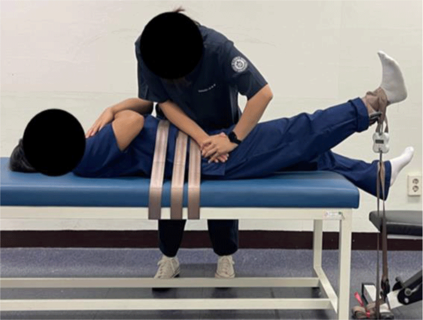
In this experiment, subjects were selected using 8 evaluations, following the KEMA approach (Figure 1).2 The subjects were positioned on a table, and a non-elastic belt was used to stabilize their trunk. The strength of the hip flexor muscles was measured using a Smart KEMA tensiometer sensor (Factorial Holdings Co., Ltd., Seoul, Korea). The strap length was adjusted to allow for hip flexion of up to 25° while in a supine position. Before the measurements, examiners were trained on how to use the tensiometer sensor. The tensiometer was capable of detecting forces up to 100 kg, with a resolution and accuracy of 0.1 kg. The isometric maximal voluntary contraction strength was then measured for the dominant side during ASLR, both with and without FAGT, in a randomized order within each test session. During the measurements, both limbs were extended at the knees while performing ASLR with 25° of hip flexion. To measure the strength of hip flexion during ASLR without manually fixing the femoral head, participants were instructed to flex their hip joint without fixation of the femoral head. For the task of manual fixation of the femoral head, they were asked to perform the task with the exa-miner’s manual fixation of the femoral head around ASIS during ASLR.13,14 During the ASLR, the pressure was applied to a specific region on the femoral head without interrupting limb movement.13,14 Following the sequence, the participants provided 5 seconds of maximal hip flexor strength measurements for each side, three times, with 2 minutes of rest between the tasks. After a 2-minute rest period, the same procedure was repeated.
During the study, the participants were assessed for several physical evaluations.2
The first was the presence of numbness or pain in the groin area, which was checked while they were standing and the examiner tapped around the groin. If the participant reported feeling either numbness or pain, it was considered a positive response.
The second condition was the posterior pelvic tilt posture, which was measured by determining the angles between ASIS and PSIS using a PALM. The degree of pelvic tilt was then calculated, with a tilt of approximately 15° being considered normal. If the participant exhibited a posterior pelvic tilt posture below 15 degrees, it was considered a positive response.
The third condition was weakness of the iliopsoas muscle. Participants were positioned in a supine position with a 90° flexion in the dominant-side hip and knee joint. The examiner then placed their hands 5 cm below the participant’s knee joint and pulled the leg down towards them. If the participant was unable to resist the force applied by the examiner and exhibited posture changes, it was considered a positive response.
The fourth condition was a limitation of hip flexion, which was assessed while the participant was in a supine position. Both hands were used to pull the dominant-side leg towards their chest while the lumbar spine area was supported by a towel to prevent compensatory movements. If there was a limitation in hip flexion below 120°, it was considered a positive response.
The fifth condition was the muscle shortness of the hamstring, which was assessed while the participant was in a supine position. They actively flexed their hips to a 90° angle and then performed maximal knee extension without ankle dorsiflexion. If the hip joint flexed to 90° and the knee joint extended beyond approximately 80°, it was considered within the normal range. If the range was limited below the normal range, it was considered a positive response.
The sixth condition was an anterior glide of the femoral head during ASLR in a supine position. Participants per-formed a 10° hip flexion while in a supine position. The examiner then applied pressure to the greater trochanter (GT) of the participant’s femur to detect any anterior or anteromedial movement of the GT. If there was an anterior or anteromedial movement of the GT during the ASLR, it was considered a positive response.
The seventh condition was the weakness of GM. Participants were positioned in a prone position and flexed their knee joints to a 90° angle. The examiner then measured the strength of the GM by fixing the participant's lumbar spine with one hand and applying resistance to the thigh with the other hand. If the participant was unable to overcome the examiner's resistance, it was considered a positive response.
The final condition was an anterior glide of the femoral head during prone hip extension. Participants performed a 10° hip extension while in a prone position, and the examiner applied pressure to the GT to detect any anterior or anteromedial movement. If the GT moved in an anterior or anteromedial direction during hip extension in the prone position, it was considered a positive response.
The statistical analysis of the collected data was con-ducted using SPSS Version 20.0 software. To assess nor-mality, the Shapiro-Wilk test was used. A two-way mixed analysis of variance was conducted to determine significant differences between groups (with vs. without FAGT: between factors) and conditions (with vs. without manual fixation: within factors). The level of statistical significance was set at α=0.05.
RESULT
The data demonstrated a normal distribution (p>0.05). In subjects with femoral anterior glide type, without fixation of the femoral head, the mean hip flexor strength was 13.72± 3.98 kg, whereas, with fixation of the femoral head, it was 10.84±3.58 kg. It was confirmed that there was a significant difference in muscle strength change depending on whether the femoral head was fixed, with a change of 2.88 kg (p=0.014) (Table 2). However, in subjects without femoral anterior glide type, there was no significant difference in hip flexor muscle strength between conditions with and without fixation of the femoral head (Table 2).
DISCUSSION
This study aimed to compare the strength of hip flexors during ASLR with and without the fixation of the anterior glide of the femoral head between subjects with and without FAGT. The results showed a significant strength difference of 2.88 kg (20.99%) in a group with FAGT, based on whether the femoral head was fixed or not. When the femoral head was not fixed, the strength was measured at 13.72 kg, while it was 10.84 kg when fixed (p=0.014). In the group without FAGT, the strength difference based on whether the femoral head was fixed or not was found to be 0.4 kg (2.83%).
The study identified two main factors that contributed to these results. Firstly, the decrease in hip flexor strength when the femoral head was fixed can be attributed to the fact that, in the FAGT group, the central axis of the femoral head was unable to maintain neutrality within the acetabulum during ASLR. This might affect the reduction in hip flexor strength. Previous studies have indicated that fixing the femoral head to prevent deviation from the central axis of the hip joint during ASLR can result in a more significant restriction, especially when the Ham is shortened.6 Consequently, increased stiffness in the Ham can limit hip flexion during ASLR.6 Therefore, Ham tension influenced the decrease in hip flexor strength. Other previous studies have reported that increased Ham tension, which opposes hip flexion, is a contributing factor to the reduction in hip flexor strength during ASLR.17,18 In cases of Sahrmann, Ham stiffness influences posterior pelvic tilting, which restricts anterior pelvic tilting.6 Consequently, The muscle weakness of IP leads to decreased hip flexion strength and muscle imbalance. Previous studies involving soccer players have indicated that hamstring shortness contributes to limitations in hip flexion and knee extension during soccer ball striking.18,19 Hamstring muscle fiber shortness and changes in viscoelasticity, which result in shortened muscles, can impair the performance of the Ham and IP muscles.18,19 Ultimately, while direct comparisons with previous studies are challenging, it is widely believed that shortness or increased stiffness of the Ham affects hip flexor strength.20
Second, the increase in hip flexor strength when the anterior glide of the femoral head was not fixed can be attributed to a decrease in the contribution of IP strength. This causes other hip flexor muscles like RF, TFL, and SAR to co-contract together as compensation.6,9 In the FAGT group, a decrease in IP strength might be observed due to the elongation of the IP tendon caused by the anterior glide of the femoral head.1 This elongation resulted in a decrease in strength because of the length-tension relationship, and other muscles like the AL, TFL, and SAR had to compensate for the decreased strength. In contrast, in the subjects without FAGT, the elongation of the IP tendon might be minimized by the maintenance of the femur head in the acetabulum during hip flexion. This factor might contribute to the slight difference between hip flexor strength with and without the manual fixation of the femoral anterior glide during ASLR. In addition, previous studies have shown that subjects with anterior hip joint pain during hip flexion use TFL more actively as it acts as a compensatory muscle.6 Therefore, RF, TFL, and SAR work together through compensatory actions during hip flexion, leading to an increase in hip flexor strength when the anterior glide of the femoral head is not fixed.21,22
This study has several limitations. Firstly, we did not measure the distance of the forward glide of the femoral head during ASLR. Secondly, we did not directly measure the increased length of the iliopsoas (IP) resulting from the anterior glide of the femoral head. Thirdly, we did not individually measure and evaluate the electromyographic activity of the hip flexor muscles.
CONCLUSIONS
In conclusion, when comparing hip flexor strength in subjects with and without FAGT, with the femoral head fixed and not fixed, we observed a decrease in strength when the femoral head was fixed in subjects with FAGT. The anterior gliding of the femoral head may provoke inaccuracies when clinically measuring hip flexor strength during the ASLR test in subjects with FAGT. Therefore, it is recommended to accurately measure hip flexor strength in subjects with FAGT by preventing the anterior glide of the femoral head during the ASLR test.