INTRODUCTION
Patellofemoral pain syndrome is one of the most common knee joint disorders observed in clinical practice. According to Collins et al., research into patellofemoral pain syndrome should be expanded to reduce the impact of knee injuries.1 According to some studies, patellofemoral pain syndrome accounts for 25% to 40% of knee patients, although the actual incidence is still unclear.2 Various studies have shown that patellofemoral pain syndrome affects women more than men, with a ratio close to 2:1.3,4 The prevalence of patellofemoral pain syndrome in adolescent patients exceeds 20%. If not treated for an extended period of time, this is associated with a poor prognosis and a high rate of invalidity.5
Patellofemoral pain syndrome is a common type of knee pain that affects adults and young people as well as active and healthy workers.5 The main characteristic of patellofemoral pain syndrome is pain in or around the anterior knee that worsens with activities that increase patellofemoral loading, such as stair climbing, sitting with the knee bent, kneeling, and squatting. Pain caused by patellofemoral pain syndrome is often worsened by prolonged sitting or walking up and down stairs. This has a significant impact on the patient’s normal life.6 A meta-analysis found that the presence of pain when squatting was the most sensitive physical examination judgment for patellofemoral pain syndrome.7 The cause of patellofemoral pain syndrome is unknown, but it is most likely the result of a combination of factors, including training methods. Six anatomical areas are known to be involved: subchondral bone, synovial membrane, meniscus, skin, nerves, and muscles.8 According to research, four main factors contribute to the condition: lower extremity and/or patella imbalances, lower extremity muscle imbalances, overuse/overload, and trauma.9 Of these four factors, overuse appears to be the most important.10
A systematic review also found that decreased quadriceps strength is associated with a significantly increased risk of patellofemoral pain syndrome due to patellar instability.11 Other causes of patellar instability, such as knee sprains, can also contribute to patellofemoral pain syndrome.12 We used the Q-angle created by the anterior superior iliac spine and tibial tubercle, the center of the patella, to calculate quadriceps force.13 Because the outward force acting on the patella increases with increasing Q-angle, it has long been assumed that a high Q-angle increases the risk of patellofemoral pain syndrome; however, recent studies have not found a high Q-angle to be a clear cause of patellofemoral pain syndrome.14,15,16 Patella orientation and alignment can also affect knee joint stability. If the patella is misaligned, it can cause overuse/overload (overpressure) in that part of the femur, resulting in pain, discomfort, or irritation. This deviation could be caused by a variety of factors.2
Dynamic valgus is another mechanism associated with patellar femoral pain syndrome. The knee collapses medially due to severe supination, internal and external rotation, or both in dynamic valgus.17 When a person stands, the area where the sole of the foot hits the ground is anatomically divided into four parts: medial forefoot, lateral forefoot, medial rearfoot, and lateral rearfoot.18 This increases the outward force on the patella, resulting in hallux valgus. Female athletes are more likely to have dynamic hallux valgus, which may explain why women have a higher incidence of patellofemoral pain syndrome.19 Foot abnormalities, such as hindfoot pronation and supination cause internal rotation of the tibia, which can further contribute to dynamic hallux valgus.20
Patellofemoral pain syndrome is normally treated conservatively, with the goal of reducing pain, improving patellar monitoring, and restoring previous levels of function. Several studies support exercise as a recommended treatment for individuals with patellofemoral pain syndrome.6,21 Exercise is the most comprehensively researched treatment for patellofemoral pain syndrome, and it can specifically address the dynamic hallux valgus malalignment that many individuals have.22 The forward lunge is a common weight-bearing exercise used by athletes and people with healthy knees to train the hip and quadriceps muscles.23,24 Physical therapists use the forward lunge and other weight-bearing exercises to rehab individuals with knee injuries and pathologies, including patellofemoral rehabilitation for patients with patellofemoral pain syndrome.25 Forward lunges have been performed with a variety of techniques, including changes in hip position.
Numerous studies have indicated that improving hip posture and quadriceps exercise can help people with patellofemoral pain syndrome.26,27 Strengthening the hip adductors increased the therapeutic benefit of patellofemoral pain syndrome. Strength in the hip adductors lengthens the vastus medialis oblique (VMO), changing its length tension characteristics and allowing it to perform better with more positive contractile force.28 However, other studies in the squat position have shown that recruiting hip flexors weakens the hip vastus lateralis (VL), has a more severe effect on patellofemoral pain syndrome, and induces higher adduction rotational forces.29,30
Many studies have been conducted to investigate the effectiveness of hip adductor strength exercises in treating patellofemoral pain syndrome.31,32,33,34,35 There is a lack of research comparing the effectiveness of hip adductor strength training with hip neutral position strength training for patellofemoral pain syndrome, and the forward lunge is considered as a good therapeutic exercise for the condition.23 However, there has been little research on how muscle activity varies in the forward lunge with hip adduction. Therefore, in this study, we aimed to determine the changes in muscle activity and plantar pressure during two hip positions, neutral and adduction, with the forward lunge to provide a basis for future therapeutic exercises for patellofemoral pain syndrome. We hypothesized that the plantar pressure and the muscle activity of VMO and VL of forward lunge with hip adduction (ADD) were larger than that of forward lunge with hip neutral position (NEU).
METHODS
Sample size estimation was obtained using the G-power software ver. 3.1.9.7. In a pilot study on four participants, the testing power at 0.95, effect size at 8.04, and significance level at 0.5 led to a sample size of 3. Considering the drop-out rate at 20% and validity of this study, the number of participants in this study was determined as 20. (Table 1). Participants were excluded if they had a current or previous history of knee injury or patellofemoral pathology; if they had any history of knee pain during any recreational or daily living activities, such as forward lunge within the last 3 months; or if their body mass index was greater than 30. All respondents read and signed the university-approved human subjects’ authorization form before participating. Prior to the investigation, participants signed a consent form and were informed of their right to withdraw. The investigation was approved by the Institutional Review Board of Daegu University (approval number: 1040621-202207-HR-068).
| Characteristics | Mean±SD |
|---|---|
| Age (yr) | 23.7±2.51 |
| Height (cm) | 169.4±7.8 |
| Weight (kg) | 64.3±12.7 |
| Body mass index | 22.26±2.48 |
Prior to the study, participants performed a 5-min standardized warm-up consisting of forward lunges with multiplane hip movements and were given instructions to become familiar with the measurement protocol and asked to practice plantar pressure and muscle activity of the VMO and VL measurements to perform appropriate movements. Then, during the forward lunge with hip neutral position and the forward lunge with hip adduction, plantar pressure and muscle activity of the VMO and VL were obtained. To avoid muscular fatigue, the measurement protocol began with plantar pressure measurements, followed by the maximal voluntary isometric contraction measurements and then the VMO and VL’s muscle activity measurements were performed. The order of the forward lunge with hip neutral position and forward lunge with hip adduction was randomized, and each measurement was repeated three times. When assessing muscular activity, subjects held each measurement trial for 5 s and rested for 1 min between repetitions. To avoid muscle fatigue, a 5-min rest interval was included between the muscle activity assessments for the VMO and VL.
In this study, a gait checker (GHW-1100, GHiWelCo., Ltd., Yangju-si, Gyeonggi-do, Korea) was used to measure the plantar pressure of the study subjects. To measure the muscle activity of the subject’s VMO and VL, data were collected using a wireless surface electromyography (TeleMyo DTS, Noraxon Inc, Arizona, USA). With the notable exception of the first and last, each muscle’s maximum voluntary isometric contraction value was performed three times for a total of 5 s, followed by the middle 3 s. The amount of either signal was averaged across seconds.
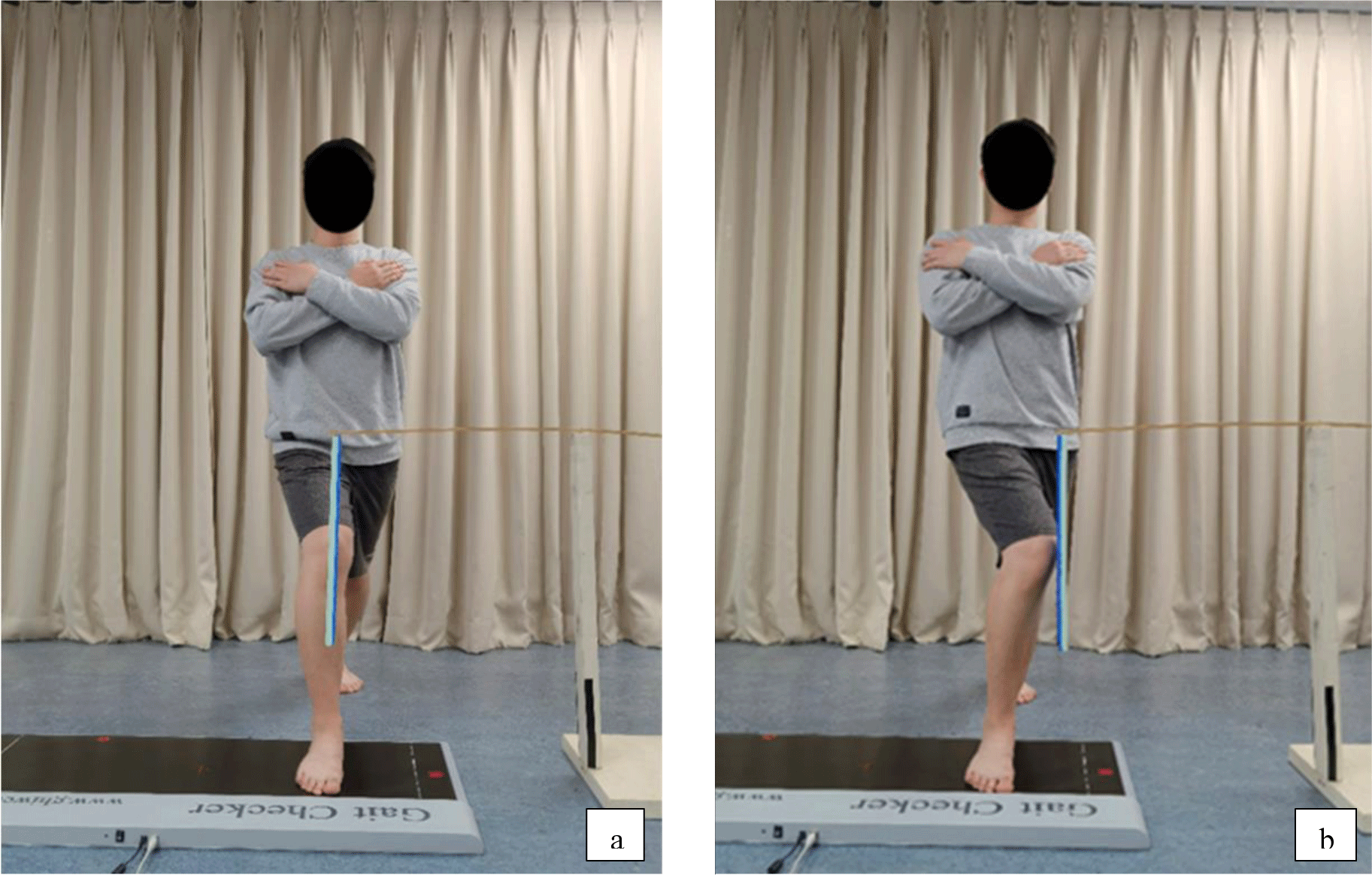
The plantar region was divided into four areas: the medial forefoot, the lateral forefoot, the medial rearfoot, and the lateral rearfoot. The line from the second toe to the heel divided the plantar area into medial and lateral areas. The vertical line between the most concave part of the arch and the midline divided the plantar area into fore and rear parts.18 While performing the forward lunge, the subject’s front leg rests on top of gait checker walking mat. Allowing the experimental subjects to perform multiple peak hip adduction motions prior to the measurement and inserting a guide plate at the knee joint will help in determining the peak hip adduction distance. During the measurement, the participant can contact the guide plate (the same as neutral position) to reach the peak hip adduction distance. Forward lunges with hip neutral position and forward lunges with hip adduction begin and end in 5 s. To prevent fatigue, repeat each action three times, with each measurement 1 min apart. Five seconds of two movements were recorded for three times (Figure 1).

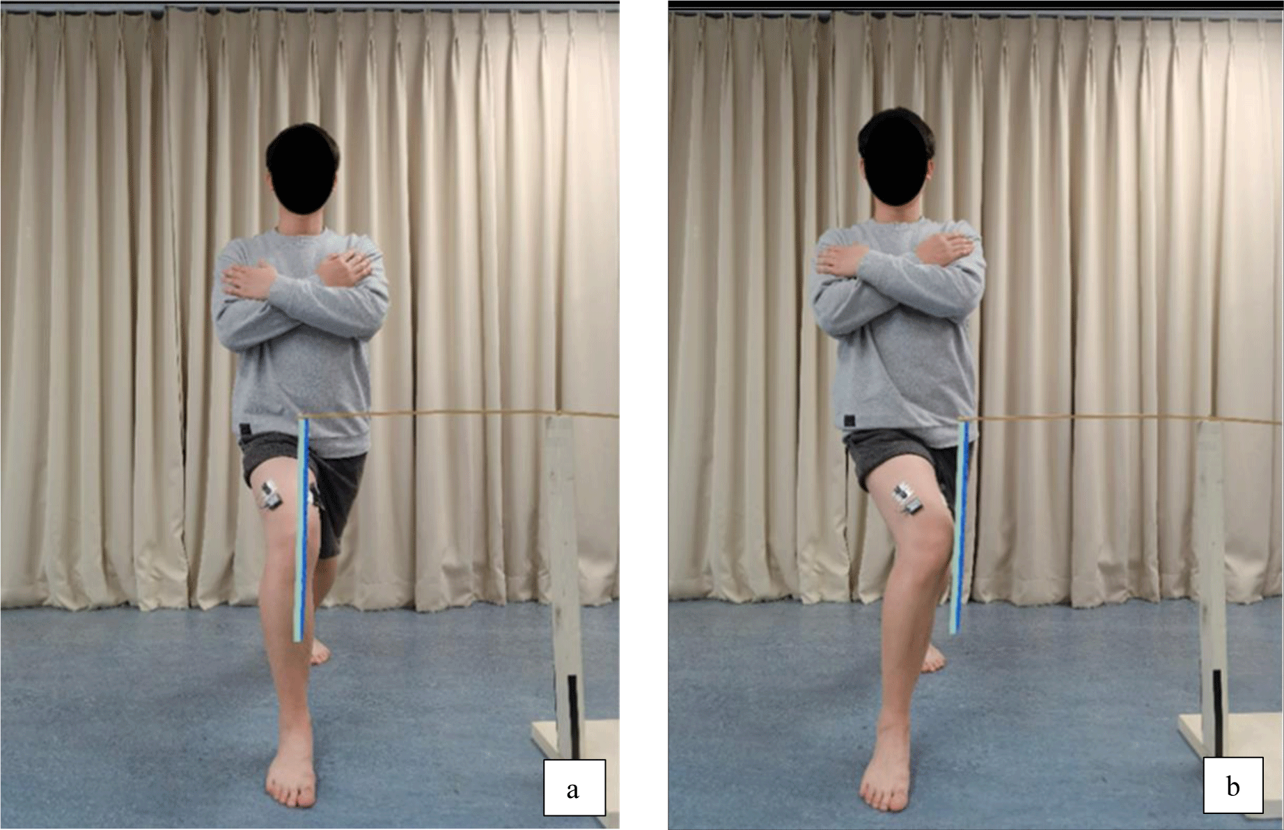
A maximal voluntary isometric contraction (MVIC) test was performed before the two postures to consistently measure the EMG signals caused by the two postures. An electrode patch was placed 10 cm from the patella on the line connecting the anterior superior iliac spine and the outside of the patella to measure the VL. An electrode was placed 4 cm from the patella on a line that forms a 50° angle with the parallel line connecting the outer edge of the patella to the anterior superior iliac spine.36 Popliteal fossae were positioned against the edge of the platform at a 90° knee flexion angle, and participants were instructed to sit on a high platform without touching the floor. They were told to lift the affected legs to a knee flexion angle of 60° and then straighten them completely. The examiner’s hand was placed on the test leg’s ankle to oppose it and cause a maximal isometric contraction of knee extension. When the subject is standing on both legs, the commonly used lower limb takes a step forward and flexes the knee joint 60°, keeping the lower leg perpendicular to the ground with peak hip adduction displacement and no pelvic movement. Allowing the experimental subjects to perform multiple peak hip adduction motions prior to the measurement and inserting a guide plate at the knee joint will help in determining the peak hip adduction distance. The participant can contact the guide plate during the measurement to reach the peak hip adduction distance. Keep the other leg straight and heel on the ground. Hold both hands in front of the chest, keep the upper body perpendicular to the ground, and look straight ahead (Figure 2). The test was repeated three times with a 1-min break between repetitions to avoid muscle fatigue.37 After the MVIC test, participants received a 5-min break before being trained in two postures. Each movement was performed three times with a 1-min break in between. Between each session, the participant took a 5-min break.

In this study, data were expressed as mean±standard deviation. All data were tested for normal distribution by using the Kolmogorov-Smirnov test. To compare the plantar pressure and EMG of VMO and VL, between the conditions analysis (comparison between ADD and NEU positions) was performed using paired t-tests. Statistical analysis was performed using the Statistical Package for the Social Sciences (SPSS) version 20.0 for Windows software (SPSS, IBM, USA). The statistical significance level was set at 0.05.
RESULTS
The plantar pressure of the forward lunge with hip adduction differed significantly from that of the forward lunge with hip neutral position in the medial forefoot and lateral rearfoot areas (p<0.05, Table 2). The plantar pressure of the forward lunge with hip adduction was not significantly different from that of the forward lunge with hip neutral position in the lateral forefoot and medial rearfoot areas (p>0.05, Table 2). The muscle activity of the VMO differed significantly between the forward lunge with hip adduction and the forward lunge with hip neutral position (p<0.05, Table 3), and the muscle activity of the VL was not significantly different between the forward lunge with hip adduction and the forward lunge with hip neutral position (p>0.05, Table 3).
| NEU | ADD | t | p | |
|---|---|---|---|---|
| VMO | 34.88±9.48 | 58.3±14.42 | –6.068 | 0.000* |
| VL | 33.79±11.56 | 40.98±12.64 | –1.875 | 0.069 |
DISCUSSION
Patellofemoral pain syndrome is becoming more common in everyday life and affects the normal life of people. The forward lunge can help with the patellofemoral pain syndrome. The purpose of this study was to determine the safest posture from the forward lunge with hip neutral position and forward lunge with hip adduction by comparing plantar pressure and the muscle activity of the VMO and VL.
The results of this plantar pressure study showed statistically significant differences in individual four-foot areas during the forward lunge with hip adduction and the forward lunge with the hip neutral position. On the middle zone, the plantar pressure of the forward lunge with hip adduction was evidently greater than the plantar pressure of the forward lunge with the hip neutral position. Forward lunge with hip adduction induced more adduction of the knee, resulting in the most plantar pressure on the knee, which could cause knee instability and potentially knee valgus.38,39 The plantar pressure of a forward lunge with hip neutral position was the same as the plantar pressure of a forward lunge with hip adduction in the lateral area. Consequently, at the plantar pressure point, the forward lunge with hip adduction performed no better than the forward lunge with hip neutral position.
There was significant difference in the EMG of the VMO between two postures. The forward lunge with hip adduction produced higher muscle activation than the forward lunge with hip neutral position. The adductor complex includes the three adductor muscles (longus, magnus and brevis).40 The distal part of the VM muscle was found to originate from the adductor magnus tendon in all cases in the presented research.41 The present study concludes that the VM has two parts, VML and VMO, based on the variation in the angle of muscle fiber orientation.41 Thus, when adducting the hip, the VMO participated in the adducting motion. Hence the greater muscle activity of the VMO was seen in the forward lunge with hip adduction. The muscular output of the VL generated by hip adduction created a lateral force that resulted in excessive lateral tracking of the patella.32 The VMO has a specific role in medial stabilization of the patella.41 Therefore, the increased VMO muscle activity means to counteract the lateral force produced by the VL and reduce the excessive lateral tracking of the patella to help stabilize the patella. According to Kumar et al.,32 hip delivery serves two functions in the rehabilitation of patients with patellofemoral pain syndrome. First, by selectively activating the VMO, the hip supply lowers lateral traction on the kneecap. Second, strong hip adapters provide a stable origin for the VMO. According to Miao et al.,42 adding hip delivery dilates the VMO muscle, changing its tension-length characteristics and resulting in a greater contraction force.
At the same time, there was no significant difference in VL muscle activity between the two postures. However, the muscle activity of the VL in the forward lunge with hip adduction was more effective than the VL muscle activity in the forward lunge with hip neutral position. Muscular co-contraction helps to stabilize the motion practice so that the agonist muscle can withstand resistance and produce the movement. In this study, forward lunge with hip neutral position was the best movement among two forward lunge postures for VMO and VL muscle activity.
In this study, the forward lunge with hip adduction increased muscle activity in the VMO more than the forward lunge with hip neutral position significantly. Strengthening the VMO muscle may also help patients with patellofemoral pain syndrome improve patella stability; however, the forward lunge with hip adduction caused an imbalance of the patella tracking, which is not helpful for patellofemoral pain syndrome. Meanwhile, the hip adductors can modulate the femur’s frontal plane position, influencing knee conditions, such as knee valgus.34,43 According to the results of this study, a forward lunge with hip neutral position may be more effective in strengthening the hip adductor muscles31,32 than the forward lunge with hip adduction.
Meanwhile, the limitations of this experiment should be recognized. This study did not confirm the motion of the hip joint. The gluteus medius, tensor fasciae latae, adductor brevis, and adductor longus may affect frontal plane stability of hip motion; however, muscle activity in the hip frontal plane muscles was not evaluated in this study. Furthermore, because the study’s subjects were young, healthy, and in sufficient numbers, the conclusions may not be generalized to other populations. Because this was a cross-sectional study, more long-term studies are required to determine the effects of forward lunge with hip neutral position and forward lunge with hip adduction on quadriceps muscle strength and knee stability in patellofemoral pain syndrome patients and normal subjects.
CONCLUSION
In this study, although the muscle activity of VMO in the forward lunge with hip adduction was significantly larger than that of VMO in the forward lunge with hip neutral position, the risk of low knee stability and intra-articular stress on the patellofemoral joint would be on the basis of the significantly larger medial plantar pressure of the forward lunge with hip adduction. Therefore, forward lunge with hip adduction was not suitable for patellofemoral pain syndrome patients to exercise, although they were good for the muscle activity of the VMO in this study. A forward lunge with hip neutral position may be beneficial in considering the physical characteristics of reduced plantar pressure for patellofemoral pain syndrome patients and normal subjects.


