INTRODUCTION
Scapular upward rotation is performed by the upper trapezius (UT), lower trapezius (LT), and serratus anterior (SA) muscles.1 These muscles are force-coupled to one another; when the balance between them is not maintained well, the dysfunction of the scapula may occur and act as a cause of shoulder impingement.2-7 Previous studies reported that the UT is frequently hyperactive, and the LT and SA are weakening muscles.8-10 In the shoulder with impingement, the ratios of the SA to UT activities and the LT to UT activities are lower than those of normal subjects.11-14
In previous studies aimed to strengthen the SA and LT, prone arm lift exercise has been reported as a way to significantly increase the activities of the SA and LT.15-17 The SA activity increases proportionally to the increase in the humeral elevation angle, but LT activity increases steeply in the end range.18 Therefore, humeral elevation in the end range may be effective in simultaneously increasing the activities of both muscles.19-21 In most studies, the activities of the SA and LT significantly increase as the humeral elevation angle increases.20, 21 Several other factors may affect the muscle activity of scapular upward rotators. Scapular upward rotators may change muscle activities depending on the position of the head (forward head or neutral head posture) and the exercise plane.22 The muscle activity of scapular upward rotators is affected by the posture and inclination of the trunk.23-25
Robert-Lachaine et al.26 reported that the scapulohumeral rhythm in full-can exercise is significantly higher than that in empty-can exercise. These findings suggest that scapular and humeral movements are affected by the position of the arm. The muscle activity of scapular upward rotators varies depending on arm posture. These factors should be considered important in an exercise process for the treatment of shoulder dysfunction or shoulder impingement. However, studies investigating the muscle activity of scapular upward rotators during arm lifting have kept the posture neutral in most cases,16, 21, 22, 25 and no studies have focused on the effect of arm position.
Therefore, in this study, we investigated the effect of arm position on the muscle activity of scapular upward rotators during prone arm lift exercise. We hypothesized that the muscle activity of scapular upward rotators would significantly differ depending on the arm position during prone arm lift exercise.
METHODS
G*Power software (version 3.1.2; Franz Faul, University of Kiel, Kiel, Germany) was used to estimate the necessary sample size. Sample size was calculated a priori for a power of .95, effect size of .80, and alpha level of .05. This calculation indicated that at least 28 subjects were needed to investigate significantly different muscle activities of scapular upward rotators at the three arm positions during a prone arm lift exercise. A total of 32 healthy subjects were included in this study (28 male, 4 female; mean age: 23.54± 3.53 years; height: 173.4±.90 cm; weight: 75.38±13.01 kg). Exclusion criteria were neuromuscular disorder of the shoulder and upper extremity, limit of motion of the shoulder joint (shoulder flexion with internal rotation, neutral, and external rotation), fracture of the shoulder girdle, pain on the neck and shoulder. Prior to the study, the investigator explained the experimental procedures to the subjects who participated voluntarily in this study. All participants gave informed consent, and the Institutional Review Board (IRB) of Joongbu University approved this study.
A TeleMyo 2400T EMG instrument with a wireless telemetry system (Noraxon, Scottsdale, AZ, USA) was used to collect EMG data. The sampling rate was 1000 Hz, and the raw signal was filtered using a bandpass digital filter (Lancosh FIR) between 20 and 400 Hz. Root-mean-square (RMS) values were calculated with a moving window of 50 ms. The EMG data were analyzed with MyoResearch Master Edition 1.06 XP (Noraxon, Scottsdale, AZ, USA). For the collected EMG activity, the skin at the electrode sites was prepared by sanding and cleaned with rubbing alcohol. The EMG activities of four muscles (SA, UT, LT, and IS) were measured. The EMG signals were collected during prone arm lift exercises in three arm positions (palm down [PD], neutral [NT], palm up [PU]) and then expressed as percentages of the calculated mean RMS of MVIC (% MVIC).
Polhemus Liberty™ (Polhemus, Colchester, VT, USA) was used to calculate scapular motion (distance of the C7 spinous process to the acromion) during a prone arm lift exercise. The electromagnetic motion tracking system was firmly attached to the skin on C7 and the acromion with adhesive tape to diminish sensing motion artifacts. The orientation of the electromagnetic tracker system was defined with a +X parallel line to the medio-lateral axis, a +Y line parallel to the anterior–posterior axis during shoulder motion. In this study, the C7 and acromion sensors in the +X and +Y lines were used to measure the distance of the C7 to the acromion.
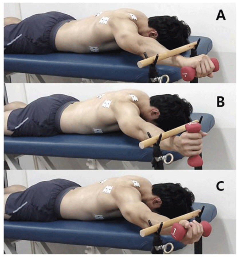
Testing conditions (PD, NT, and PU) were randomly assigned and all tests were performed by the participants using their dominant arm.27 The EMG activities of the UT, LT, SA, and IS and the kinematic data of scapular motion were tested during prone arm lift exercises in three arm positions (PD, NT, and PU; Figure 1). The experiment was conducted in a prone position while holding a 1 kg dumbell. The subject’s humerus and scapulae were hand positioned by the examiner. Then shoulder abduction and flexion were held with maximal PD, NT, and PU, and the scapula was passively posterior tilted until the end range. The arm of the subjects was aligned diagonally overhead by using a goniometer with a shoulder abduction of 145°. After the target bar was placed on the subject’s wrist, the subjects were asked to avoid dropping the target bar. Three trials on the three exercises held for 5 s were performed to measure the EMG activity of the four muscles and the scapular position. A 10 min rest was allowed between conditions (PD, NT, and PU) to minimize the testing effect and fatigue.

The maximum voluntary isometric contraction (MVIC) of the UT, LT, SA, and IS was measured in the manual muscle testing position recommended by Kendall et al.28 to normalize EMG data. EMG data were collected for 5 s. The first and last second of EMG data were discarded, and the remaining 3 s of data were used to determine the mean amplitude of MVIC. The mean of the three trials for MVIC measurement was calculated for data analysis. The means of the three trials for each exercise were then used for data analysis. The collected EMG amplitude of the UT, LT, SA, and IS muscles during each exercise was expressed as a percentage of the mean MVIC (% MVIC).
To obtain the kinematic data of the scapula during shoulder motion, measure the distance of C7 to acromion, The Polhemus Liberty™ was used. The electromagnetic motion tracking system was firmly attached to the skin on the C7 and acromion. According to arm position (PD, NT, and PU), the distance of the C7 to the acromion was collected as the mean of the three trials of each exercise.
One-way repeated-measures ANOVA was conducted to assess the statistical significance of the EMG activities of the UT, LT, SA, and IS and the distances of the C7 spinous process to the acromion. If a significant difference was found, post hoc paired t-tests were performed. Kinematic data were analyzed using an independent t-test. The level of statistical significance was set at α=0.05. In a post hoc paired t-test, Bonferroni correction was performed and set at 0.017 (p=0.05/3=0.017). Statistical analysis was performed using SPSS statistical software.
RESULTS
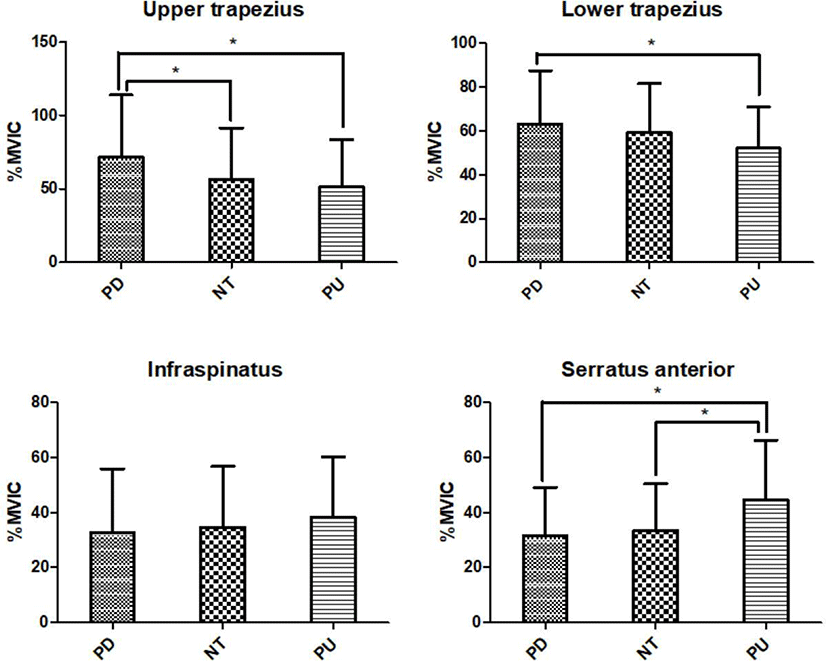
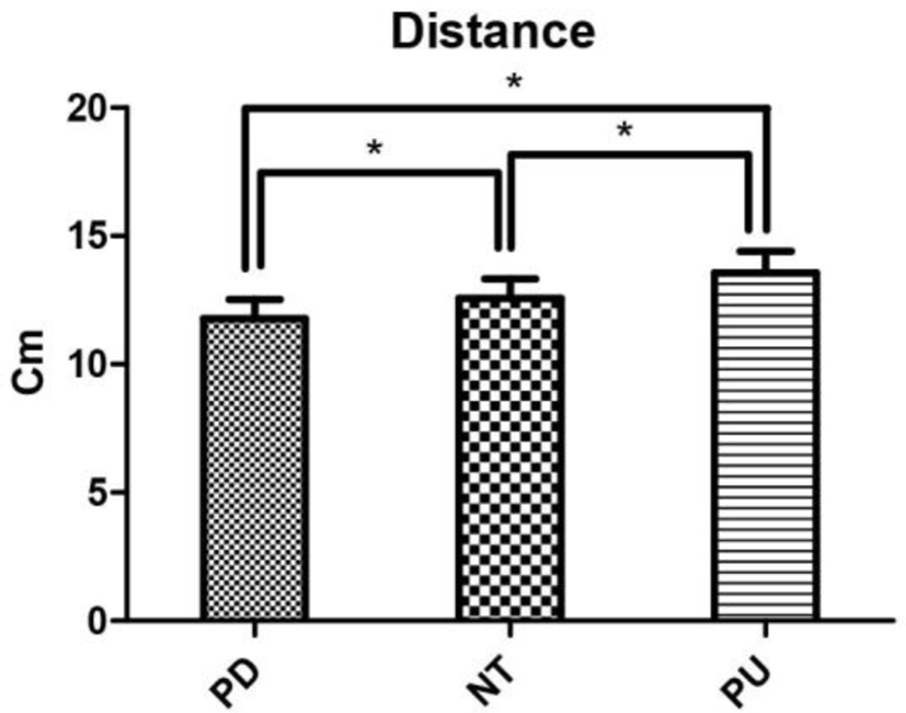
The EMG activities of the UT, LT, and SA significantly differed among the three conditions (PU vs. NT vs. PD; p<0.05). However, the activity of the IS was not significantly different among the three conditions (p>0.05). The post hoc test showed that the muscle activities of the UT and LT were significantly higher in the PD than in the PU (p<0.017). Similarly, the muscle activity of the SA was significantly higher in PU than in PD and NT (p<0.017; Table 1, Figure 2). In addition, the distance from the C7 to the acromion according to the arm position significantly increased in the NT compared with that in the PD; likewise, this distance in the PU increased compared with that in PD (p<0.017; Table 2, Figure 3).

| Arm position | F | p | |||
|---|---|---|---|---|---|
| PD | NT | PU | |||
| Distance (cm) | 11.77±0.75 | 12.57±0.75 | 13.55±0.86 | 6.582 | <0.01* |

DISCUSSION
This study was conducted on 32 subjects to investigate the muscle activity of the shoulder muscles and the motion of the scapula according to three arm positions (PD, NT, and PU) during a prone arm lift exercise. As a result, the activity of the SA was significantly higher in the PU arm position than in the two other positions (p<0.017). The activities of the UT and LT were significantly the highest in the PD position (p<0.017). However, the IS muscle activity had no significant difference according to the three arm positions (p>0.05). The distance from the C7 spinous process to the acromion of the scapula was significantly different among all three conditions, and the distance was the smallest in the PD arm position and the largest in the PU position (p<0.017).
Several potential mechanisms may explain why the activities of the SA, UT, and LT significantly differed according to the three arm positions. First, the direction of the moment may influence the activities of the SA, UT, and LT. In this study, the humerus internally rotated to take the PD position and externally rotated to have the PU position. The SA is the abductor of the scapula, and the UT and LT are the adductors of the scapula. The direction of the humeral internal rotation moment is similar to that of the scapular adduction moment, and the direction of the humeral external rotation moment is similar to the direction of the scapular abduction moment. For this reason, the UT and LT, which are scapular adductors, are more activated in the PD position; the SA, which is a scapular abductor, is activated in the PU position. The distance from the C7 spinous process to the acromion of the scapula was significantly shorter in the PD position than in the PU position. Therefore, scapular abduction occurred in the PU position, where the distance from the C7 to the acromion was the longest, and scapular adduction occurred in a PD position.
The significant difference in the activities of the SA, UT, and LT according to arm position can be explained by the length–tension relationship.29 The position of the scapula is affected by the humeral position during the prone arm lift exercise. The scapula is adducted to the PD position more than that to the two positions other; the UT and LT had a shorter range than that in the PU position. Therefore, the muscle activities of the UT and LT in the PD position and the muscle activity of SA in the PU position were higher than those in the other positions.
The muscle activities of the UT, LT, and SA significantly differed according to arm position possibly because of the change in the mobility and flexibility of the scapula and glenohumeral joint. Robert-Lachaine et al.26 reported that the full-can exercise increases the scapulohumeral rhythm, and empty-can exercise increases the movement of the scapula during rehabilitation exercises of the shoulder. They explained that the increased motion of the scapula during the empty-can exercise compensates for the limitation of the glenohumeral joint movement. In this study, the distance between the C7 and acromion and the muscle activities of the UT and LT increased in the PD position requiring humeral internal rotation.
The IS muscle activity had no significant difference according to three arm positions possibly because the change in muscle length according to the three arm positions was smaller than that in other muscles. The SA, UT, and LT are thoracoscapular muscles, and the IS is a scapulohumeral muscle. Therefore, unlike the UT, LT, and SA originating from fixed structures such as the spine or rib, the IS is from the scapula, a non-fixed structure, and is thought to have less changes in the length than other muscles. Because changes in muscle length were small, the difference in muscle activity would have been small.
Several limitations should be considered in this study. Since this study was performed with young normal adults, care should be taken in generalizing the results. This study was conducted to investigate the effect of three arm positions on the activities of the shoulder muscles during prone arm lift exercise only. In further studies, various exercises for training the shoulder complex should be explored.
CONCLUSION
Arm position affects the muscle activity of shoulder muscles during a prone arm lift exercise. The muscle activity of the SA increased in the PU position, and the activities of the UT and LT increased in the PD position during prone arm lift exercise. Therefore, we recommended that the arm in the PU position should be taken to strengthen the SA muscle, and the arm should be in the PD position to strengthen the LT or UT.


