


INTRODUCTION
The gluteus maximus (GM) is a powerful hip extensor and external rotator.1,2 Of the hip extensors, the GM plays an important role in functional activities of daily living, including sit-to-stand, stair climbing, and maintaining an upright posture while walking.3 However, people with decreased GM activity often have uncontrolled pelvic and lumbar movements during these activities.4 Furthermore, changes in pelvic posture can affect the length–tension relationship of the GM and reduce its stabilization capacity.5 Improving GM activation through strengthening exercises is important to prevent injury.6
The Pilates method is recognized as the best way to improve spinal stability by increasing trunk and GM muscle activity using mat exercises.7 Pilates exercises are designed to improve strength and posture with special emphasis on the trunk muscles.8,9 Bergson et al. demonstrated that variation in postural changes was due to augmented activation patterns of the multifidus (MF), GM, and oblique muscles during four Pilates exercises.10 Kim et al. also reported that three Pilates exercises significantly affected GM and MF muscle activity.11 Many studies have examined the effects of Pilates exercises on the trunk and GM muscles, but evidence for the most effective exercise for improving GM muscle activity is lacking.
Although Pilates exercises, including swimming (SW), one leg kick (OLK), shoulder bridge (SB), and leg pull front (LPF), strengthen the hip muscles, few studies have objectively quantified changes in hip strength during these exercises. Also, there is few information on lumbo-pelvic motion during Pilates exercises for the GM. This information is not well known, so it is very important for effective strengthening exercises and injuries prevention programs. Therefore, additional objective data are essential to be able to personalize GM exercises to meet patient needs.
In this study, we quantified the activity of the trunk and GM muscles and pelvic rotation angle during the OLK, SW, LPF, and SB. Our results provide valuable information about trunk and GM muscle activation during Pilates exercises without unwanted lumbo-pelvic motion and may aid clinical decision making and help personalize injury-prevention programs.
Methods
Twenty healthy women (mean age 27.53±5.97 years; mean height 163.27±5.24 cm; mean weight 48.67±6.28 kg) volunteered for this study. Individuals with known medical problems, past spinal or abdominal surgery, or episodes of back, shoulder, or hip pain requiring treatment during the previous 6 months were excluded.11 Before beginning the study, the principal investigator explained all of the procedures and the subjects provided written informed consent. The study protocol was approved by the institutional review board of Inje University, Korea (INJE 2022-02-009-001).
A Delsys Trigno Wireless electromyography (EMG) system (Delsys, Boston, MA, USA) with a Trigno EMG sensor was used to collect surface EMG data. Analog signals recorded from each muscle were converted into digital signals and processed using DELSYS EMG Works acquisition software on a personal computer. The EMG signal sampling rate was set at 2,000 Hz and the band pass filter was set at 20–450 Hz. The EMG signals for each muscle were analyzed based on the root mean square (RMS). The site for each electrode was shaved and then cleaned with cotton and alcohol to reduce skin impedance. The dominant leg was determined by asking the subject to kick a soccer ball, and the kicking leg was determined to be the dominant leg.12 All participants were right-leg dominant. To record bilateral transverse abdominis/internal oblique (TrA/IO) activity, EMG sensors were placed at a location approximately 2 cm medial and inferior to the anterior superior iliac spines.13 For the bilateral external oblique (EO), the electrodes were placed inferiorly and laterally to the 8th rib.14 EMG sensors were attached at a location 2 cm lateral from the L5 spinous process to record bilateral MF activity.15 For the GM, the electrodes were located halfway between the greater trochanter and second sacral vertebra in the middle of the muscle belly, at an oblique angle at or slightly above the level of the trochanter.16 The maximum voluntary isometric contraction (MVIC) was measured in a manual muscle testing posture to normalize the EMG values for each muscle.1 Data were collected for three trials for 5 s each. The first and last second were excluded and 3 s of mean EMG signal data were used as the %MVIC. The subjects rested for 1 min between trials to prevent muscle fatigue.
To measure the pelvic transverse rotation angle, a smart phone was connected to the wooden holder of a smart phone-based measurement tool (SBMT). Previous research has shown that pelvic rotation measurements using the SMBT have excellent reliability.17 Before measuring pelvic rotation, an inclinometer application (Clinometer level and slope finder; Plaincode Software Solutions, Stephanskirchen, Germany) was calibrated by placing the SBMT on a flat surface. The base of the SBMT was located at both the anterior superior iliac and posterior superior iliac spines and the inclinometer application was used to record the pelvic rotation angle for each exercise.
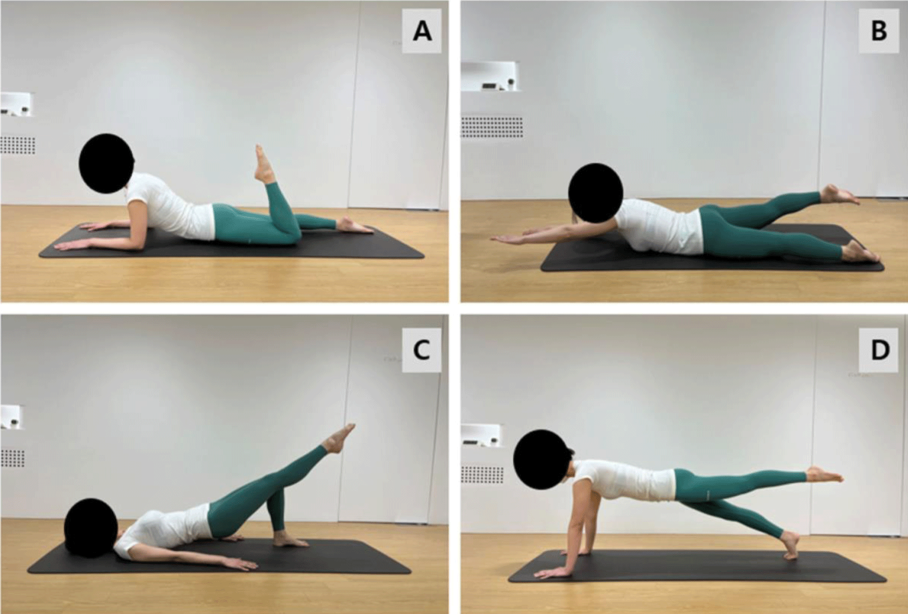
Each participant performed the OLK, SW, SB, and LPF (Figure 1). All exercises were explained by a trained Pilates instructor. The subjects learned how to perform the four exercises through 10 min of instruction. The OLK exercise started with lumbar static hyperextension (e.g., maintaining a static lordotic lumbar posture for the entire exercise) with the hands placed on the mat. The subjects alternated leg flexion and extension, kicking the heel toward the buttock and then extending the leg (Figure 1). For the SW exercise, the subjects assumed a prone position, with arms extended overhead and the trunk and legs lifted (Figure 1). For the duration of each exercise, the subjects maintained their neck in alignment with the spinal column (a Pilates principle). The SB exercise used unilateral hook-lying, as described for the bilateral bridge, except that the contested lower limb remained on the table (0° at the hip and knee). The subject then pushed the foot into the table with the tested limb to raise the pelvis until 90° of knee flexion was achieved ipsilaterally (Figure 1). For the LPF exercise, the subjects assumed hip extension in the plank position (Figure 1). The subject was asked to perform a single leg lift to a predetermined target bar. The target bar was placed at the level of 10° hip extension, as measured using an inclinometer.18 The Pilates principles of body alignment, breathing control, and abdominal muscle control were emphasized throughout the session. Each subject, in random order, performed the four Pilates exercises three times for 5 s each, with a 1 min rest between trials.
The Kolmogorov-Smirnov test was used to determine that each variable was normally distributed. Muscle activity and the angle of pelvic rotation during each Pilates exercise were analyzed using a repeated measured analysis of variance (ANOVA). When necessary, post hoc analyses were performed using the paired t-test. All statistical analyses were performed using the SPSS ver. 18.0 (SPSS, Chicago, IL, USA) with statistical significance set at p<0.05.
RESULTS
Right-side GM activity was highest during the LPF followed by the SB (p=0.016) and SW (p=0.002) exercises. The activity of the left-side TrA/IO was significantly lower during the SW exercise than during the SB (p=0.001) and LPF (p=0.006) exercises. However, right-side TrA/IO activity did not significantly differ during any of the four exercises. Bilateral MF activity was significantly lower (p=0.0001 for both) during the LPF exercise than during the SB, SW, and OLK exercises. Right-side EO activity was highest during the LPF exercise, followed by the OLK (p=0.003) and SW (p=0.003) exercises. Left-side EO activity was significantly increased during the SB compared to the SW (p=0.011) exercises (Table 1).
The angle of pelvic rotation was significantly increased during the LPF exercise compared to the SW (p=0.008) and OLK exercises (p=0.004) (Table 2).
| Pilates exercise | p | ||||
|---|---|---|---|---|---|
| One leg kick | Shoulder bridge | Swimming | Leg pull front | ||
| Pelvic rotation angle (°) | 2.44±1.36a) | 4.98±2.73 | 3.58±1.56 | 6.49±3.66b,c) | 0.000* |
DISCUSSION
We compared the EMG activity of the trunk and GM muscles and pelvic rotation angle during four Pilates exercises and found that the LPF facilitates TrA/IO and GM muscle activities, while the OLK minimizes pelvic transverse rotation. This suggests that the four exercises should be recommended for selective trunk and GM muscle activation in exercise programs to minimize unwanted lumbo-pelvic motion.
During the OLK and SB movements, all trunk and GM muscles showed relatively high muscle activity (20% MVIC). Sekendiz et al. previously showed that the OLK and SB exercises increased strength and endurance of the trunk muscles in healthy women.19 In our study, participants were able to perform alternating leg flexion and extension contractions during the OLK and SB exercises without uncontrolled pelvic movements. Instability during the OLK and SB exercises causes co-contraction of all trunk and GM muscles to maintain balance. Therefore, we recommend the OLK and SB to increase trunk stabilization and GM muscle activity.
We observed significantly greater TrA/IO and EO activity during the LPF than during the other Pilates exercises. This is probably because the LPF has a smaller base of support than the other movements. As the base of support becomes unstable, more abdominal muscles are recruited to maintain balance, stability, and load on the lumbar spine. When the upper or lower limbs are lifted from the ground, the base of support and stability are decreased, and trunk stabilization muscles are contracted to maintain spinal posture.20 In accordance with this, our results demonstrate that the TrA/IO and EO were more activated during the LPF than during the other Pilates exercises.
MF activity was significantly greater during the SW than during the other Pilates exercises. There were no significant differences in MF activity between the SW and OLK exercises (p=1.000). Our results agree with Maryela et al.,21 who reported that MF activity was highest during SW among the prone Pilates exercises (SW, double leg kick, and OLK). SW is a dynamic movement in which one arm and opposite leg are lifted simultaneously. Previous studies have shown that back muscle activity gradually increases when the arms and legs are far from the axis of rotation of the lumbar spine.15,22,23 Furthermore, during SW, the MF is activated to maintain spinal alignment by resisting the torsional forces created by lifting the opposing upper and lower extremities.24 Therefore, MF activity during SW would be expected to be higher than during the other Pilates exercises because SW has a longer lever arm and requires more muscle activity to maintain spinal alignment against the greater rotational force on the lumbar spine.
We also found that GM activity was highest during the LPF, but it was not significantly different from the LPF and OLK exercises. This may be because the LPF and OLK have a smaller base of support than the other exercises.25 Because the trunk is in an unsupported position during the LPF and OLK, GM activity is increased to provide adequate support and stability.25 Therefore, GM activity would need to increase to compensate for the smaller bases of support during the LPF and OLK. Based on our results and those of previous studies, the LPF and OLK may be effective methods to increase activation of the GM.
The pelvic angle was the smallest during the OLK and largest during the LPF. The OLK has a greater base of support than the other Pilates exercises because both elbows, the lower extremities, and pelvis are on the mat. However, the LPF creates more instability because it has a smaller base of support than the other Pilates exercises, which may increase pelvic rotation by adding hip extension. The OLK had the lowest pelvic rotation angle, therefore, the OLK is recommended to reduce unwanted pelvic movements while increasing trunk muscle activity.
There were several limitations to this study. First, our results may not be generalizable to other populations because the subjects of this study were young healthy individuals. Second, these results apply only to traditional Pilates mat exercises. Third, we could not confirm whether the observed muscle activity was bilateral. Future studies should include more subjects, including patients with lower back pain.
CONCLUSIONS
This study investigated how four Pilates exercises affect trunk and GM muscle activity and pelvic rotation angle. Lt. TrA/IO, EO, and Rt. GM of muscle activation was the highest during LPF. Rt. TrA/IO of muscle activation was the highest in OLK. MF of muscle activation was the highest in SW. For GM Muscles, LPF and OLK exercise, and MF target are SW exercise, and if the abdominal muscle is a target, it is recommended to do LPF and OLK exercise.