INTRODUCTION
Shoulder dysfunction and pain are common musculoskeletal problems among elite athletes as well as in the general public, with a steady increase in prevalence with aging.1-4 While various muscles contribute to shoulder movements, an inadequate level of lower trapezius (LT) muscle activity accompanied by an excessive level of upper trapezius (UT) muscle activity has often been reported.5,6 Some factors may be involved in an increase in the forward tilt of the scapula and thoracic joints and in an excessive elevation of the clavicle in the chest, while causing a rotator cuff impingement in lifting. Impingement syndrome exhibits the highest prevalence among shoulder problems, accounting for 36%.7,8 In a previous study on patients with impingement syndrome, excessive UT and low LT muscle activities were observed in these patients compared with that noted in the control group;9 moreover, the patients showed changes in movement patterns, including reduced scapular external rotation, upward rotation, and posterior tilt.10,11
Reduced shoulder muscle strength has been previously suggested to be the main factor leading to imbalance in the shoulder stabilization system and ultimately to inappropriate dynamics between the shoulder and scapula.12 Meanwhile, muscle imbalance and abnormal muscle activity ratio have been reported as other main factors in recent studies.13,14 Changes in shoulder muscle activities have been shown to reduce upward rotation and narrow the subacromial space to cause subacromial impingement syndrome (SIS).15 Hence, well-coordinated activities of the UT, LT, and serratus anterior muscles are thought to be required in the upward rotation of the scapula. A previous study examining the relative scapula activities found that patients with SIS showed higher UT/middle trapezius (MT) and UT/LT ratios than those of a control group.16,17 In another study, arm lifting led to a decline in LT activation and a consequent elevation in UT activity pertaining to scapula contraction, the result of which was a higher UT/LT ratio.18 Thus, exercise that can stimulate low UT activity and high MT and LT activities without narrowing the subacromial space is predicted to assist in treating patients with shoulder pain.19
An ideal shoulder rehabilitation program should comprise an exercise focused on muscle activation during scapula upward rotation (serratus anterior, UT, and LT).20 In contrast, the activation of muscles during scapula downward rotation should be minimized, and the intervention should effectively increase the subacromial space during the overhead movement of the arm.18,21 However, most conventional rehabilitation exercises use overhead positioning,18,22-24 which has been shown to induce a potential compressive force on the subacromial space and consequently increase the probability of subacromial surface compression on the rotator cuff; therefore, shoulder muscle strengthening exercise with arms lifted at an angle ≥90° should not be prescribed.25,26 Alternatively, a scapula retraction exercise wherein the strengthening of scapula stabilization muscles is promoted but the posture that may cause an impingement is avoided, is recommended in the early phase of rehabilitation.27-30
Generally, rowing and scapula retraction exercises are known to enhance the performance of shoulder muscles through activation of trapezius muscles.16,31 Consequently, these exercises have been widely applied by clinicians. Additionally, the retracted position of the scapula has been shown to improve muscle functions and reduce impingement symptoms in patients.30,32 The general pattern of movements in scapula retraction exercise is external rotation and posterior tilt, both of which increase the subacromial space and are generally applied in patients with impingement syndrome.18,33,34 The seated cable row exercise (SCRE) is most frequently prescribed by specialists for simultaneous improvement in conditioning and fixation of imbalance based on increased shoulder muscle strength through scapula stabilization. However, the SCRE is a basic multi-joint upper body exercise for improving the angles of the posterior shoulder, back, and elbow extensor muscles and can be performed by athletes and non-athletes.35-37 The modified SCRE (MSCRE) is a revised form of SCRE, which selectively increases the LT and MT muscle activities. The MSCRE uses a narrow grip,38 wherein the position of the weight cable should be at 90° of the elbow and the elbow in the last phase of pulling should not part from the side of the trunk or move behind the trunk.14,18,39
In previous studies on scapula contraction exercises, the trapezius muscle activities were compared among various contraction exercises, mostly performed in the prone, sitting, and standing postures.38,40,41 However, the effects of varying trunk angles on trapezius muscle activities in scapula contraction in a sitting posture remain unknown. It is predicted that the variation in trunk angle could induce changes in scapula dynamics and the ratio of different trapezius muscle activation.42,43 Therefore, this study aimed to determine the changes in UT, MT, and LT muscle activities and the UT/MT and UT/LT ratios according to the trunk angle during the MSCRE in individuals without shoulder pain.
METHODS
Sample size estimation was obtained using the G-power software ver. 3.1.9.7. In a pilot study on five participants, the testing power at 0.8, effect size at 0.69, and significance level at 0.5 led to a sample size of 21. Considering the drop-out rate at 20%, the number of participants in this study was determined as 25; however, a total of 23 individuals participated in this study, excluding two individuals who complained of pain in the elbow and shoulder during practice. The participants were recruited from the J Gym in Ulsan, South Korea (Table 1). Inclusion criteria were as follows: healthy adults aged 20–39 years without pain or angular restriction in the GH and SC joints and without any neurological damage. In terms of sex ratio, the selected participants were 11 male and 12 female adults. Exclusion criteria were as follows: individuals with shoulder pain or a history of shoulder surgery within the past six months; individuals with damage or pain in the lower limbs or trunk within the past three months; and individuals with musculoskeletal or cardio-pulmonary disease. Any individual who complained of discomfort during the experiment was also excluded. Details on participant demographics, such as age, height, and weight, were collected at the time of recruitment. Signed written consent was obtained from each participant prior to the experiment. This study was approved by the Institutional Review Board of Daegu University.
| Variables | Mean±SDa | Range |
|---|---|---|
| Age (yrs) | 29.8±7.7 | 20–39 |
| Height (cm) | 165.1±9.1 | 159–181 |
| Weight (kg) | 65.14±8.2 | 56–85 |
| Gender(male/female) | 11/12 | |
The surface electromyography (sEMG) Noraxon DTS system (Noraxon Inc, Scottsdale, AZ, USA) was used to measure the UT, MT, and LT muscle activities. The muscle activity data from each muscle type were collected and analyzed using the Noraxon Myo Research XP 1.06 software, where the EMG sampling rate was set at 1,500 Hz.44 The intra-rater reliability of sEMG was high (intraclass correlation coefficient = 0.75–0.98).45
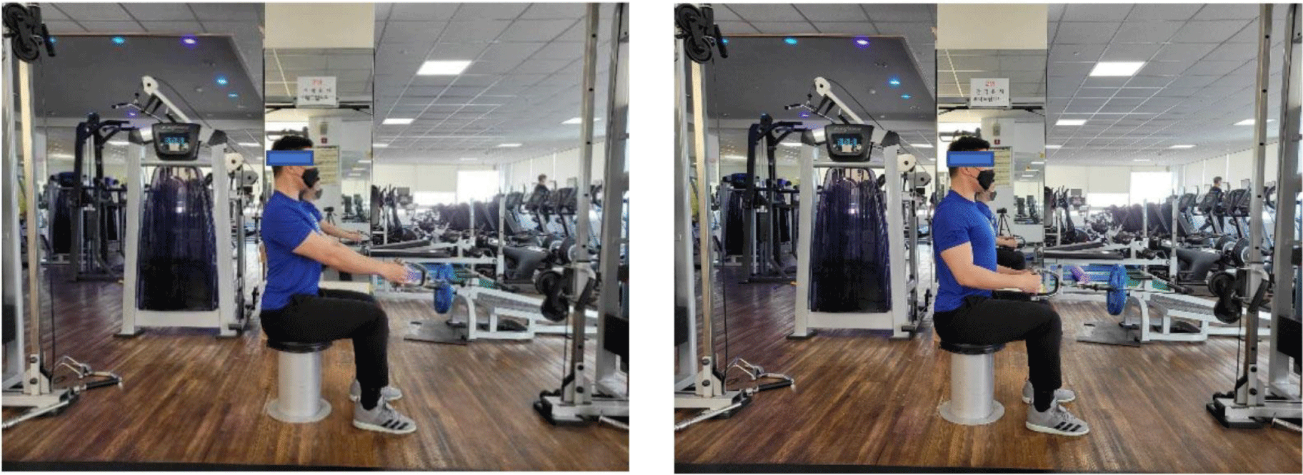
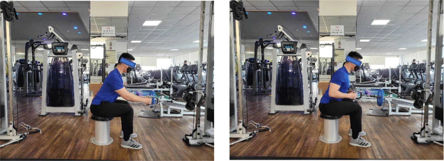
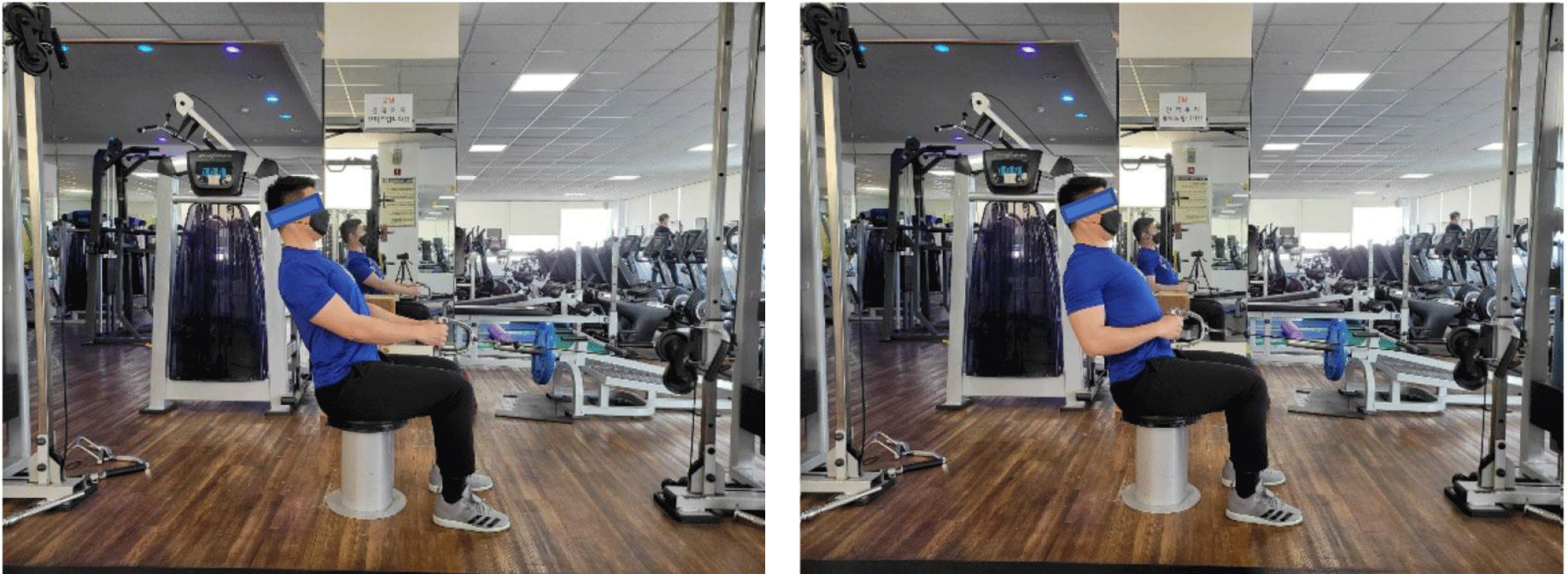
This study involved a cross-sectional study design. The experiment was performed on the dominant hand used by the participants for eating and writing. Each participant was given a 30-min orientation on the exercise method, followed by a process of familiarization with the practice of motions in the exercise. To minimize the bias or personal judgment of each participant regarding the experiment, the Excel program (Microsoft Corp., Redmond, WA, USA) was used to randomize the order of motions in the exercise. Prior to the experiment, the participants were guided to perform adequate stretching and warm-up exercises. A metronome set to 60 beats per min was used to ensure 5-s eccentric, 5-s isometric, and 5 s eccentric contractions,46 and the weight was set to 65% of 1 RM for standardization. The MSCRE was divided into the starting position, pulling phase, and lowering phase. In the starting position, the participant aligns the feet to the same width as the shoulders in a relaxed sitting posture. The cable with a handlebar was positioned at the cable column at elbow height and a narrow grip was applied. The elbow was in complete extension, while the handle bar was maintained immediately above the legs. The head and neck were maintained in an alignment immediately above the shoulders, trunk, and hip during the exercise, while the participants gazed forward at a fixed point. All repeated motions started and ended with this position. In the pulling phase, the trunk alignment was maintained, while the chest was thrust upward and outward and the scapula showed contraction or abduction. The participants were asked to bend their elbows to pull the bar backward but not behind the trunk. In the lowering phase, the trunk alignment was maintained as in the previous phase, and the handlebar was controlled in return to the starting position. The muscle activity and ratio at the following three trunk angles during the MSCRE were analyzed in this study: upright posture (UP) 0°, forward lean (FL) 30°, and backward lean (BL) 20° (Figure 1, 2 and 3).
The experiment was performed by one rater and one researcher with a clinical career in physical therapy spanning three years. The rater recorded and analyzed the EMG findings, and the researcher supervised the participants’ postures and controlled the study flow. Two electrodes were attached to the center of the muscle belly for the UT, MT, and LT muscles, parallel to the muscle fiber. In the area of electrode attachment, the hair was shaved and the skin was disinfected with alcohol to lower the resistance. For the UT, the electrode was attached to the middle point between the acromion of the scapula and seventh cervical vertebra. For the MT, the electrode was attached to the middle point between the medial border of the scapula and spine. For the LT, the electrode was attached to a point approximately 5-cm below the spine of the scapula. The distance between the electrodes was fixed at 2 cm. The sEMG electrodes were arranged according to the suggestions by Criswell (2010).47 During the last motion in the pulling phase, the UT, MT, and LT muscle strengths were measured at maximum values during the 5-s isometric contraction, and the mean of triplicate measurements was obtained. The signal of each muscle type was simultaneously analyzed by the root mean square, and the mean of the middle 3 s of the 5 s, excluding the first and last seconds, was obtained. Using the manual muscle test for the UT, MT, and LT, as suggested by Kendall et al. (2005),48 the muscle activities at the maximal voluntary isometric contraction (MVIC) were analyzed and the activities were converted to percentage (%MVIC), according to the measured motion. A rest period of approximately 3 min was allowed to minimize muscle fatigue across different motions.
All data in this study were statistically analyzed using SPSS 26 (SPSS Inc., Chicago, IL, USA). Shapiro-Wilk tests were used to determine whether data followed a normal distribution to approve the use of parametric techniques. The UT, MT, and LT muscle activities and the UT/MT and UT/LT ratios according to the three trunk angles during the MSCRE were compared using one-way repeated measures (RM) analysis of variance (ANOVA). For descriptive statistics, the mean and standard deviation were presented, and in a case of significant variation, the Bonferroni post-hoc test was performed to indicate the value of follow-up. The level of significance was set at α=0.05.
RESULTS
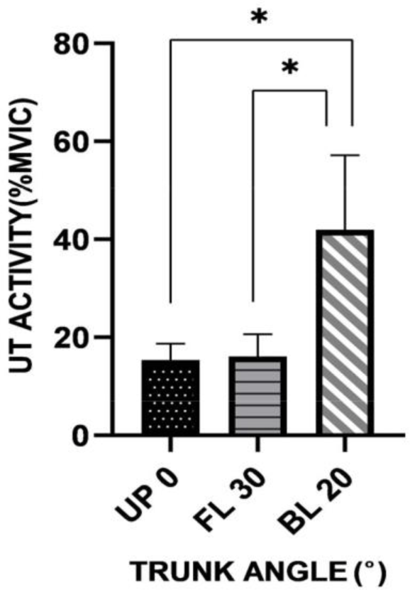
The muscle activity of the UT according to trunk angle is shown in Table 2. The highest UT activity was observed at BL 20° and the lowest was observed at UP 0°. The one-way RM ANOVA of the UT activity showed a significant difference in angle (p<0.05), and the post-hoc analysis showed a significant difference in posture between UP 0° and BL 20° and between FL 30° and BL 20° (p<0.0167) (Table 2) (Figure 4).
The muscle activity of the MT according to trunk angle is shown in Table 3. The highest MT activity was observed at UP 0° and the lowest was observed at BL 20°. The one-way RM ANOVA of the MT activity showed a significant difference in angle (p<0.05). The post-hoc analysis showed no significant difference across all the angles (Table 2).
| Trunk angle | UP 0° | FL 30° | BL 20° | F(P) |
|---|---|---|---|---|
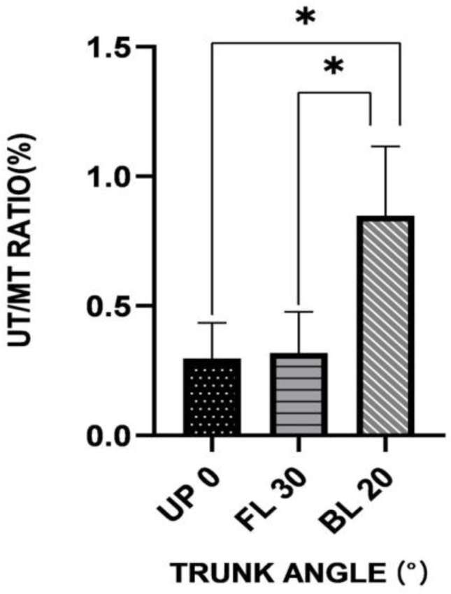
| UT/MT ratio | 0.30±0.13 | 0.32±0.15 | 0.84±0.27 | 74.999 (.000) |
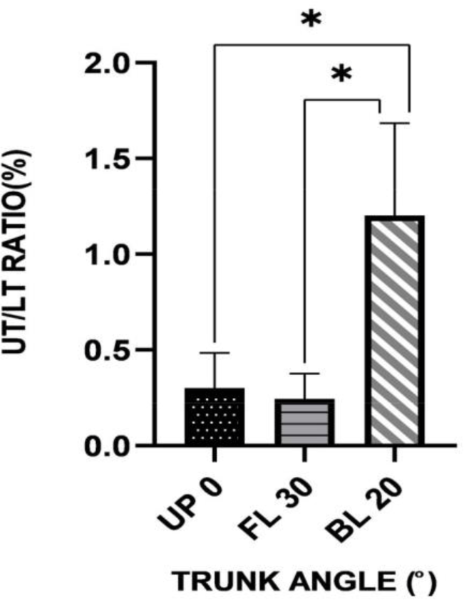
| UT/LT ratio | 0.30±0.18 | 0.21±0.13 | 1.20±0.48 | 98.956 (.000) |
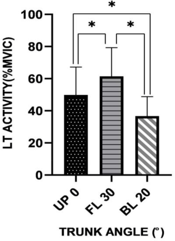
The muscle activity of the LT according to trunk angle is shown in Table 4. The highest LT activity was observed at FL 30° and the lowest was observed at BL 20°. The one-way RM ANOVA of the LT activity showed a significant difference in angle (p<0.05), and the post-hoc analysis showed a significant difference in posture between UP 0° and FL 30°; UR 0° and BL 20°; and FL 30° and BL 20° (p<0.0167) (Table 2) (Figure 5).
The UT/MT ratio according to trunk angle is shown in Table 5. The ratio was the highest at BL 20°, and the lowest at UP 0°. The one-way RM ANOVA of the UT/MT ratio showed a significant difference across the trunk angles (p<0.05), and the post-hoc analysis showed a significant difference in posture between UP 0° and BL 20° and between FL 30° and BL 20° (Table 3) (Figure 6).
The UT/LT ratio according to trunk angle is shown in Table 6. The ratio was the highest at BL 20° and the lowest at FL 30°. The one-way RM ANOVA of the UT/LT ratio showed a significant difference across the trunk angles (p<0.05), and the post-hoc analysis revealed a significant difference in posture between UP 0° and BL 20° and between FL 30° and BL 20° (Table 3) (Figure 7).
DISCUSSION
This study determined changes in the muscle activity and ratio of the UT, MT, and LT according to the three trunk angles during the MSCRE and also identified the trunk angle at which the muscle activity was the highest for each muscle type. The key finding of this study is that the trunk angle has a significant influence on the activity of specific trapezius muscles and on the ratio of trapezius muscle activities during the MSCRE. The sEMG during the MSCRE showed that the UT activity was the highest at a trunk angle of BL 20° but the lowest at FL 30°. The UT exhibited 73% muscle activity at BL 20° for the highest MVIC. For the LT, the activity was the highest at FL 30° and the lowest at BL 20°, while the LT exhibited 62% muscle activity at FL 30° for the highest MVIC. This is presumed to be due to the variation in the activated muscle according to the trunk angle in line with the variation in the vector for the line of gravity. At UP 0°, the MT showed a slightly higher muscle activity than the other trapezius muscles, but the lack of significant difference in the MT activity across the trunk angles implies that the variation in the vector was insufficient to cause significant variation in the MT activity.
According to McCan et al. (1993), an intermediate level of activation requires 21–50% MVIC, while a high level of activation requires ≥50% MVIC, suggesting that the MVIC in this study should be regarded as a high level of activation.49 In another study, an intermediate level of muscular activation (20–40% MVIC) was claimed to be appropriate for retraining the control of dorsal scapular nerve roots.1,50 In this study, the result for FL 30° showed a low level of UT activity (12–20% MVIC) and a high level of LT activity (43–78% MVIC; an intermediate level of activation), suggesting an appropriate role in each phase of the nerve root control and rehabilitation and muscle strengthening exercise protocols.49,51 Thus, the increase in LT activity associated with low levels of UT activity at UP 0o and FL 30° is considered to be necessary to reduce UT activity to relieve pain and excessive UT activity when patients have shoulder pain.
In a previous study by Larsen et al., the UT, MT, and LT activities were measured and each muscle type was selectively activated in participants with different occupations.52 The study reported that the MT or LT activity in patients with SIS was lower than that in healthy individuals. Additionally, Lopes et al. showed that the UT activity in arm lifting weight exercise increased by 12% in patients with SIS and scapula dyskinesis than that noted in healthy individuals.53 The study suggested that the increase in UT activity was a pattern of compensatory activation to assist with arm lifting or the control of scapula dyskinesis. While the exercise in this study varied from that used in previous studies with regard to the methodology and the fact that the participants were not patients but healthy individuals, the UT and LT activities agreed with the previously described higher UT and low LT activities observed in patients with SIS than in controls52,53. Thus, for shoulder rehabilitation rather than selective UT muscle strengthening, the backward trunk tilt during the MSCRE seems unsuitable, while for selective LT muscle strengthening, the trunk angle of FL 30° during the MSCRE is likely to be ideal.
In this study, the lowest UT/LT ratio (0.21) was observed at FL 30° during the MSCRE, while the lowest UT/MT ratio (0.30) was observed at UP 0°, and the highest UT/LT ratio (1.20) was observed at BL 20°. In terms of the balance of trapezius muscles, a high ratio indicates an imbalance and excessive activation of the UT.54 While the MSCRE is not considered an optimum exercise for scapular balance rehabilitation based on the low UT/LT ratio, the observed values indicated the utility of the MSCRE in the nerve root control rehabilitation rather than the utility of the trapezius muscle strengthening exercise.55 The ratio at FL 30° reflecting a high level of LT activity in association with a low level of UT activity is ideal for rehabilitation exercises. In a previous study evaluating shoulder muscle activity, a higher UT/LT ratio (in agreement with the result in this study for the trunk angle of BL 20°) was observed in patients with impingement syndrome than in participants of the control group.18,21 According to the latest trend in rehabilitation, kinetic chain exercise is recommended throughout the rehabilitation program rather than independent strengthening of shoulder muscles, with subsequent gradual integration into other parts of the body.56 To summarize, as the UT/LT and UT/MT ratios are viewed as critical factors in shoulder stability based on the synergistic rather than the independent activities of scapula muscles to produce a controlled set of scapula movements, care should be taken regarding the backward tilt of the trunk for the purpose of shoulder rehabilitation. Furthermore, an appropriate ratio should be selected for the trunk angle during exercise for strengthening of all the three trapezius muscles rather than of that of just a specific trapezius muscle.52,57
This study has several limitations that warrant discussion. First, all the participants were young healthy adults without any shoulder pain or dysfunction. It is unclear whether patients would exhibit the same level of muscle activity during the scapula contraction exercise, and it should be noted that each patient may display varying levels of UT, MT, and LT activities during the MSCRE. Thus, generalization of the study results to patients requires precautions, and further studies should investigate the effects of the MSCRE on the UT/LT and UT/MT ratios in patients with SIS or scapula dyskinesis. Second, the contribution of other shoulder muscles, such as the serratus anterior and rhomboids muscles, was not considered. The results of this study indicate the balance and imbalance of the trapezius muscles at specific trunk angles. However, as the shoulder muscles exhibit synergistic effects, the functions of the serratus anterior and rhomboids muscles should be considered simultaneously and should not be interpreted independently from the functions of other shoulder muscles. Third, the trunk angles were varied only as far as three angles; hence, the large gap among the three angles prevents the accurate prediction of muscle activity at other angles.
CONCLUSIONS
This study determined the muscle activity and ratio for the UT, MT, and LT based on three trunk angles during the MSCRE in healthy male and female participants in their 20s and 30s. The angle at which each muscle exhibited the highest activity was identified, and the results showed that the UT, MT, and LT showed the highest activity at different angles. The UT/LT ratio was the lowest at FL 30°, and the UT/MT ratio was the lowest at UP 0°, while the UT/LT ratio was the highest at BL 20°. At present, several studies suggest the use of various exercises to strengthen or increase the activity of a specific muscle type, but the findings in the present study and several other notable studies predict that the therapeutic goal should be recovery of the normal and balanced activities of the trapezius muscles in rehabilitation for shoulder pain. Further studies are warranted to develop exercise protocols for the recovery of trapezius and serratus anterior muscle balance and to investigate the simultaneous and balanced activation of other shoulder muscles and muscle activities according to more diversified trunk angles.


