INTRODUCTION
Stress urinary incontinence (SUI) is involuntary urinary loss on sneezing, coughing, or physical exertion.1,2 SUI contributes to quality of life regarding to psychosocial factors as well as physical limitations.3 The association between SUI and pelvic floor muscles (PFM) function is well reported.4-6 The PFM are constituted of the urogenital diaphragm and the pelvic diaphragm. The levator ani muscles affect to continence by decreasing hypermobility of the bladder neck as dorsocaoudal movement,7,8 while the sphincter is contribute to increase urethral close pressure.7,8 Thus, for mild to moderate SUI, PFM training is a first guide-line treatment for stress and mixed urinary incontinence.
However, the PFM alone could not be able to produce enough force or tension to resist intra-abdominal pressure and maintain continence during coughing, sneezing and jumping, based on the PFM architecture.9 This represents that other anatomical structures, such as trunk or hip joint muscles (HJM) may be important for maintaining normal PFM function.9-11 The trunk or HJM could be important structures to consider in sacroiliac joint dysfunction and incontinence, because of the anatomical location.11 The trunk or HJM may facilitate co-contractions of PFM and may be appropriate target muscles for non-invasive treatment or rehabilitation of SUI.11
PFM training was generally guided while selected or combined with contractions of the trunk and HJM, such as hip extensor, adductors, abductors, and external rotator muscles.12 Previous studies suggest strong relationship between PFM and core/hip region that could support the intervention in PFM training. Hip adductor, gluteal muscle and abdominal muscle contraction could give synergistic muscle contraction with the PFM in healthy subjects.13 Also, magnetic resonance imaging and electromyography (EMG) have investigated that the pubococcygeus, the iliococcygeus, the fossa ischioanalis, and the gluteus maximus are anatomically and functionally linked. Other studies also reported a coactivation between PFM and abdominal muscle by surface EMG during PFM contractions.14,15 Another study determined that not combined with hip-adductors contraction, but only gluteal-muscle co-activation with PFM contraction resulted in bladder-neck elevation as continence mechanism.16 Thus, there is still no consensus on the role of HJM contraction on the PFM functions and continence mechanism.
It has been supposed that the HJM are related to the continence mechanism and that HJM weakness or deficiency could undermine the normal function. There is a lack of studies confirming the association between PFM and HJM strength and incontinence symptom in women with SUI. For establishing an accurate target for non-invasive treatment or rehabilitation of SUI, it is necessary to determine whether there is a correlation between the HJM strength, such as hip extensor, abductor, and adductor, and quality of life (QoL) of incontinence symptoms.
Therefore, the purpose of our study was to confirm the relationship between QoL of incontinence symptoms measured by incontinence quality of life questionnaire (I-QOL) and PFM strength, HJM strength in women with SUI. We hypothesized that not only PFM muscle strength, but also the HJM strength would be associated with QoL of incontinence symptoms in women with SUI.
METHODS
From August to December 2018, the present study was performed in the laboratory setting. G*power calculated sample size for a power of 0.80, an α level of 0.05 and an effect size |ρ| of 0.41, as performed by pilot data (33 subjects) about coefficient between I-QOL score and HJM strength. The sample size was needed at least 41 subjects. Recruitment of participants was performed by advertisements that noted a phone contact; all participants were requested for visiting urogynecology clinic for diagnosis of SUI and were screened for the inclusion and exclusion criteria. Inclusion and exclusion criteria display Table 1.
The forty-two subjects were recruited [age: 42.9±8.1, body mass index (kg/m2): 23.0±3.4, and deliveries: 1.8±0.9]. All participants were informed about experimental protocol and gave written informed consent form approved by the Institutional Review Board of Yonsei University Mirae Campus (approval no. 1041849-201806-BM-056-02), before the present study.
The I-QOL assessed incontinence-specific quality of life.17 I-QOL consists of three factors: avoidance and limiting behaviors, psychosocial impacts, and social embarrassment for incontinence specific quality of life. We applied I-QOL translated Korean version.18 The I-QOL includes 22 items, and a total score is calculated by adding each three factors score. Total score was converted to a 0 to 100 scale, with the higher scores presenting greater QoL.17
Perineometer measured the PFM strength in hook-lying position.19,20 The perinometry probe is linked to a microprocessor through latex tubing that transmits pressure generated by compressing vaginal wall. The baseline pressure as mmHg was measured in resting PFM and was zeroed. Participants were requested for contracting their PFM with maximum voluntary contraction (MVC) for three seconds. Participants were requested for pulling their PFM up and in as much as possible when participants did not contract abdominal and HJM and examiner monitored the compensation movement and abdominal and hip muscle contraction.21 PFM strength as recorded mmHg was measured for the pressure from the baseline to the maximal vaginal pressure which calculated as the mean of two MVC.22
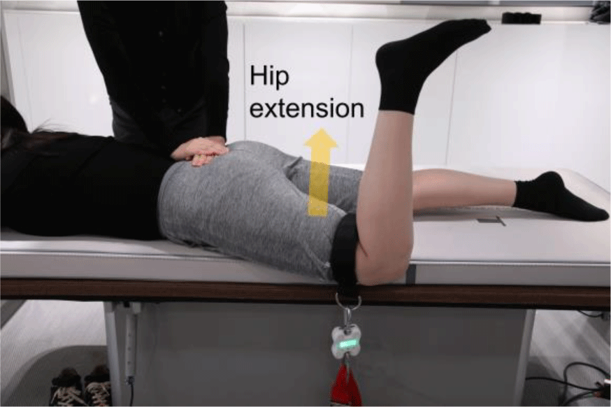
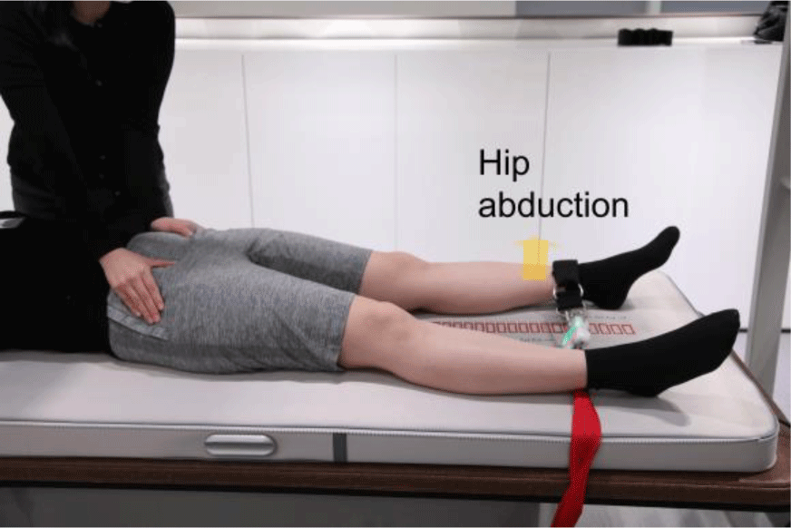
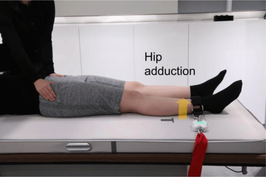
The HJM strength was measured by Smart KEMA strength sensor (KOREATECH Co., Ltd., Seoul, Korea) presented excellent intra- and inter-rater reliability (ICC> 0.95).23 The two ends of the tension sensor were connected to the distal end of the leg by a strap and to a fixation using two adjustable belts. The strength sensor recorded the force data for MVC of the HJM strength. For measuring the HJM strength, the belt length was modulated to set initial tension of belt by 3kgf in start position. Participants were asked to sustain maximal voluntary isometric contraction for 5 s and mid 3 s was analyzed for average. The mean values of three trials were applied for data analyses. The HJM strength data were normalized using participant’s weight and were calculated as the mean value of both sides.
Hip extensor strength was measured in prone position (Figure 1).20 The tester restricted the participant’s pelvic anterior tilting and rotation during hip extension with knee 90° flexion. Participants performed hip extension against a strap on the distal thigh. Hip abductor strength was measured in supine position (Figure 2).20 The tester restricted the participant’s pelvic lateral tilting during hip abduction with hip and knee 0° extension. Participants performed hip abduction against a strap on the ankle. Hip adductor strength was measured in supine position (Figure 3).20 The tester prevented the participant’s pelvic lateral tilting during hip adduction with hip and knee 0° extension. Participants performed hip adduction against a strap on the ankle.
The I-QOL and PFM strength were measured at urogynecology clinic and HJM strength were measured at laboratory setting. HJM strength were measured by physical therapist in random order generated by www.randomization.com. There was resting time 30 s after each HJM strength trial and for 5 min among measurement of HJM strength to inhibit muscle fatigue.
The normality of the data distribution was confirmed by the Kolmogorov–Smirnov Z-test. Pearson’s correlation coefficients were calculated to determine the associations between I-QOL and PFM and HJM strength. A correlation coefficient (r)>0.75 was considered “Good to excellent”, 0.50–0.75 was “Moderate to good”, 0.25–0.50 was “Fair”, 0.00–0.25 was “Little or no” relationship.24 All statistical analyses were performed using SPSS software (ver. 18.0; SPSS Inc., Chicago, IL, USA) with alpha set at 0.05.
RESULTS
There are the correlation coefficients between I-QOL score included three factors and total score and PFM strength and HJM strength. There were significant correlations between avoidance and limiting behaviors factor in I-QOL and PFM strength (r=0.524), hip adductor strength (r=0.356) and hip abductor strength (r=0.352) in the order (Table 2). The psychosocial impacts and social embarrassment factors in I-QOL were not correlated with the PFM strength and HJM strength. The total score in I-QOL was correlated with PFM strength (r=0.368) (Table 2).
DISCUSSION
Our main findings support our hypothesis, which suggest that both PFM muscle and HJM strength would be related to QoL of incontinence symptoms in women with SUI. Previous study have demonstrated the relationship between PFM strength and QoL of incontinence symptoms.25 Also, there is little scientific evidence and the peer-reviewed literature that HJM strength is associated with QoL of incontinence symptoms in women with SUI. Though study design was an observational cross-sectional and, thus, it was not difficult to determine causality, we carefully deduced that the decreased PFM and HJM strength may be associated with decreased QoL of incontinence symptoms. Therefore, when designing which muscles to train for improving QoL of incontinence symptoms, the HJM as well as the PFM could be considered.
Regarding the PFM strength, we found statistically significant relationship between avoidance and limiting behaviors factor in I-QOL and PFM strength (r=0.524) and between total score in I-QOL and PFM strength (r=0.368). PFM contraction affect urethral closure by elevating the bladder neck and compressing the urethra.16,26 Because of biomechanical role, there is justification for the non-invasive treatment of urinary incontinence by PFM training. Underwood et al. (2012) confirmed that there was negatively correlation between ability of repeated PFM contractions and International Consultation on Incontinence Questionnaire–Short Form ratings (r=–0.329, p=0.022).27 Previous study determined that there was positively correlation between PFM contractility and maximum urethral closure pressure (r=0.24, p=0.001).28 However, another study found no statistically significant correlation between the PFM function and QoL measured by the King’s Health Questionnaire in postmenopausal women with and without pelvic floor dysfunction.29 The PFM strength may be weaker in patients with more severe incontinence symptoms. Although it is difficult to determine overall QoL for PFM function, the presentation of incontinence symptoms assessed by I-QOL defines how much the QoL will be impaired.
For the hip adductor strength, the present study confirmed statistically significant relationship between avoidance and limiting behaviors factor in I-QOL and hip adductor strength (r=0.356). Peschers et al. suggested that only the gluteal-muscle co-contraction with PFM contraction affected the continence mechanism, but not when co-contracted with hip adductors and PFM.16 Also, the co-contraction of the PFM and the hip adductors did not improve efficiency for increasing or holding PFM strength during the period evaluated.30 However, there is anatomical evidence suggesting that structures surrounding the hip and pelvis to continence.31 Myers suggested that the fascia integrates whole body structure that continuously transmits forces.32 The deep front line among the several fascial lines connected between PFM and adductor magnus and minimus and played in providing lumbopelvic stability and positioning changes to the trunk structure.32 Thus, despite the still controversial suggestion, it may be a chance for a various approach to PFM training and the muscles surrounding the hip regions.
Regarding the hip abductor strength, we found statistically significant relationship between avoidance and limiting behaviors factor in I-QOL and hip abductor strength (r= 0.352). Previous study confirmed that hip abductor strength was not significantly correlation with International Consultation on Incontinence Questionnaire–Short Form ratings (r=–0.282, p=0.052).27 However, the absolute difference in hip abduction strength between groups with and without SUI represented a 12% relative weakness.27 The obturator internus, as hip external rotator, linked to a fascial connection with the iliococcygeus, and may contributed to integrate for PFM function.11 The gluteus medius was comprising part of the hip external rotator, despite of hip abductor.33 Thus, when planning rehabilitation for improving QoL of incontinence symptoms, the hip abductor muscles could be considered.
The present study had some limitations. First, the present study design was cross-sectional. Therefore, it is need to confirm effect of hip muscle strengthening on QoL of incontinence symptoms in women with SUI. Second, the sample size may be too small to generalize. Thus, further study is needed to generalize the results. Third, we only confirm correlation between HJM strength and QoL of incontinence symptoms. It is a need to further study the correlation between more various muscles, such as trunk muscles, and QoL of incontinence symptoms.
CONCLUSIONS
PFM strength was significantly correlation with avoidance and limiting behaviors factor and total scores in I-QOL. Hip abductor and adductor strength were significantly correlation with avoidance and limiting behaviors factor in I-QOL. Although it was not possible for confirming causality, the weakness of PFM and HJM strength could be related to decrease QoL of incontinence symptoms. Therefore, not only PFM but also the HJM could be considered, when targeting and designing which muscles to train for improving QoL of incontinence symptoms.


