


INTRODUCTION
Hallux valgus (HV) is a common deformity of the toe and foot affecting approximately one-third of people over the age of 65 years.1 HV deformity is characterized by the progressive hallux deviating laterally and protruding inside of the first metatarsophalangeal joint protruding medially.2,3 Although complex factors involved in the development of HV deformity are known, the accurate pathogenesis of HV deformity has not been verified yet.4,5 HV development is related to functional disabilities, such as osteoarthritis (OA) of the ankle and first metatarsophalangeal joint, foot pain, and abnormal gait and balance ability.6-8 Ankle osteoarthrosis is one of major musculoskeletal disorders that can cause ankle pain and disability and most commonly occurs as a result of trauma.9 The prevalence of ankle arthrosis associated with HV is difficult to determine. Hagedorn et al.10 verified the relationship between foot disorders and foot function in a large population. They reported that participants with a pronated foot were more likely to have HV and overlapping hallux and second toe. Abnormal ankle and foot postures such as pronated foot, supinated foot, and pes planus are more frequently noticed in individuals with HV than in healthy control.11-13
Objective and reliable biomechanical investigation plays an important role in attaining successful outcomes for patients with musculoskeletal dysfunction such as HV and ankle osteoarthrosis.2,4,13 However, many previous studies reported that the effects of interventions such as conservative physical therapy, orthotic correction, and surgical treatment on pain or clinical outcomes used a simple measurement in patients with HV deformation.3,14,15 This simple measurement can be problematic to ensure the accuracy of curative effects of the interventions.15 Although previous studies reported the kinetic and kinematic effects of various interventions on the lower extremity joints and segments in individuals with HV deformity using a high quality three-dimensional (3D) motion analysis system, most of the studies have mainly focused on the hip, knee, and first metatarsophalangeal joints or pelvic complex rather than ankle joints.1,4,17 Therefore, studies investigating the effect of HV correction on the kinetics and kinematics of the ankle joints during freely gait in individuals with HV deformity are limited and rare.
Among treatment and management methods for HV deformity, non-operative physical intervention such as toe-foot orthoses for correcting the position of the first interphalangeal and metatarsophalangeal joint are always preferred.18 Many previous studies on biomechanical effects of various types of foot-toe orthoses in patients with HV deformity have only verified changes in joint pain, gait parameters, ground reaction force, or range of motion of hip and knee joints.2,5,18 Therefore, the aim of this study was to verity effects of foot-toe orthoses (soft- and hard-type) on 3D moments and range of motion of both ankle joints known to contribute to ankle arthrosis in individuals with HV using a motion analysis system during gait. It was hypothesized that when wearing the foot-toe orthosis conditions, the participants would walk with improved kinetic and kinematic variables of ankle joint compared to those without an orthosis condition.
METHODS
Participants were twenty-six individuals (8 males and 18 females) with a HV deformity. Inclusion criteria were: 1) a clinically confirmed HV angle of more than 15° in both feet measured using a universal goniometer; and 2) no history of hallux pain or foot surgery.2 The exclusion criteria of the study participants were those who had osteoarthritis, rheumatoid arthritis, or any neurologic deficit of the lower limb joints, or those who were taking any medications that might affect their walking. All participants voluntarily participated and were given a precise explanation of the measurement process. The participants were asked to submit a written informed consent and did not have any other health problems when performing gait trials wearing the foot-toe orthoses for HV correction. The Institutional Review Board of Jeonju University approved the study design (jjIRB-180905-HR-2018-0904). General characteristics of participants are shown in Table 1.
| Characteristics | Mean±SD |
|---|---|
| Gender (M / F) | 8 / 18 |
| Age (yrs) | 28.7±4.2 |
| Height (cm) | 163.5±7.1 |
| Weight (kg) | 61.8±10.6 |
| Gait speed (m/s) | 1.3±0.3 |
| Step length (cm) | 124.5±9.4 |
| Step width (cm) | 10.4±2.7 |
Two force platforms (AMTI, Watertown, MA, USA) based on a Vicon Motion Analysis System (Vicon Inc., Oxford, England) with eight cameras (T10 model) was used to acquit 3D kinetics moment data of the ankle joints. The force platforms were placed in the middle of a 6-m walkway while the sampling rate was set at 500 Hz.19 The kinetic moment data were low-pass filtered with a fourth-order Butterworth filter to process the pure moment variables of the ankle joints and a cutoff frequency of 15 Hz.13 A 7.5 cm T-frame wand was used to calibrate the motion analysis system and a calibration reference object was used to identify the 3D X-Y-Z lab origin. The captured kinetic moment data of the ankle joints were processed using Nexus 1.8.5 software (Vicon Inc., Oxford, England).
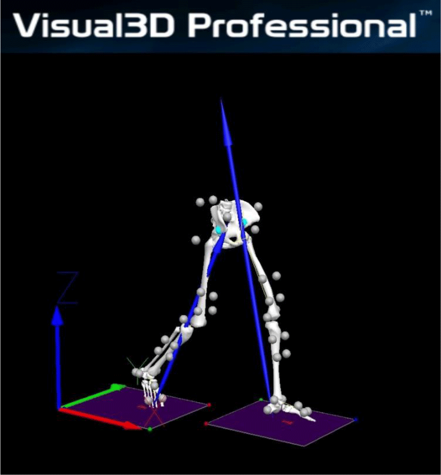
A Visual3D motion analysis software (C-Motion, Rockville, MD, USA) was used to process the final moment results and graphical reports of the ankle joints following the data obtainment and processing using the force platforms and the Nexus software program. Visual3D produced virtual lower limbs segments of each participant in laboratory space based on a set of attached reflective markers that enabled relevant 3D knee moments and motions to be calculated in the total gait cycle (Figure 1).
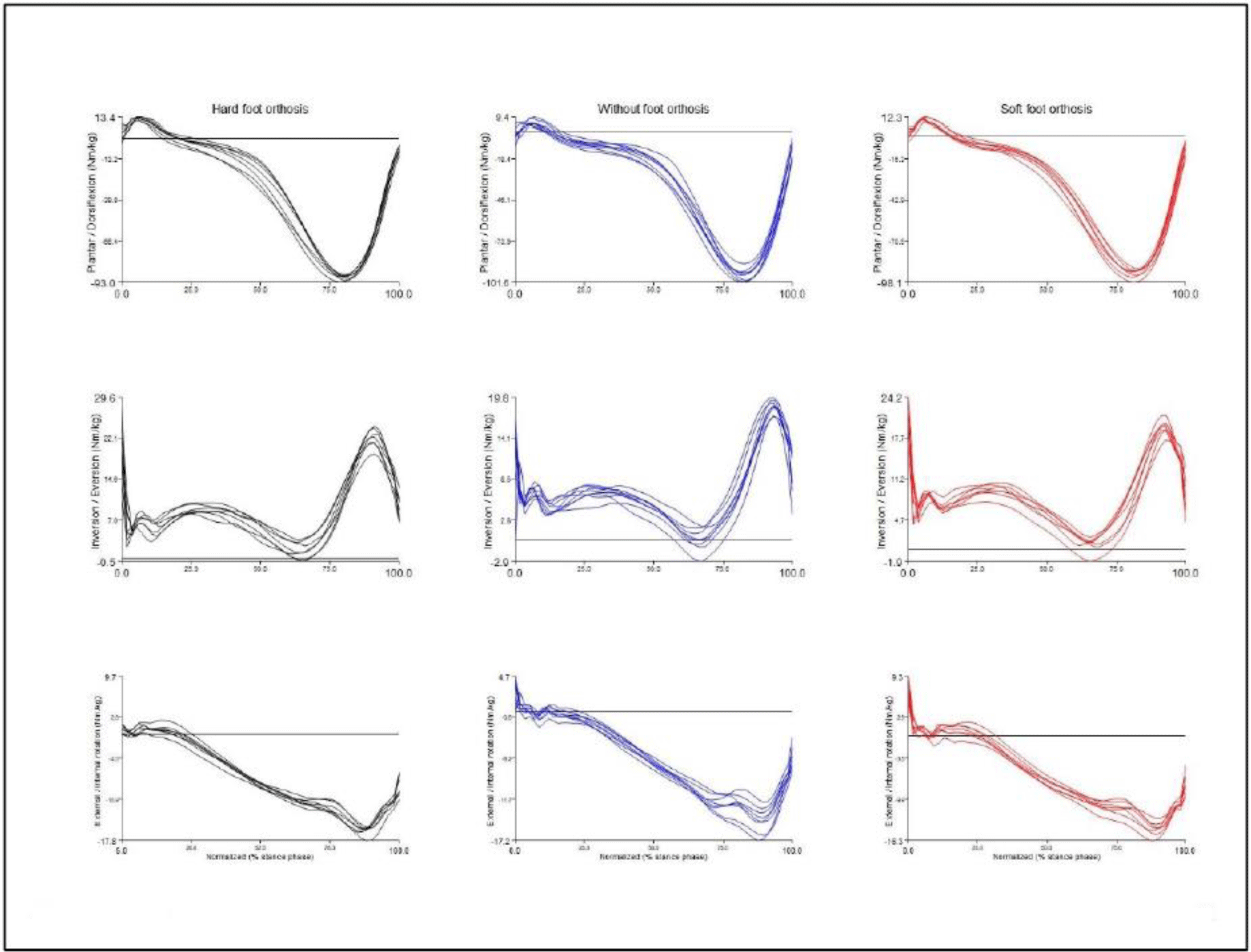
To acquire kinetic moment data based on motion analysis, 40 retroreflective markers (14 mm) were attached to both lower limbs and pelvic to forefeet, rearfoot, midfeet, malleoli, femur epicondyles, greater trochanters, anterior and posterior superior iliac spines. Four-marker clusters were attached bilaterally to the calf and thigh segments according to six degrees of freedom (6DOF).19 Initial static calibration was captured to set a musculoskeletal model for dynamic gait trials from each subject. The calibrated anatomical system technique was used to notify kinetic moment variables that developed in the ankle joint under the two different foot-toe orthoses and without orthosis conditions: hard foot orthosis (Hallufix AG, Grünwald, Germany), soft foot orthosis (Sanshin Enterprises Co., Tokyo, Japan), and without foot orthosis. The hard foot orthosis was consisted of a hard plastic hinged splint and side gel cushion attached some straps. The soft foot orthosis is composed of a soft silicone toe separation pad with a cloth strap. Following the measurement setup for gait trials, participants were asked to walk along the walkway at a free speed. A total of 8 to 10 walking trials were executed for each orthosis condition and both limb kinetic moment values of the ankle joints were measured during the gait cycle (Figure 2). The experimental order of each foot-toe orthosis condition was randomly assigned by dice throwing.
The Kolmogorov-Smirnov test was used to investigate that the kinetic data of the ankle joints were distributed normally. Repeated-measures analysis of variance (ANOVA) with Bonferroni’s adjustment was used to compare 3D moment data of the ankle joint according to foot-toe orthosis conditions and lower limb sides. If the main effect (orthosis condition or limb side) was significant, post-hoc testing was used to verify pairwise comparison based on ANOVA results. All analyses were conducted using SPSS version 26.0 (IBM Corp., Armonk, NY, USA). Differences were considered significant at α=0.05 level.
RESULTS
Means and standard deviations of general characteristics and gait parameters of subjects were shown Table 1. The mean age, weight, and height of all subjects were 28.7±4.2 years, 61.8±10.6 kg, and 163.5±7.1 cm, respectively. In addition, the mean gait speed, step length, and step width were 1.3±0.3 m/s, 124.5±9.4 cm, and 10.4±2.7 cm, respectively.
There were significant differences in some ankle peak moment variables between foot-toe orthosis conditions during gait (p<0.05) (Table 2). The plantar flexion moment peak (F=6.724, p=0.004), eversion moment second peak (F=5.871, p=0.005), and external rotation moment peak (F=4.697, p=0.007) that occurred at the 50%-100% stance phase during gait differed significantly according to the foot-toe orthosis condition (Table 2). The interaction effects between foot-toe orthosis conditions and lower limb sides for any ankle peak moment values were not found (p>0.05) (Table 2). As a result of the post-hoc test, there were significant differences in the plantar flexion moment peak, eversion moment second peak (F=5.871, p=0.005), and external rotation moment peak of the ankle joint between without foot orthosis and hard foot orthosis conditions (p<0.05) (Table 3). In addition, there were significant differences in plantar flexion moment peak between without foot orthosis and soft foot orthosis conditions (p<0.05) (Table 3). However, most peak moment values of the knee joint showed no significant difference regarding foot-toe orthosis conditions (p>0.05) (Table 3).
DISCUSSION
This study investigated 3D ankle moments with or without foot-toe orthoses for HV correction using two force platform based on the motion analysis system during gait in individuals with HV deformity. The results of the study showed significant differences in some knee moment variables in the without-HV-corrected orthosis condition compared to when wearing foot-toe orthoses for correcting HV. In order to conduct an accurate examination of effects of foot-toe orthoses for correcting HV, it is essential to understand biomechanical characteristics that occur in joints of the lower limbs during gait in individuals with HV deformity.4,13,16 This study analyzed kinetic moment data of the both ankle joints during walking using a force platform system, widely selected as the most objective measurement method in biomechanics.19,20
The study results indicated that HV deformity affected ankle moment during gait in the hard foot-toe orthosis condition compared to the without foot orthosis condition. Significant changes were found in the first peak eversion, external rotation, and plantar flexion moment of the ankle joint during the 50%-100% of the stance phase in the hard type orthosis condition than the without orthotic condition. Although our study results cannot be directly compared to outcomes of previous studies, Golightly et al.11 investigated the relationship between foot disorders including HV, overlapping toes, hammer toes, or claw toes and foot function related over-pronated and over-supinated. They reported that an over-pronated foot was related with HV (adjusted odds ratio 1.36), and an over-supinated foot was inversely associated with HV (adjusted odds ratio 0.85).11 Additionally, a prior study reported that the HV patients showed significantly higher mean pronator and external rotation moments at toe-off during stance phase than the control.1 These results showed that HV deformity negatively affects the moments occurred general motion plane of the ankle joint at the late stance phase during gait. Similarly, the present study was also found that the ankle moments observed from applying hard foot-toe orthosis were significantly decreased compared to without orthosis condition. The plantar flexion moment peak of the ankle joint, which developed at 50%-100% stance phases, was significantly decreased in the hard foot orthosis condition compared to without foot-toe orthosis conditions. A previous study reported that the center of pressure on the plantar surface of the foot in participants with HV compared to that in control group during walking using an electric pressure sensor mat.20 The plantar pressure on the first metatarsal head in healthy control group was significantly higher than that of the HV group at the terminal stance,20 indicating that the hard type foot-toe orthosis for HV deformity could promote more efficient transfer of the plantar pressure, which resulted in more positive moment of the ankle joints in the terminal stance phase during walking in this study.
In biomechanics, malalignment of the foot and toe joints affect joint power and moment values occurred at the ankle.12 In particular, foot disorders such as pes planus, pronated foot, and overlapping toes are known to be associated with HV that may contribute to the evolution of secondary abnormal alignment and degenerative arthrosis of the ankle joints of the lower extremity.9,11,12,21 In addition, over-pronated or over-supinated foot elicits the further movement of the GRF line in the medial or lateral side from the ankle axis, creating an increased ankle coronal moment. Increased peak eversion moment of the ankle joint during gait can affect the development of over-pronated foot and ankle osteoarthrosis on the lateral talocrural compartment.22,23 The intervention of the hard type foot-toe orthosis for HV correction decreased ankle eversion and rotation moments compared to other orthosis conditions in this study, indicating that it could have a positive effect on the prevention and management of ankle osteoarthrosis.
This study has some strengths. First, this study examines two types of foot-toe orthosis for HV deformity to confirm their effects on moments of the ankle joints using a force platform and a quantitative high technology of 3D motion analysis system. Second, this study showed a potential relationship between ankle osteoarthrosis and HV deformity through an increase of the ankle moment values when applying the without foot-toe orthosis condition in the terminal stance phase. However, some limitations can be drawn from this study as well. Though all participants had mild to moderate HV severity, most of them had no musculoskeletal discomfort such as pain in their feet or toes. So, it is difficult to generalize results of this study to all HV individuals with and without musculoskeletal disorders. Future studies are needed to examine the characteristics of patients with HV between with and without musculoskeletal problems including pain using an objective and reliable measurement method.
CONCLUSIONS
This study was executed to verify the effects of two types of foot-toe orthoses on 3D ankle moments using a high quality evaluation system. The results indicated the potential relationship between HV deformity and ankle osteoarthrosis through increasing 3D ankle moments at the terminal stance under the without foot-toe orthosis condition. Therefore, the application of a foot-toe orthosis like a hard supportive correction for HV deformity could contribute to treatment and prevention of ankle osteoarthrosis as well as HV correction. Maintenance of normal 3D moments of the ankle joints during walking is necessary to decrease ankle loading and improve gait function.