


INTRODUCTION
Patellofemoral pain syndrome (PFPS) is one of the most common forms of knee pain around the anterior aspect of the knee.1 Activities such as prolonged sitting, descending stairs, and squatting often exacerbate pain in patients with PFPS because these functional activities increase the compressive force on the patellofemoral joint.2 Especially, greater hip adduction or knee abduction in the frontal plane may increase the stress to the patellofemoral joint during walking and single leg standing, leading to PFPS.3 People with PFPS had greater angle of knee valgus during squat than asymptomatic people.4 In prospective study, people who developed PFPS had greater hip adduction during single leg squat than people who did not develop PFPS, suggesting that excessive hip adduction in the frontal plane may be considered as one of the causes of PFPS.5
A set of reflective markers and inertial sensors have been used for investigations via a kinematic analysis system.6 However, these markers are rarely used for three-dimensional (3D) motion analysis in clinics due to their significant financial, spatial, and temporal costs.7 Markerless motion-capture techniques can expand the applicability of human motion-capture systems and do not require special equipment for motion tracking or laborious processing, thus leading to a considerable decrease in preparation time in terms of recording and analyzing motion data.8,9 Microsoft Kinect and Intel RealSense systems with low-cost depth cameras are often used in markerless 3D motion analysis due to their high accuracy.10
New advances in markerless motion analysis have been achieved using cameras in mobile smartphones, tablets, and PCs; thus, an extra 3D depth camera is not required.11 An open-source system named OpenPose has been launched on TensorFlow, an open-source platform for machine learning. OpenPose can automatically identify anatomical points and segments of the human body using an artificial-intelligence engine that includes robust machine-learning algorithms and a pre-trained human motion-tracking algorithm.9 A previous study suggested that OpenPose can be used as a two-dimensional (2D) markerless motion-analysis system for musculoskeletal assessment that can calculate ranges of motion and human postures without manually marking key points on the body.9 If so, an OpenPose-based motion-analysis system will be valuable for evaluating the frontal range of hip and knee motion in individuals with PFPS, which is usually challenging.
Measurements of the frontal hip and knee angles and the functionality of the hip abductor during hip abduction, step-down, and squat activities have been used to identify risk factors for PFPS.12,13 However, the accuracy of the OpenPose-based motion-analysis system has not yet been fully validated for individuals with PFPS undergoing knee rehabilitation. Therefore, the purpose of this study was to assess the validity of the OpenPose-based motion-analysis system compared with 3D motion analysis for measuring the frontal range of the hip and knee joints during weight-bearing activities in individuals with PFPS.
METHODS
Eight subjects with PFPS participated in this study. The inclusion criteria were knee pain intensity of >3 points on the visual analog scale (0–10 points), Western Ontario and McMaster Universities Osteoarthritis Index (WOMAC) score of >30 points, presence of retropatellar or anterior knee pain, and pain exacerbation with at least two of the following activities: prolonged sitting, squatting, ascending or descending stairs, and kneeling.14-16 Participants were excluded if they had a history of surgery or musculoskeletal disorder within the previous year; had diseases that affect balance or walking (vestibular and neurological disorders), osteoarthritis, or rheumatoid arthritis; or could not walk independently without a walking aid.17 All subjects consented to participate in this study and provided informed consent. This study was approved by the Jeonju University Institutional Review Board (jjIRB-191115-HR-2019-1108).
The Vicon 3D motion-analysis system (Vicon MX System; Oxford Metrics, Oxford, UK) was used to investigate the validity of the TensorFlow-based application. The sampling rate was 100 Hz. Six high-speed cameras were set up in the laboratory and sixteen 16-mm reflective markers were secured using double-sided tape to the skin or with tight-fitting pants on the anterior superior iliac spine, posterior superior iliac spine, lateral thighs, lateral femoral epicondyles, lateral shanks, lateral malleoli, calcaneus, and the dorsal surface of the second metatarsal of the feet. After calibration, Vicon Nexus version 1.8.5 software was used to capture kinematic parameters while the participants performed standing hip abduction, semi-squat, and step-down movements (Figure 1). Raw kinematic data were filtered using fourth-order Butterworth filters with a cut-off frequency of 6 Hz and exported as a CSV file for statistical analysis.
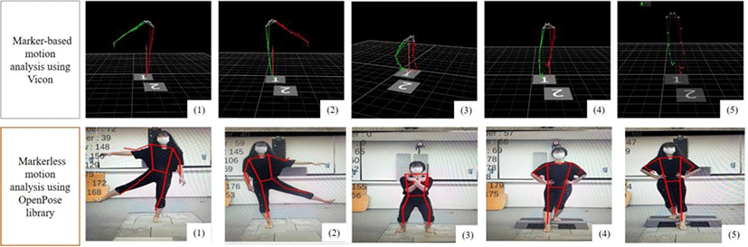
OpenPose is a popular open-source 2D pose-estimation system that can identify joint centers with a web camera or smartphone camera using a convolutional neural network and supervised learning. The OpenPose system can recognize joint positions such as the nose, neck, eye, ear, shoulder, elbow, wrist, hip, knee, and ankle, and estimate joint kinematics automatically without markers.18 To measure the frontal angle of the hip and knee, markerless motion capture was performed using a smartphone camera (Samsung Galaxy S8, Samsung Electronics Co., Ltd., Suwon, South Korea) on the tripod without tilting, which was placed on a 20-cm tripod at a height of 100 cm in the frontal plane of the subject. The frontal plane angles of the hip and knee joints were analyzed based on the OpenPose library in the Python environment. The sampling rate was 30 Hz. The raw kinematic data were smoothed using a zero-lag fourth-order Butterworth low-pass filter. The cut-off frequency of the filter was 2 Hz. The OpenPose system automatically calculates the relative angle between two vectors during hip abduction, semi-squat movements, and step-down movements. The feature points of the hip and knee joints were estimated using OpenPose during weight-bearing activities recorded using a smartphone camera (Figure 1).
Before data collection using Vicon and OpenPose, the participants reported their height, weight, age, medical history, leg length, knee width, ankle width, PFPS intensity in both knees on a visual analog scale (0–10 points), PFPS duration, and level of disability using the WOMAC (Table 1).
| Characteristic | Value |
|---|---|
| Gender (M/F) | 2/6 |
| Age (years) | 25.3±4.1 |
| Height (cm) | 167.0±9.9 |
| Weight (kg) | 63.9±20.3 |
| WOMAC | 54.3±9.6 |
| VAS (cm) | 4.0±1.1 |
Participants randomly performed three weight-bearing activities at a self-selected speed in a standing position (hip abduction with both legs, semi-squats, and stepping down with both legs). Each participant practiced the weight-bearing activities three times for familiarization and were allowed a rest period of 1 minute after each activity. Three trials of each activity were recorded simultaneously using both Vicon and OpenPose. The more symptomatic leg (MSL) and less symptomatic leg (LSL) were divided based on the VAS. For standing hip abduction with the MSL and LSL, the starting position was standing with both arms and legs straight while looking straight forward. The participant was asked to abduct the arm of the test side to 90° to make the activity more unstable than hip abduction alone. The participant was then asked to open their leg as wide as they could. To perform semi-squats, the participant started in a standing position with their arms folded across the chest and the feet shoulder-width apart, looking straight ahead. The participant was then asked to flex both knees until an angle of 45° was reached. To perform step-down movements with the MSL and LSL, the participant stood with their feet shoulder-width apart and their arms folded and positioned their toes toward the front end of the step, which was set at a height of 20 cm regardless of the length of their leg. A previous study showed no significant difference in the intensity of knee pain between three step heights (8, 14, and 20 cm) in patients with PFPS.19,20 The participant was asked to flex the knee of the tested side until the heel of the non-tested limb touched the floor without putting weight on the heel and then return to the starting position.
To investigate the validity of the measurements, kinematic data for the hip and knee in the frontal plane were recorded simultaneously using Vicon and OpenPose during the three weight-bearing activities in the aforementioned laboratory setting (Figure 1). The peak angles of hip and knee joints in the frontal plane were calculated and the mean values of three trials of weight-bearing activities were used for the analyses.
The data were tested for normality using the Shapiro-Wilk normality test.21 Pearson and Spearman correlation analysis was used to assess the validity of the OpenPose-based motion-analysis system by comparing measurements of the hip and knee joint angles in the frontal plane between OpenPose and Vicon. The correlation coefficients were interpreted based on Swinscow’s classification, as follows: 0.00–0.39, very weak to weak correlation; 0.40–0.59 fair to moderate correlation; 0.60–0.79 good correlation; and 0.80–1.0 strong correlation.22 Statistical analyses were performed using SPSS software (ver. 26.0; IBM Corp, Armonk, NY, USA). The significance threshold was set at p<0.05.
RESULTS
Correlation coefficients ranged from 0.04 to 0.61 for the MSL and 0.02 to 0.88 for the LSL when participants were performing the three weight-bearing activities (Table 2).
When performing standing hip abduction with the MSL (unsupported side), the validity of the measurements of hip abduction and knee abduction for the MSL was fair and weak, respectively. For the LSL (supported side), the validity of the measurements of hip abduction and knee abduction was weak and good, respectively (Table 2).
When performing standing hip abduction with the LSL (unsupported side), the validity of the measurements of both hip abduction and knee abduction for the LSL was fair. For the MSL (supported side), the validity of the measurements of both hip abduction and knee abduction was weak (Table 2).
When performing semi-squats, the validity of the measurements of hip abduction and knee abduction for the MSL was weak and fair, respectively. The validity of the measurements of both hip abduction and knee abduction for the LSL was fair (Table 2).
When stepping down with the MSL (unsupported side), the validity of the measurements of hip abduction and knee abduction for the MSL was good and weak, respectively. The validity of the measurements of both hip abduction and knee abduction for the LSL (supported side) was weak and strong, respectively (Table 2).
When stepping down with the LSL (unsupported side), the validity of the measurements of hip abduction and knee abduction for the LSL was weak and fair, respectively. The validity of the measurements of both hip abduction and knee abduction for the MSL (supported side) was fair (Table 2).
DISCUSSION
The purpose of the current study was to investigate the validity of measurements provided by an OpenPose-based motion-analysis system for the angles of the hip and knee joint in the frontal plane during standing hip abduction, semi-squat, and step-down movements in individuals with PFPS. We found that the validity of the measurements ranged from weak to strong. Although markerless motion-analysis systems are less accurate than marker-based motion-analysis systems, the former system is more suitable for use in clinical rehabilitation and sports, where it is difficult to perform marker-based motion analysis.23
A previous study reported that markerless motion analysis provided measurements with low validity, as tracking using an automated skeleton and relatively low camera sample rates was difficult.24 In addition, a recent systematic review reported that the outcomes of low-cost video-based motion-analysis and 3D motion-analysis systems exhibited “poor” (r=0.025) to “strong” agreement (r=0.992).25 Current results also showed measurement validity ranging widely from weak (r=0.02) to strong (r=0.88). OpenPose was shown to provide measurements of sagittal angles and frontal angles of the hip and knee in a gait analysis of healthy participants that exhibited good and poor agreement, respectively, with measurements provided by 3D motion-analysis systems.26 The low accuracy of the current results, which is consistent with that in a previous study, may be because we measured frontal angles of the hip and knee when the participants were performing weight-bearing and functional activities. In addition, our participants comprised a patient group with PFPS. Although we did not compare hip and knee angles between people with and without PFPS, we confirmed that measurements of the LSL were more accurate than those of the MSL. The genu valgum angle was larger for the MSL than for the LSL. With a larger genu valgum angle, the sides of the knees are closer together, making it difficult to detect key points of the hip and knee using video-based OpenPose programming. By using 3D depth cameras built into smartphones in the future, we should be able to address this technical limitation and produce more valid measurements for PFPS patients that will complement the measurement of frontal hip and knee angles performed by clinicians. However, further studies are required to verify if this is possible.
We obtained measurements of the angle of hip abduction with good and fair validity for the MSL and LSL, respectively, during standing hip abduction. OpenPose measurements of the frontal plane of hip abduction/adduction in walking healthy participants were previously found to be poorly correlated with Vicon measurements, although measurements of the hip angle in the sagittal plane were strongly correlated.27 OpenPose captures motion data using only one digital camera, so angles in a frontal or sagittal plane with a transverse plane rotation were not measured with high accuracy in a previous study.27 In the current study, although we asked participants to perform hip abduction, it is difficult for participants with PFPS to perform only hip abduction without hip rotation or flexion/extension. These complex motions in multiple planes during standing hip abduction might have decreased the accuracy of the measurements compared to those from Vicon.
When performing semi squat and step down, weak to good validity for the angle of hip adduction and knee abduction of MSL and LSL sides were showed in current study. PFPS patients demonstrated excessive adduction and internal rotation of hip during weight-bearing activities such as single leg squat and step down.13,28 We found the occurrence of hip internal rotation when performing semi squat and step down based on the data obtained by Vicon. Motion in transverse plane may lead to lower accuracy in OpenPose motion analysis, compared to 3D motion analysis system during semi squat and step down. When performing semi squat, more trunk forward bending with hip flexion was occurred than step down and standing hip abduction, which make difficulty to detect the hip joint as the key points when using OpenPose library, leading to low validity. Interestingly, when performing step down, fair and strong validity were showed for angle of knee abduction in each supported side. Clinicians used to measure the angle of genu valgus in order to confirm the risk factors of knee injury and give feedback when applying valgus control intervention in weight bearing position.29 Thus, it is meaningful that OpenPose system with fair and strong validity of knee abduction during step down can be used for patients with PFPS.
Our study had some limitations. The participants were relatively young. Therefore, our findings may not be applicable to older populations. Additionally, only eight participants with PFPS were analyzed to assess the validity of the OpenPose-based motion-analysis system. Thus, we suggest using markerless motion analysis with caution when highly accurate assessments of kinematic variables are necessary until further studies with a larger sample size are available to assess the validity of such systems. Although the validity of most measurements was not strong in the current study, the OpenPose motion-analysis system has been demonstrated to have almost perfect reliability.30 If the OpenPose motion-analysis system can sufficiently discriminate between people with and without PFPS or reveal pre- and post-intervention clinical differences despite producing measurements with weak to strong validity, the markerless system can be clinically useful.
CONCLUSIONS
Our results support the use of the OpenPose-based motion-analysis system as a physical function assessment tool to investigate the frontal hip and knee angles of patients with PFPS due to its cost- and time-effectiveness, ease of use outside the laboratory, and utility for remote rehabilitation. However, to enhance the utility of the OpenPose-based motion-analysis system, the errors between the true values and data obtained using OpenPose programming should be reduced. Based on our findings, future studies could assess whether the OpenPose-based motion-analysis system can be used to discriminate between patients with and without PFPS while they are performing functional activities or monitor the outcomes and disease progression of PFPS.