


INTRODUCTION
The thoracic spine consists of 12 vertebral segments located between the neck and the lumbar spine, and it is composed of a rigid structure connected to the ribs. The spine has a stable structure with a complex ligament, such as a radiant ligament attached to the vertebral body, intervertebral discs and ribs, and strong intercostal fascia.1
Physiological movements of the spine are caused by coupled motions occurring in the intervertebral joints and rib joints, and structural changes, such as changes in the spine bend of these joints, limit the movement of the spine.2 In the spine, each vertebral torso forms a bony slope of 3.8° from the back to the front and forms an average of about 20°–40° of spine bend.3 Spine curvature increases with increasing age and is more increased in women than in men.4,5
Hyperkyphosis refers to the state in which the curvature of the spine area is severely bent backward.6 If there is a structural deformation of the spine itself or a persistent bad posture, thoracic kyphosis may increase when the balance of the trunk muscles is broken. The weakening of the hair roots may be the cause.7 This increased spine curvature adds to the load on the spine, which contributes to pain and disability, resulting in severe stress to the bones, joints, ligaments, and muscles. Tension occurs, causing muscle weakness and stretching support for the spine.8 A previous study confirmed the reduction of thoracic kyphosis by applying renal and muscle strengthening exercises to the individual with thoracic kyphosis.9
Deformity of the spine leads to the narrowing of the rib cage due to growth disorders, compression of the lungs, and reduced mobility, resulting in decreased swelling and contraction of the lungs and thus in reduced muscle activity in the chest and weakening of the respiratory muscles.10 A small deformation of the spine does not significantly affect the cardiopulmonary function, but as the deformation gradually increases, cardiopulmonary function disorder occurs.6,11
Research on the cardiopulmonary function of patients with spinal curvature is still insufficient. Thus, this study investigated the changes in spine bends and respiratory function when the self-extension exercise and the muscle energy technique of trunk muscles are applied to individuals with thoracic kyphosis of over 40°.
METHODS
This study selected 30 subjects (24 males and 6 females) who fully understood the purpose of the study among college students attending Baekseok University in Cheonan and who agreed to participate in the study. The selection criterion for the study subjects was individuals with thoracic kyphosis of 40° or more,12 and the exclusion criteria were those who did not experience spinal surgical treatment, those without rheumatoid disease and arthritis, and the respiratory system. Those with no disease, no neurological diseases, and no other diseases that could affect the experiment were selected.
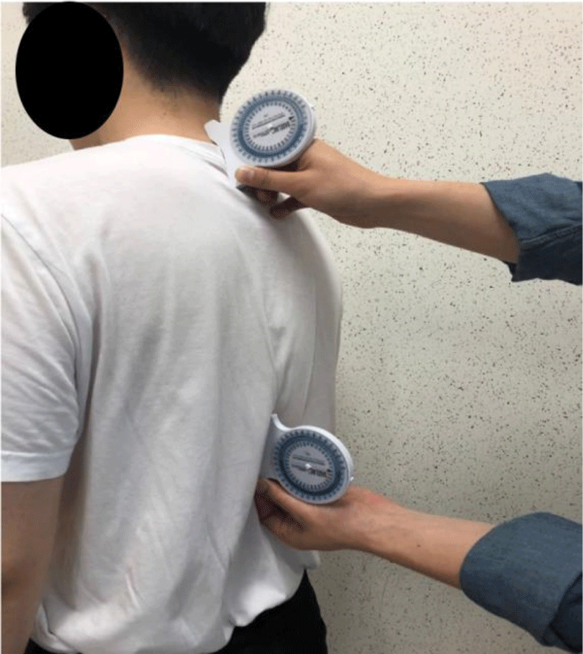
The subject faced the front and walked in place for 10s. Then, the subject stood in a comfortable position with the feet spread over the shoulder width.13 A previous study reported that the method of combining the bubble incli-nometer and observation to confirm thoracic kyphosis could show the correct posture.14 The areas of the seventh cervical vertebra and the first lumbar vertebra were found and marked. The lowest rib in the right position was found to determine the first lumbar vertebra, and then the twelfth vertebra and the first lumbar vertebra were shown. The positions of the twelfth thoracic vertebra and the first thoracic vertebra were indicated by attaching markers.
To use the inclinometer, the subject stood in a comfortable position with eyes staring in front, resting the arm and fixing the posture. The angle of inclination of the seventh cervical vertebra and the first vertebra shown through observation was measured using an angle gauge, followed by that of the twelfth vertebra and the first lumbar vertebra.6 The total measurement was performed three times, and the average value was used as the final value (Figure 1). Inclinometers are easy to use and have reduced potential errors in measurement.15
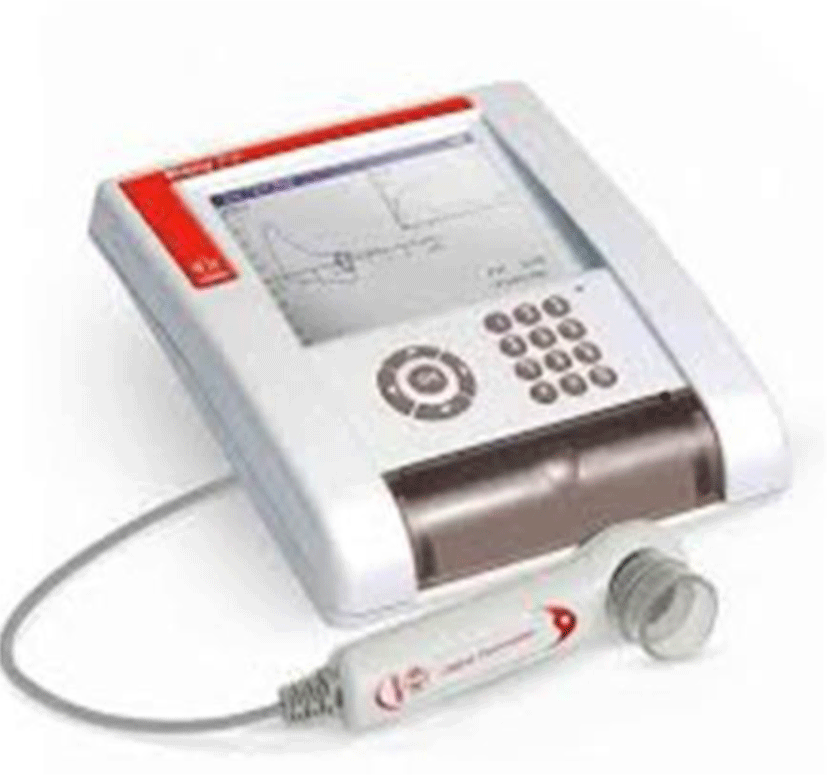
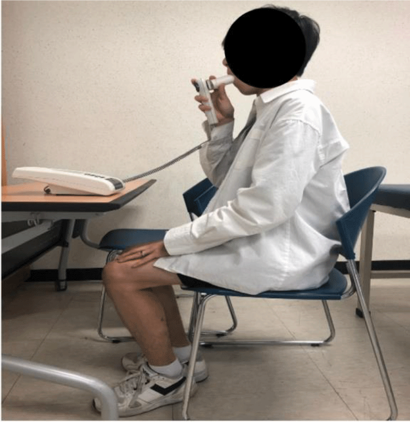
The respiratory function of the subjects was measured using a respiratory function meter (Pony FX, Cosmed Inc., Korea) (Figure 2). The Pony FX used to measure respiratory function has been reported to have a very high confidence level of 0.99.16 The measurement indicators measured the forced vital capacity (FVC), forced expiratory volume in 1 second (FEV1), and peak expiratory flow. Effortful waste is an indicator of the total exhaled volume from one breath during maximum exhalation. The expiratory volume per second is the index indicating the expiratory volume discharged during the first 1 s of the maximum expiratory expiration.17 These measurement indicators are the basic measurements in evaluating respiratory function, and the measured values may be measured differently depending on the posture of the experimenter. In general, the lying down posture in a normal person is measured to have a lower pulmonary function than the sitting posture. The reason is that in the lying position, the measured value may be low because the organs or muscles of the abdomen press against the diaphragm.18 Therefore, in this study, the hip and knee joints were bent at 90°, and the waist and chest were positioned in a straight posture to accurately measure respiratory function. In the measurement, each index was measured three times, and the average value was used as the final value.
The control group was allowed to sit back and rest with the knee and hip joints bent at 90° for 12 min, which is the same as the intervention time in the other two groups. Before and after resting, the degree of spine bending and respiratory function were measured.
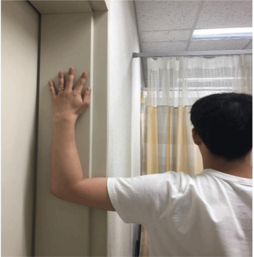
In the self-extension exercise group, before starting the exercise, training was conducted on the exercise programs, and the exercises were learned through a direct motion demonstration. The subject stood shoulder-to-wall, with the shoulders bent at a 90° angle and the palms facing the wall (Figure 4). In this position, one step forward was made, and stretching was performed by stretching the small pectoral muscle on the wall.19 The exercise was maintained for 10 s in both left and right movements and repeated 10 times. Three sets were performed, and the resting time between each set was 30 s.
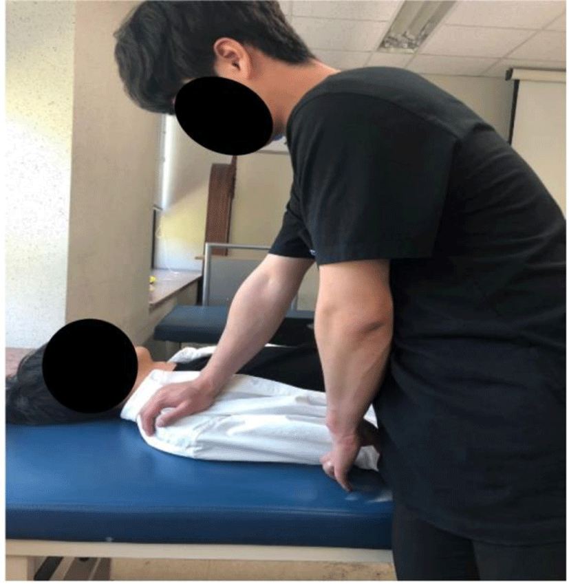
The group of muscle energy techniques underwent training on the exercise programs before starting the exercise, and the subjects learned the exercise through a direct movement demonstration. The subject lies on the treatment table, with both feet together and both knees bent. The researcher tries to press the subject’s scapular down the treatment table, while the subject resists it. The position is held for 10 s, and relaxation is repeated (Figure 5).20 The exercise was maintained for 10 s in both left and right movements and repeated 10 times. Three sets were performed, and the resting time between each set was 30 s.
To compare the difference between thoracic kyphosis in the group and the respiratory function according to each intervention technique, a paired t-test of the SPSS ver. 20 program was used. One-way ANOVA and post-analysis Scheffe test were used to compare the changes in bending and respiratory function. The statistical significance level was set to 0.05.
RESULTS
The differences in thoracic kyphosis and the respiratory function of the subjects in each group according to each intervention technique were compared. In the control group, no significant differences were found in kyphosis angle, effortful aeration, 1 s effortful expiratory volume, and maximum expiratory speed (Table 1). In the self-extension exercise group, thoracic kyphosis significantly decreased, but it significantly increased in the effortful waste amount and effortful expiratory amount in 1 s (Table 2). In the group of muscle energy technique, thoracic kyphosis significantly decreased, but it significantly increased in effortful waste (Table 3).
We compared the thoracic kyphosis and respiratory function between the groups according to the three intervention techniques (Table 4). Kyphosis angle significantly decreased in the self-stretching exercise group compared with the control group, but it significantly increased in the effortful waste amount and effortful exhalation amount in 1 s [Table 4]. Spine curvature was significantly decreased in the muscle energy technique group compared with the control group, but it significantly increased in effortful waste (Table 4). The 1 s effortful expiratory volume increased more in the self-extension exercise group than in the muscle energy technique group. The maximum exhalation speed slightly increased in the muscle energy technique group compared with the self-extension exercise group.
DISCUSSION
Most lesions are caused by the lack of exercise, persistent stress, and incorrect posture.21 The spine, which transmits forces between the upper and lower body of an individual, is an important segment that requires stability and mobility for optimal function.22,23 In humans, an increased spine curvature is considered to be caused by structural changes and poor habits, and anatomical changes and decreased mobility significantly increase spine curvature.4,24 When thoracic kyphosis increases, it causes many problems, such as changes in the musculoskeletal system, functional limitations of the body, falls, and increased mortality.6 In changes in the musculoskeletal system, as the mobility of joints decreases, stress is applied to other joints because of compensatory effects, and the functional limitations of the upper and lower extremities cause negative effects on daily activities or life.25,26 Moreover, excessive spinal curvature decreases respiratory function and increases mortality due to cardiopulmonary problems.27,28 Recently, the importance of the effects of active exercise on patients with thoracic kyphosis has been demonstrated.29,30
This study aimed to investigate the effects of self-extension and muscle energy techniques on kyphosis angle and respiratory function. The results showed that spine bending significantly decreased in the self-strength exercise group and the muscle energy technique group, which were the experimental groups. Spine bending was also observed to increase significantly. These findings are consistent with those of the reduction in thoracic kyphosis by applying muscle strengthening in previous studies.19,24 In the self-extension exercise group and the muscle energy technique group, the thoracic kyphosis significantly reduced, and the respiratory function significantly increased. Increased kyphosis angle causes the shortening of the pectoral muscles. In this study, kyphosis angle had a significant effect on thoracic kyphosis because self-stretching exercises and muscle energy techniques were applied to relax the pectoral muscles.31 Therefore, this study shows a link between spinal hyperflexion and respiratory function, and it can serve as a reference for the development of an exercise therapy program for patients with spinal hypertropia. The limitations of the study are that the small number of subjects, subjects’ age limited to younger age groups, and short duration of the intervention. Future studies should include a larger number of subjects with more age ranges.
CONCLUSIONS
This study investigated the effects of self-extension and muscle energy techniques on the kyphosis angle and respiratory function of 30 subjects (24 males, 6 females) with excessive spine curvature. The results showed that self-extension exercise significantly increased the kyphosis angle, effort disposal, and 1 s effort exhalation but did not affect the maximum exhalation speed. The muscle energy technique showed a significant decrease in spine deflection and effort waste but did not affect the 1 s effort expiratory volume and maximum expiratory speed. Thus, the self-extension exercise and muscle energy technique reduced the spine curvature and positively affected the pulmonary function of effortful waste and 1 s effort. This study can be used as a reference for the development of respiratory and exercise therapy programs for patients with spinal deformity.