


INTRODUCTION
There are two causes of shoulder impingement syndrome (SIS), first described by Neer: the primary cause being the narrowing of the interarticular space due to soft tissue infection or bone growth and the secondary cause being the upward movement of the humeral head because of the weakness of the muscles or the imbalance of the force among the muscles around the shoulders.1,2,3 Secondary impingement is mainly caused by the force imbalance among the shoulder muscles generating the force couple. The imbalance or co-activation of the deltoid and rotator cuff muscles and the imbalance of the scapular upward rotators are known to be the main causes of SIS.4,5,6
When an imbalance occurs between the force couple of the deltoid and rotator cuff muscles, the subacromial space narrows, and the humeral head slides upward while rising the arm, causing the impingement of the acromion and humeral head.7 The scapular downward rotation syndrome, which lacks the scapular upward rotation because of the predominance and shortening of the downward rotators of the scapula, may also cause SIS.8,9 In addition, people with SIS symptoms had increased upper trapezius action and reduced lower trapezius and serratus anterior action than normal subjects when elevating their arms, thus resulting in the decreased upward rotation and increased scapular anterior tilt.8,10,11 These factors reduce the space between the acromion and humerus, resulting in SIS. The weakening of the external rotators of the glenohumeral joint may also cause the impingement of the greater tubercle of the humerus and the acromion during arm elevation. Therefore, to prevent secondary shoulder impingement,10,11,12 it is important to activate the weakened scapular upward rotators and rotator cuff muscles so that the subscapular articular cavity is properly maintained during arm movement.6,13,14
Among the scapular upward rotators, the weakened muscles are mainly the serratus anterior and lower trapezius muscles.10,15,16,17,18 Exercise methods for strengthening the weakened serratus anterior and lower trapezius muscles include the push-up plus exercise, protraction exercise, prone arm lift exercise, and modified cobra exercise.19,20,21 Methods for resolving the imbalance between the deltoid and rotator cuff muscles are as follows: to increase the length of the shortened deltoid muscle and decrease the activity of the deltoid muscle and to activate the rotator cuff muscle.8,22,23 To do so, Sahrmann proposed to reduce the load on the deltoid by bending the elbow joint during arm elevation.8 Huston and Adam suggested methods for activating the rotator cuff muscle by rotating the upper arms inward or outward. In addition, a method for activating the infraspinatus and teres minor muscles (both of which are rotator cuff muscles) by rotating the upper arm outward and elevating arm through wall sliding with the ulnar side of the hand was introduced.8,24
Previous studies investigated various factors affecting muscle activity as well as exercise methods for strengthening the scapular upward rotators or the rotator cuff muscles. For example, it was found that the muscle activity of the rotator cuff muscles changed depending on the head position (forward vs. neutral head position) or visual feedback.9,25 It was also reported that the muscle activity of the scapular muscles varied depending on the weight loaded on the hand, grip strength, and the angle of bending of the glenohumeral joint.10,13,26,27
According to Xavier et al., the scapular upward rotation was increased and the mobility of the glenohumeral joint was decreased during the full-can test (arm elevation with the thumb pointing upward) compared to during the empty-can test (arm elevation with the thumb pointing downward).28 The internal or external rotation of the glenohumeral joint was suggested as a way to activate the rotator cuff muscles.8,23 These results indicate that the muscle activity of the scapular upward rotator and rotator cuff muscles is affected by the upper arm positions.
Muscle activity of the scapular upward rotators, rotator cuff muscles, and deltoid muscles is an important factor for SIS.4,8,10,23 Nevertheless, to our knowledge, no studies have focused on the muscle activity of the scapular upper rotator and rotator cuff muscles according to the position of the upper arm. Therefore, this study aimed to investigate the effect of upper arm position and the shoulder flexion angle on the activity of the scapular upward rotators, infraspinatus, and deltoid muscles, which are the major muscles associated with SIS.
METHODS
G*Power software (version 3.1.2) was used to estimate necessary sample size. Sample size was calculated a priori for a power of 0.95, and effect size of 0.80, and alpha level of 0.05. This calculation indicated that the necessary sample size was 16 subjects for the study. 23 healthy adults at the Joongbu University participated in this study, and were selected according to the selection criteria. The purpose, method, and procedure of this study were explained to each subject, and they voluntarily participated in the experiment after providing written informed consent. This study was approved by the Institutional Review Board of the Joongbu University.
In this study, the following participants were excluded: those who expressed pain in the past or during clinical examination, those who had functional issues due to restricted shoulder joint motion, those who experienced instability in the shoulder joint during activities of daily living, those who had neurological symptoms around their necks, and those who had experienced numbness or loss of sensation in the upper limb in the six months before the experiment. The general characteristics of the subjects are shown in Table 1.
| Variable | Mean±SDa |
|---|---|
| Age (yrs) | 22.91±1.53 |
| Height (cm) | 163.96±9.42 |
| Weight (kg) | 57.35±10.97 |
The muscle activity of each muscle was measured using surface electromyography (Noraxon TeleMyo 2400 system, Noraxon Inc, Scottsdale, AZ, USA). Noraxon MyoResearch 1.06 XP was also used to collect and analyze the muscle activity data of the muscles being assessed. Sampling of the electromyogram signal was set to 1,000 Hz, and a band pass filter of 10–450 Hz was applied to remove the electrocardiogram signal, which is a biological signal noise.
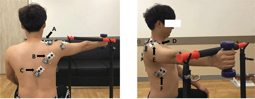
The participants underwent depilation and keratin removal for the attached part before application of the electromyographic electrodes (Ag/AgCl surface electrodes). Their skin was cleaned with alcohol using cotton, and the electrodes were attached according to the standardized position for each muscle. The gap between the electrodes was fixed at 2 cm, and they were attached parallel to the direction of muscle fibers.29 The electromyographic electrodes were attached to the upper trapezius, lower trapezius, serratus anterior, infraspinatus, anterior deltoid, and middle deltoid muscles. The electrodes of the upper trapezius were attached parallel to the direction of the muscle fibers at the middle point between the spinous process of the seventh cervical vertebra and the posterolateral acromion. The electrodes of the lower trapezius were attached to the middle part of the muscle located at an angle of 55° from the lower angle of the scapula.
The electrodes of the serratus anterior were attached to the parallel muscle of the serratus anterior, which is located at the same height as the inferior angle of the scapula, in front of the latissimus dorsi muscle under the axilla. The electrodes of the infraspinatus muscle were attached parallel to the muscle fibers at the midpoint of the infraspinatus fossa.29 The electrodes of the anterior deltoid muscle were attached to the muscle belly of one finger based on the acromion while comfortably lowering their arms. The electrodes of the middle deltoid muscles were attached parallel to the direction of the muscle fibers at the most convex point on the line extending the acromion and lateral epicondyle of the cubitus (Figure 1).30
The experiment was conducted by two measurers who had at least three years of clinical experience in physical therapy. One measurer operated the computer, and the other controlled the participants’ position and experimental process and did not allow the participants to know the data of their muscle activity displayed on the computer. The two measurers thoroughly understood all the procedures and made it impossible for the measurers and participants to predict the test results and know the meaning of the test results. The participants were given preliminary experiments with detailed explanations of measurement positions and methods so that they could become familiar with the experiment. The participants were assessed in a random order. The experiment was performed in a sitting position on a chair without armrests, and during the experiment, they maintained the correct sitting position.
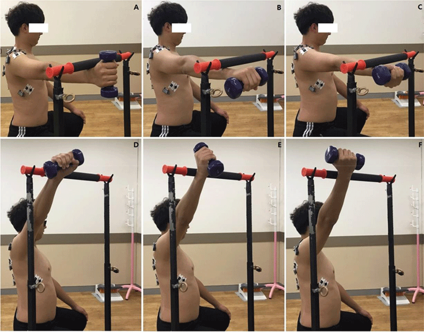
The EMG signals collected in the following positions: palm down (PD) position, neutral position (NT), and palm up (PU) position with a 2 kg dumbbell on hand while sitting. The participants raised their hands up to the pre-set target-bar at the angle of 90° and 120° with the above positions and maintained their position for 5 sec. At this time, the movement of the arm was generated in the scapular plane considering the stability of the shoulder joint (Figure 2).31 The experiment was carried out three times with two flexion angles of the shoulder joint (90°, 120°) and in three positions (PD, NT, PU). Between two consecutive measurements, a two-minute rest was allowed to prevent muscle fatigue.
The Kolmogorov-Smirnov test was employed to assess normality of the distribution. The data collected through the experiments were analyzed using SPSS ver. 21 for Windows. According to the upper arm position and shoulder flexion angle, a two-way repeated ANOVA was used to determine the change in muscle activity of the shoulder muscles, and the significance level was set at 0.05. When there was a significant difference in this analysis, post-hoc analysis was performed using the paired-sample t-test with Bonferroni correction. The significance level at this time was 0.017 (0.05/3).
RESULTS
Table 2, Table 3, and Figure 3 show the change in the muscle activity of the upper trapezius, lower trapezius, serratus anterior, infraspinatus, anterior deltoid, and middle deltoid muscles according to the upper arm position and the flexion angle of the shoulder joint during arm elevation.
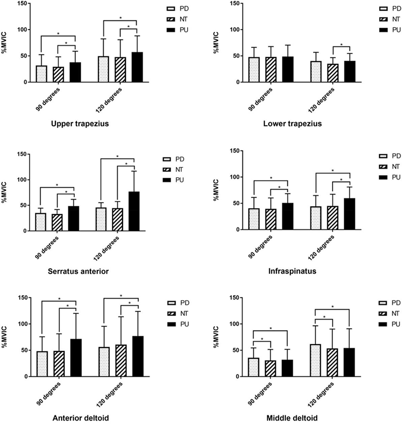
For the all muscles of upper trapezius, lower trapezius, serratus anterior, infraspinatus, anterior deltoid, and middle deltoid, the main effects were significant for shoulder flexion angle (p<0.05). And for the all muscles except lower trapezius, the main effects were significant for upper arm position (p<0.05). There was also no interaction between the two factors in all muscles (p>0.05).
According to the post-hoc analysis, muscle activity of the upper trapezius and serratus anterior muscles in the PU position were significantly higher than the other humeral positions at both angles (p<0.017). And muscle activity at the 120° flexion angle in all three humeral positions showed significantly higher value than 90° flexion angle (p<0.017). Muscle activity of the lower trapezius muscle at the PU position was significantly higher than the other two humeral positions only at a 120° flexion angle (p<0.017).
In the PU position, the muscle activity of the infraspinatus and anterior deltoid muscles was significantly higher than the other two positions at both angles (p<0.017), and only in the PU position, it showed a significantly higher muscle activity at a 120° flexion angle than the a 90° flexion angle (p<0.017). The muscle activity of middle deltoid muscle displayed a significantly higher in the PD position compared to the other two positions at both angles (p<0.017), and showed significantly higher values at a 120° flexion angle compared to a 90° flexion angle in all three positions (p<0.017).
DISCUSSION
This study aimed to investigate the effect of upper arm position and shoulder flexion angle on the activity of major muscles associated with SIS during arm elevation. The muscle activity of the scapular upward rotators, infraspinatus, and deltoid muscles was significantly different according to the upper arm position and the shoulder flexion angle (p<0.05). There was a significant difference in muscle activity of upper trapezius, serratus anterior, infraspinatus, anterior deltoid, and middle deltoid muscles except for the lower trapezius muscle depending on the upper arm position (p<0.05). There was also a significant difference in muscle activity of all muscles according to the flexion angle of the shoulder joint (p<0.05).
All muscles except for the lower trapezius showed higher muscle activity at 120° depending on the arm elevation angle (p<0.017). This is because of the length–tension relationship.32 The scapular upward rotators, infraspinatus, anterior deltoid, and middle deltoid muscles are shortened at 120° rather than 90°, thereby showing a higher muscle activity. Previous studies on shoulder muscles also showed increases in muscle activity as the flexion angle of the shoulder joint increased.4,10,13,33 In this study, there was no significant difference in the lower trapezius muscle activity according to the angle. The lower trapezius muscle activity sharply increased in the end range during arm elevation, thus resulting in a small difference between the 90° and 120° bending angles of the shoulder joint.34
In this study, muscle activity of the upper trapezius and serratus anterior muscles was significantly higher value in the PU position compared to NT and PD positions at both 90° and 120° angles (p<0.017). And there was no significant difference between the NT and PD positions in this regard. There are two reasons for the muscle activity being significantly higher in the PU position than in the NT and PD positions.
First, this may have resulted from changes in the mobility of the scapula and the glenohumeral joint due to the upper arm position.28 To obtain the PU position, an external rotation of the glenohumeral joint occurs, and the contraction of the infraspinatus and teres minor muscles that occurs at this time limits the mobility of the glenohumeral joint, resulting in an increase in the movement of the scapula. Consequently, the muscle activity of the upper trapezius and serratus anterior muscles may have been further increased due to the length–tension relationship.8,11
Second, the passive tension of the teres major muscles may have affected the result. Passive insufficiency of movement due to the teres major muscles may occur while raising the glenohumeral joint in the external rotation to achieve the PU position, thereby acting as a resistance to the scapular upward rotators. 8,11
In addition, the muscle activity of all the muscles measured in this study did not show a significant difference between the NT and PD positions at both angles; this may be due to the compensation of the forearm and elbow joint. To obtain the PU position, the external rotation of the humerus is essential, but the humerus position in NT and PD is greatly affected by the pronation or supination position of the forearm, thereby leading to no significant difference.
The muscle activity of the infraspinatus was also significantly higher in the PU position than in the NT and PD positions (p<0.017), which may be the inevitable result of the external rotation of the glenohumeral joint to achieve the PU position.
The muscle activity of the anterior deltoid muscle in the PU position was a significantly higher value compared to the NT and PD positions (p<0.017). And the middle deltoid muscle activity was significantly higher in the PD position than in the NT and PU positions (p<0.017). These observations may have resulted from the change in the direction of motion and the arrangement of muscle fibers by the external rotation of the glenohumeral joint in the PU position.
In this study, the muscle activity of the middle deltoid muscle was significantly increased in the PD position than in the NT and PU positions (p<0.017). When the action of the middle deltoid muscles is dominant over that of the rotator cuff muscles, the humerus head easily moves upward, causing shoulder impingement.28 In the PD position, the greater trochanter of the humerus moves in front of the shoulder, and the space under the shoulder becomes narrower, thereby increasing the possibility of shoulder impingement due to the shearing force.7 Therefore, to prevent shoulder impingement during arm elevation, it is preferable to assume the PU position wherein the middle deltoid muscle activity is lower than it is in the PD position and the greater trochanter of the humerus rotates outward.
The serratus anterior is the muscle that rotates the scapula upward, and when the muscle activity is decreases, the upward rotation of the scapula becomes difficult and causes shoulder dysfunction, such as winging and SIS.35,36 Strengthening the serratus anterior muscle is important to address abnormal scapula movement and prevent shoulder dysfunction.10,18,20,37,38 According to the results of this study, the muscle activity of the serratus anterior muscle was significantly higher at 120° than at 90° in the PU position than in the PD and NT positions. Therefore, elevating the arm at an angle of 120° and maintaining the position may be suggested as an exercise for increasing the muscle activity of the serratus anterior.
There are some limitations of this study. First, this study was carried out on healthy adults who had no abnormality in shoulder function; thus, its applicability to individuals with compromised shoulder movements is questionable. In this study, only the muscle activity of the infraspinatus muscle was measured among the rotator cuff muscles; therefore, the results could not explain the changes in the muscle activity of the rotator cuff muscles. Follow-up studies should be conducted on study subjects with SIS and impaired movement of supraspinatus, teres minor, and subscapularis depending on the various positions and flexion angles of shoulder joints.
CONCLUSION
The muscle activity of the scapular upward rotators was significantly higher when the upper arm was rotated outward. The muscle activity of the infraspinatus and anterior deltoid muscles was also high. Therefore, in order to prevent SIS, it is recommended to perform in an external rotated position of the upper arm when exercising for strengthening the scapular upward rotators, rotator cuff muscles, and anterior deltoid muscles.