


INTRODUCTION
The pelvic floor is composed of muscles, ligaments, and fascia that span the area underneath the pelvis.1 The pelvic floor muscles (PFMs) include the endopelvic fascia, levator ani, and urogenital diaphragm, which support the pelvic organs and play an important role in urination control and sexual function.2 Weakness of the PFMs can cause common musculoskeletal disorders experienced by women such as incontinence and back pain. Interventions to address PFMs weakness include PFMs exercises, bladder training, prompted voiding, and lifestyle modifications.3 PFMs exercises improve muscle function by repeated contraction and relaxation of the PFMs.4 PFMs activation restores the normal position of pelvic organs and contribute to trunk stability.5,6
The PFMs co-contract with the transverse abdominis (TrA), multifidus (MF), and diaphragm, resulting in increased intra-abdominal pressure (IAP) and stiffness of the vertebral segments.7 Sapsford et al.8 demonstrated that co-contraction of the abdominal muscles in three lumbar spine positions while lying supine can promote PFMs function. Lee7 determined that PFMs contraction via a local stabilizing system effectively activates deep multifidus contraction. Co-contraction of local muscles during PFMs contraction may benefit trunk stabilization.
A sitting position may optimize neutralization of the pelvic girdle and lumbar vertebrae.7 The lumbo-pelvic upright sitting position increases activation of the deep-MF and internal abdominal oblique (IO) muscles compared to slumped and upright sitting positions.9 Additionally, contraction of the PFMs can be maintained in a sitting position without supporting weight on a chair or wall, with the lumbar as the neutral position, indicating that the sitting position may be optimal to activate the PFMs.10
The PFMs are not visible, and they are difficult to voluntarily contract. Biofeedback has been used to facilitate PFMs activity in clinical settings.11 Previous studies demonstrated that biofeedback training using real-time ultrasound and a perineometer is effective at activating the TrA and IO muscles during PFMs contraction.12,13 Maintaining subject participation can be problematic due to the invasive nature of the vaginal insertion of a perineometer, and the difficulty and discomfort associated with ultrasound usage in clinical settings.14
An indirect biofeedback method using a pressure biofeedback unit (PBU) is easy to apply and non-invasive. The PBU monitors the level of contraction with a visual gauge during indirect PFMs activation exercise.15 The PFMs co-activates with trunk muscles in a sitting position. No prior studies have investigated the effects of indirect PFMs activation on the trunk muscles while in a sitting position. The purpose of this study was to investigate the effects of indirect PFM activation using a PBU in a sitting position on trunk muscle activity. We hypothesized that the activity of the trunk muscles of healthy participants would increase significantly after PFM exercise.
METHODS
A total of 20 healthy adult female participants were recruited for this study. The purpose and methods of the study were explained to the participants. The exclusion criteria included musculoskeletal pain within the preceding 6 months, a history of back pain and surgery, pregnancy, or pain during an experimental posture.14 This study was approved by the Inje University Ethics Committee for Human Investigations. All study participants provided written informed consent prior to participation in the study. The participants’ characteristics are presented in Table 1.
| Characteristic | Mean±SD |
|---|---|
| Age (years) | 35.5±5.4 |
| Height (cm) | 163.1±4.2 |
| Body weight (kg) | 52.4±4.7 |
A Delsys-Trigno Wireless electromyography (EMG) system (Delsys Inc., Boston, MA, USA) was used for surface EMG data collection. A Trigno EMG sensor was used for surface EMG; analog signals recorded from each muscle were converted to digital signals and processed by DELSYS EMG Works acquisition on a personal computer. The EMG signal sampling rate was set at 2,000 Hz and the band-pass filter was set at 20–450 Hz. The EMG signals for each muscle were analyzed based on the root mean square (RMS). A PBU (Chattanooga Group Inc., Hixson, TN, USA) was used for indirect PFMs activation. The PBU was folded between the buttocks and the measured pressure was used as the reference pressure. Participants were instructed to remember the change in pressure caused by training for PFMs activation at this reference pressure. To increase the reliability of our measurements, the pressure was recorded after the first PFMs contraction training session and each measurement was taken three times. The intraclass correlation coefficient (0.93) was high.
To measure muscle activity in MF, TrA/IO, external abdominal oblique (EO), and rectus abdominis (RA), an electrode was attached by objective reference (Table 2).16 Hair was removed from the attachment area and the area was rubbed with thin sandpaper 3–4 times to minimize skin resistance to the surface EMG signal. The area was then wiped with alcohol-soaked cotton, dried thoroughly, and electrodes were attached parallel to the muscle fiber direction. The MVIC was measured in the manual muscle testing posture to normalize the EMG values for each muscle.17 Data were collected for 5 s. The first and last second were excluded, and 3 s of mean EMG signal data were used as the %MVIC.
| Variable | Position |
|---|---|
| TrA/IO | Lateral 2 cm of L4, 5 SP |
| MF | Medial 2 cm and inferior 2 cm of ASIS |
| EO | Lateral 12–15 cm of navel |
| RA | Lateral 2 cm of navel |
Participants were seated on a height adjustable chair with no backrest, the hip and knees were bent at 90°, feet set shoulder width apart, and arms resting comfortably next to the participant’s body. Eye level was directed at a point 1.5 m from the floor to maintain a normal sitting position.9 Participants were instructed to pull the inner muscles of the pelvis slowly, similar to holding one’s urine. At that time, the participant was instructed to remember the changes in pressure and maintain neutral pelvic and lumbar positions.18 Trunk muscle activity during indirect PFMs activation was measured under three conditions: before exercise, immediately after exercise, and after 10 min of exercise.
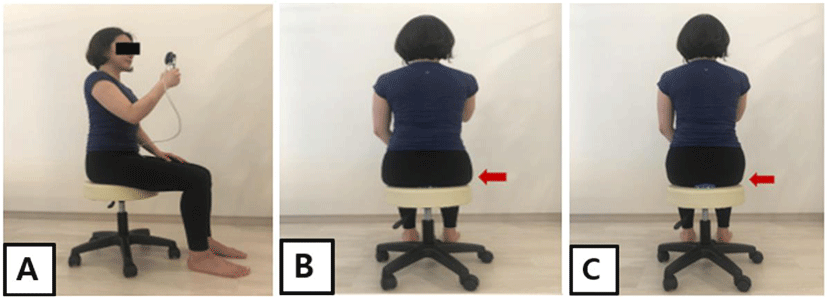
Three measurements of EMG activity were taken for 8 s prior to PFMs activation. The PBU was folded properly and placed between the buttocks of the participants. While watching the pressure gauge, maximum force was applied to the PFMs, and changes in PFMs contraction and relaxation were confirmed. PFMs contraction training using a PBU was performed for 5 min (Figure 1). During contraction of the PFMs, both gluteal muscles co-contracted and the pressure increased. At relaxation, the pressure of both gluteal muscles decreased with the relaxation of the PFMs.19 Contraction of the PFMs was confirmed by portable ultrasound when the pressure changed. Immediately following PFMs activation using a PBU, muscle activity of the PFMs was measured three times for 8 s. Measurements were taken three times for 8 s after 10 min of PFMs exercise. This measurement was used to confirm the effectiveness of the exercise after applying the PBU, and as an index to confirm whether the PFMs were correctly contracted. Training was conducted prior to the experiment to maintain the hands and feet in the designated positions to maintain a static posture during each exercise. To minimize fatigue that may occur due to continuous measurement, a 3 min rest period was included after each exercise. Each indirect PFMs exercise was performed for 8 s. The first and last second of signal measurement were excluded from analysis. A total of 6 s of muscle signal measurement for each exercise was used for data analysis.
A one-way repeated measurement analysis of variance was performed to investigate changes in activity of each muscle before exercise, immediately after exercise, and after 10 min of exercise. Bonferroni’s correction was used for post-testing. The intraclass correlation coefficient (ICC 2,1) was used to examine intra-rater reliability for the PFM activation with PBU. The reliability is regarded as acceptable if ICC>0.75. Data analysis was performed with PASW Statistics for Windows software (ver. 18.0; SPSS Inc., Chicago, IL. USA). All statistical significance levels (α) for hypothesis testing were set to 0.05.
RESULTS
The activities (%MVIC) of the TrA/IO, MF, EO, and RA muscles were measured by a PBU during indirect PFMs contraction in 20 healthy adult female participants. Use of the PBU had a significant effect on the %MVIC (Table 3). The activity of the TrA/IO muscles (%MVIC) was measured before exercise, immediately after exercise, and after 10 min of exercise. PBU application made a significant difference (TrA/IO left: F=30.93; TrA/IO right: F=43.77). A post hoc t-test (Bonferroni correction) indicated that the %MVIC of the TrA/IO muscles increased significantly immediately after exercise (p<0.01) and after 10 min of exercise compared to before exercise (p<0.01) (Table 3). MF muscle activity (%MVIC) was measured before exercise, immediately after exercise, and after 10 min of exercise. PBU application had a significant effect (MF left: F=37.32; MF right: F=51.31). A post hoc t-test (Bonferroni correction) indicated that the %MVIC of the MF increased significantly immediately after exercise (p<0.01) and after 10 min of exercise (p<0.01), compared to before exercise (Table 3). EO muscle activity (%MVIC) was measured before exercise, immediately after exercise, and after 10 min of exercise. PBU application had a significant effect on the right EO after exercise (EO left: F=30.97; EO right: F=55.57). A post hoc t-test (Bonferroni correction) indicated that the %MVIC of the EOs increased significantly immediately after exercise compared with before exercise (p<0.01). The %MVIC of the right EO increased significantly immediately after exercise and after 10 min of exercise (p<0.01) (Table 3). RA muscle activity (%MVIC) was measured before exercise, immediately after exercise, and after 10 min of exercise. PBU application had a significant effect on left RA activity after exercise (RA left: F=18.88; RA right: F=5.2). A post hoc t-test (Bonferroni correction) indicated that the %MVIC of both RA muscles increased significantly immediately after exercise, compared with before exercise (p<0.01). The %MVIC of the left RA increased significantly immediately after exercise and after 10 min of exercise (p<0.01) (Table 3).
DISCUSSION
This study investigated the effect of indirect PFMs activation using a PBU in a sitting position on trunk muscle activity. Muscle activity (%MVIC) of the TrA/IO, MF, EO and RA increased significantly immediately after indirect PFMs activation (p<0.01). After 10 min of indirect PFMs activation, significant increases were observed in TrA/IO and MF activity.
During PFMs contraction using PBU, TrA/IO muscle activity increased and was maintained before exercise (left: 15.12±4.56 %MVIC; right: 14.13±5.00 %MVIC) compared to immediately after exercise (left: 24.51±4.77 %MVIC; right: 24.29±4.80 %MVIC) and after 10 min of exercise (left: 22.80±4.22 %MVIC; right: 23.42±4.64 %MVIC). The PFMs co-contracts with the TrA, MF, and diaphragm to increase IAP, which results in trunk stability.7 The TrA/IO muscles are critical for stabilization of the trunk, and studies have investigated the effective contraction of muscles in the neutral zone of the pelvis.6,20,21 The increased activation of the TrA/IO muscles we observed may have been the result of increased IAP caused by PFMs contraction. PFMs contraction using a PBU is effective and maintains a neutral pelvic position. The contraction may also improve trunk stability. Abdominal hollowing and abdominal bracing were proposed to improve trunk stability,22,23 but most studies are related to direct contraction of the activity of the abdominal and back muscles. The PFMs are difficult to study due to the available research methods, selection of subjects, and challenging experimental methods. PFMs exercises using a PBU may help improve urinary incontinence prevention and trunk stability.
During PFMs contraction using a PBU, MF muscle activity increased and was maintained before exercise (left: 7.46±1.32 %MVIC; right: 8.57±1.76 %MVIC) compared to immediately after exercise (left: 13.09±3.13 %MVIC; right: 13.19±2.33 %MVIC) and after 10 min of exercise (left: 12.20±2.40 %MVIC; right: 12.32±2.52 %MVIC). PFMs contraction leads to sacral counternutation motion; the MF must counteract this to maintain the position of the sacrum in a sitting position.24 The MF acts as the antagonist of the PFMs to maintain a neutral pelvic posture, indicating that PFMs contraction is important in trunk stabilization when sitting and is effective at increasing MF muscle activity. Contraction of the MF is commonly used to stabilize the lumbar spine and in exercise therapy for lower back pain patients. Computed Tomography or Magnetic Resonance Imaging, used as a means of identifying MF activities, reported that static measurements were possible but dynamic studies were not possible.25 PFM contraction using a PBU is an easy and safe exercise method to improve activity of the MF through PFMs contraction in a sitting position. Additionally, the maintenance of MF muscle activity after 10 min of exercise is considered to be an effective method for activating contraction of the MF in a sitting posture using a PBU for 5 min. Training methods such as ultrasound and EMG for PFMs contraction are difficult to use by physical therapists in clinical settings. In comparison, using a PBU is an easy, convenient, and effective method.
In this study, muscle activity increased during PFMs contraction with a PBU in the EOs and RA. The trunk muscles consist of global muscles and local muscles. Global muscles balance the external load such as gravity or heavy objects. Local muscles are tonic muscles that maintain the curvature of the vertebrae and play an important role in maintaining the stability of the vertebrae.26 Local and global muscles work simultaneously to stabilize the spine when a load is applied.27-29 As a result, IAP caused by PFMs muscle contraction immediately after exercise increased muscle activity of the EOs and RA. However, this activity was not maintained after 10 min of exercise, indicating that PFMs contraction using a PBU might be a more effective exercise for the local muscles affecting trunk stability.
PFMs exercise using a PBU was effective at increasing activity of the TrA/IO and MF muscles, which are important for trunk stabilization. Increased muscle activity does not indicate an increase in muscle strength or improvement in function. Muscle activity evaluation using surface EMG is an objective and scientific method to assess muscle condition. PFMs exercise using a PBU can effectively increase activity of the TrA/IO and MF muscles in a sitting position. PFMs contraction could not be directly measured in a sitting position in this study, but this method can be recommended for use in clinical practice because of increased activity of the trunk muscles.
This study has several limitations. The sample size was small (n=20) and no control group was used. The results may not be generalizable to the entire population because only healthy female participants were evaluated. This study demonstrates an easy and convenient way to activate PFMs using a PBU; however, PFMs contraction was not quantitatively measured and was not directly confirmed. The PBU method can be applied to prevent, treat urinary incontinence, and may effectively increase trunk stability. Future studies are required to measure PFMs activity quantitatively, and to validate the applicability of the method to patients with urinary incontinence.
CONCLUSIONS
This study investigated the effects of indirect PFMs activation on the activity of the TrA/IO, MF, EO, and RA muscles in a sitting position in 20 healthy female participants. Activity (%MVIC) of the TrA/IO, MF, EO, and RA muscles increased significantly immediately after indirect PFMs activation. A significant increase in the activity of the TrA/IO and MF muscles was observed after 10 min of indirect PFMs activation. Indirect PFMs activation exercise using a PBU in a sitting position was confirmed as a method to increase the activity of the TrA/IO and MF muscles, which increase trunk stabilization. PBU-assisted PFMs activation exercise is an easy and convenient method to prevent and treat urinary incontinence and effectively increase trunk stability.