INTRODUCTION
Ankle joints provide the most basic exercise strategies for balance and posture control when standing or walking.1 In addition, ankle joint plays a key role in absorbing shock, supporting weight, and maintaining stability of legs during walking.2 Causes of instability of the ankle joint include anatomical instability of the skeletal system, weakness of muscle strength, and insufficient proprioception.3 Among them, muscle weakness acts as an active factor in the instability of ankle joint. It has been reported that weakness of tibialis anterior (TA) and gastrocnemius (GCM) muscle can cause functional ankle instability.4,5
An ankle sprain is one of the most common musculoskeletal problems that can be caused by weakening of muscles that contribute to stability around the ankle joint.6,7 Ankle sprain occurs when an external trauma or perturbation causes an over-stretching of the ankle joint beyond its normal range of motion.8 Unless appropriate therapeutic intervention to improve ankle stability against the initial ankle sprain is achieved, instability of the ankle joints could persist and become chronic.9 In fact, 40% to 50% of patients with chronic ankle sprain have symptoms such as chronic pain and re-injury.10 Therefore, stabilization of the ankle joint is important in the field of physical therapy for prevention and management of ankle sprain.
Functional training or closed-chain exercise in a program to improve ankle joint stability has been performed clinically. Closed-chain exercise can increase the stability of the ankle by increasing compression force and consistency of joints when supporting weight.11 Closed-chain exercise can induce simultaneous contraction of agonistic and antagonistic muscles and induce various combinations of isometric, eccentric, and concentric contractions, leading to more effective muscle contraction.12,13 Squat as one of closed chain exercises is a complex joint movement in which hip, knee, and ankle joints move simultaneously.14 In addition, several ankle muscles can coordinate and adjust their exercise intensity to their own physical ability.14,15 The advantage of squat exercise is that it can exert a large amount of force in a stable manner because the force is uniformly applied to both legs during the exercise. In addition, the isometric motion of squat can increase the validity of measuring muscle activity through the electromyography.16,17
Since squat is a closed chain motion with feet touching the ground, the state of the support surface can greatly affect the performance. Squatting on an unstable support surface requires more neuromuscular activation than that on a stable one.18 Many previous studies have determined the activation of pelvic and knee muscles in relation to squat performance on various support surfaces.7,19-22 However, there is a lack of research on muscle activations around ankle joints. Therefore, the purpose of this study was to investigate activity of leg muscles when performing squatting on various supporting surfaces such as bottom floor (BF), balance pad (BP), and togu (TG) and determine the most effective squat exercise method for treating and preventing instability.
METHODS
Subjects of this study were 20 healthy adults. Those who had enough leg strength and balance ability to perform a squat posture on three supporting surfaces (BF, BP, and TG) were selected. Exclusion criteria were: 1) those who had ankle and musculoskeletal disorders, and 2) those who had ankle joint problem for the last six months. All participants voluntarily participated in the experiment. They understood the experimental process after obtaining preliminary explanation before the test. This study was approved by the Institutional Review Board of Jeonju University (approval number: jjIRB-2015-1103).
Before measuring electromyography (EMG) signals and attaching electrodes, any hair on the skin to be attached was shaved. The skin was then cleaned with an alcohol swab before attaching electrodes.14 EMG electrodes were attached to TA, peroneus longus (PL), medial GCM, and lateral GCM according to published recommendations.23 A Delsys Trigno EMG system (Delsys, Inc, Wellesley, MA, USA) was used to collect EMG data. EMG signals were converted to digital signals and processed using Works Acquisition EMG analysis software for personal computers (Delsys, Inc, Wellesley, MA, USA). The sampling rate of EMG signals was 2,000 Hz. EMG frequency bandwidth was restricted to 20–500 Hz. Common mode rejection ratio was set at 110 dB.
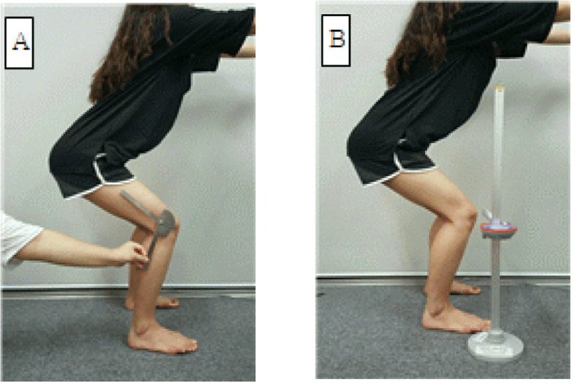
For EMG measurement, subjects were asked to start squat at a distance of two feet from shoulder width and arms extended forward with 90° shoulder flexion. For a constant squat practice, the knee joint was flexed 60 degrees with the back straight (Figure 1). At this time, the experimenter supervised to maintain the angle of the knee joint at 60° by using a universal goniometer (Goniometer SB Inc, Korea). In order to unify knee flexion direction, a target bar (Digital Backward Flexmeter 5404, Takei Inc, Japan) was installed to direct the center of the knee to the second toe direction (Figure 1). To measure the activation of lower leg muscles, supporting surface conditions including a general BF, a BP (Balance-pad AIREX Inc, Swiss; 49 × 40 × 6.5 cm), and a 35 cm diameter TG (Togu Dynair Pro Inc, Germany) were provided. Two TGs were used, one for each foot.

The sequence of application of the support surface for the squat exercise was determined randomly using a die. The experiment was started after two practice squat performances on each support surface. The squat exercise was performed for a total of 10 seconds: 3 seconds of descent in the standing posture, induction of isometric contraction for 4 seconds, and returning to the starting posture for 3 seconds. A root mean square (RMS) value of intermediate 4 seconds was used for the final analysis. The squat performance rate was 60 metronomes per minute. The mean value of activity of each leg muscle measured three times on each support surface was used for analysis. To minimize subject’s fatigue, a 30 second break between each squat exercise was allowed with a 3 minute rest interval between each support surface.
Two-way repeated measures ANOVA with Bonferroni’s correction was used to determine difference in activity among the three support surfaces and muscles of both legs during squatting. Scheffe post-hoc test was used to analyze differences in activation of each lower leg muscle among support surfaces. All statistical analyses were performed using SPSS ver. 21.0 (IBM Inc, New York, USA). Statistical significance was defined at α=0.05.
RESULTS
RMS values of leg muscles are shown in Table 1. Results of repeated ANOVA on various supporting surfaces and left and right muscle activities of each leg muscle are shown in Table 2. There were significant differences in muscle activity of TA (F(2,19)=5.207; p<0.05), PL (F(2,19)=17.165; p<0.05), medial GCM (F(2,19)=3.750; p<0.05), and lateral GCM (F(2,19)=9.757; p<0.05) according to support surface condition (Table 2). There was no significant difference in muscle activity between right and left muscles in any leg muscle (p>0.05). In addition, there was no significant difference in the activity of leg muscle according to interaction between various support surfaces and right or left leg muscle (p>0.05).
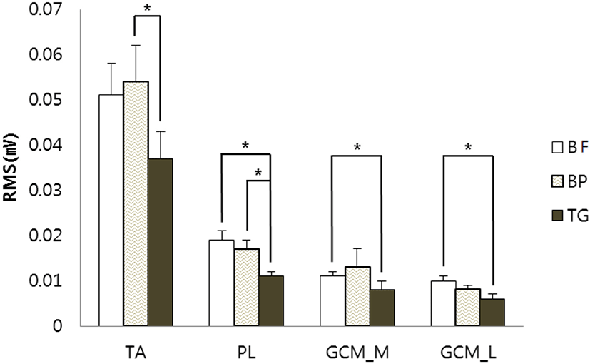
Results of multiple comparisons of RMS values according to muscle support surface conditions are shown in Figure 2. The activity of TA was significantly different between BP and TG (p<0.05). There was a significant difference in activity between BF and TG or between BP and TG (p<0.05). The muscle activity of medial or lateral GCM muscle was significantly different between BF and TG (p<0.05) (Figure 2).

DISCUSSION
Rehabilitation of ankle injuries requires activity to restore tendons, ligaments, bones, and muscles. Normal muscle tissue size, flexibility, strength, and muscle endurance should be considered.24 Especially, strengthening ankle muscles is clinically important for the treatment and prevention of ankle ligament injuries.25 The main cause of ankle instability is muscle weakness or the lack of muscle coordination.26 Balance training, proprioceptive stimulation training, and weakened muscular strength training are needed to increase ankle stability.27 Therefore, this study was conducted to investigate the effect of ankle joint on the activity of hip joint during squatting in order to increase the stability of ankle joint and to propose an effective exercise method for preventing injury and functional activity.
As a result of this study, there were significant differences in activities of lower leg muscles according to support surface during squatting (p<0.05). Activities of TA and medial GCM that provide anteroposterior stability of the ankle joint were higher in the order of BP, BF, and TG. Ankle strategy is used first when balancing the standing posture on a stable support surface condition where external perturbation is weak. The greater the degree of external perturbation, the more the hip joint strategy is used than the ankle strategy for posture control and balance.26 More muscle activity was found for TA and medial GCM muscles under BP condition which provided some instability of support surface for balance than that under BF condition. On the other hand, there was no significant difference in muscle activity of TA or medial GCM muscle in the most unstable surface, a TG. This indicated that more controlled hip joint motion was needed than ankle motion when providing greater external perturbation to maintain balance in standing.
The activity of PL and lateral GCM muscles to provide stability of the lateral side of the ankle was higher in order of BF, BP, and TG. On a more unstable support surface, the muscle activity required for stability in the anteroposterior direction of the ankle joint was increased rather than the bilateral stability of the ankle joint while muscle activities of PL or lateral GCM muscle providing lateral stability of the ankle joint were not increased significantly. However, under stable floor support conditions, instability of the anteroposterior movement of the ankle was decreased while activities of PL and lateral GCM muscle were increased.
As a result of this study, muscle activity of the lower leg muscle did not increase in proportion to the instability of support surface conditions.28 In addition, squat exercise executed to strengthen lower extremity muscles was effective when performing under a stable support surface. On the other hand, squat exercise for trunk stability was more suitable under an instable support surface condition.19 Therefore, results of the present study suggested that squat exercise for stabilization of the trunk would be recommended on a more unstable support surface condition. As a result of squat exercise on four supporting surface conditions of 10 male adults who had exercise training for 6 months or more, activities of lumbar multifidus, gluteus medius, and gluteus maximus were increased on more instable support conditions than those on a firm support surface condition during squat lowering exercise.19
The limitations of this study are as follows. First, subjects were healthy men and women in their 20s who had no ankle joint pathology. Therefore, it was difficult to generalize results of this study to various ages. In addition, our sample size was too small to generalize our findings to the general population. We did not strictly control the movement of the trunk or upper limb during squat exercise either. Therefore, future long-term studies should be done with many indi-viduals of various ages by supplementing these limitations. Further studies are needed to determine whether squat exercises under various support surface conditions can improve muscle activation of lower leg muscles for different populations.
CONCLUSIONS
The purpose of this study was to present an effective method to improve ankle joint stability by increasing activity of lower extremity muscles by squat exercise on three support surface conditions in twenty healthy adults. TA and medial GCM muscles showed the highest muscle activity in BP condition while PL and lateral GCM muscles showed the highest muscle activity in BF condition. Therefore, an unstable support surface such as a BP is recommended for anteroposterior stability of the ankle while a stable support surface such as BF is recommended for bilateral stability of the foot when performing squat exercise.


