


INTRODUCTION
The articular surfaces of the sacroiliac joint (SIJ) are relatively flat and aligned close to the vertical plane.1 Flat joint surfaces are optimal for load transfer.2 However, alignment close to the vertical plane confers vulnerability to vertical shear forces caused by gravity,3 triggering SIJ instability that is attributable to creep of the long, dorsal sacroiliac and sacrotuberous ligaments under prolonged loads.4 Sub-optimal SIJ stability is associated with lumbopelvic,5 groin,6 hamstring,7 and/or low back pain (LBP).8,9 The prevalence of sacroiliac joint pain (SIJP) is 13–30% in patients with non-specific LBP.8,10 Therefore, numerous efforts have been made to enhance SIJ stability. Effective SIJ load transfer and stability requires optimal passive, active, and neuromuscular joint control.1,11,12 These functions vary among individuals by anatomical articular stability (form closure) and neuromuscular stability (force closure) during performance of various activities.13 In clinical practice, force closure is reinforced via strengthening of pelvic and trunk muscles, including the gluteus maximus (GM), biceps femoris (BF), latissimus dorsi (LD), and erector spinae (ES).5,14 Form closure is reinforced via passive support, including a pelvic compression belt (PCBs).
Muscles engaging in forced closure are evaluated in the prone hip extension (PHE) test, which measures SIJ load transfer,15,16 strength of hip extensor,17 and the recruitment patterns of the hip extensors and trunk muscles.18,19 In previous studies, the PHE test was performed only under knee extension (PHE-KE), because the activity and re-cruitment pattern of the hip joint extensor muscles were of interest, and not muscle strength. However, as the PHE-KE test indexes both GM and BF status, it may be difficult to evaluate GM weakness because of BF compensation.20 Therefore, it is necessary to evaluate PHE-KE and PHE-knee flexion (PHE-KF) separately.
Several studies have shown that PCBs enhance form closure. Jung et al.21 found that, compared to asymptomatic individuals, the BF electromyographic (EMG) amplitude was significantly reduced in those with SIJP who used PCBs. Furthermore, the GM reaction time was significantly decreased during one-leg standing. Kim et al.22 compared healthy and LBP groups using PCBs during PHE; muscle activity of trunk was reduced significantly more in LBP subjects than in healthy controls. Mens et al.23 and Yoon et al.24 confirmed that PCBs increased strength of hip flexor and adductor, and muscle activity of abdominal. However, these studies did not explore strength of hip extensor or muscle activity of trunk.
Previous studies found that PCBs passively stabilized the SIJ via application of a compression force,25,26 thereby significantly affecting hip muscle strength.23,24,37 In particular, PCBs can be applied during PHE to evaluate SIJ load-transfer function, but we instead sought to investigate changes in muscle activity. In those with SIJP, strength of hip extensor is important because the proximal GM is attached to the SIJ, affording stability through application of compression across the joint.14 The GM is very active during abrupt limb loading; the SIJ must be stable. SIJP patients exhibit GM weakness and muscle activity delay, triggering compensation by the BF2,28 that can increase SIJP and cause LBP.29 However, no study has yet explored the effects of PCBs on strength of hip extensor. Arab et al.28 confirmed that the GM was weak in those with SIJP, but used only a pressure meter for measurement. Strength of hip extensor is preferentially measured using a hand-held dynamometer (HHD) or isokinetic dynamometer, but these instruments are difficult to employ clinically, being unreliable and expensive. Recently, tensiometers have been used to measure strength; tensiometers are both very reliable and portable.27 Therefore, we objectively measured changes in strength of hip extensor and muscle activity of trunk when SIJP patients wore PCBs during PHE.
METHODS
We enrolled 15 female volunteers with SJIP, aged 20–60 years, in Bokum general hospital of Gimhae city (Table 1). The inclusion criterion was showing at least three of a possible six positive reactions on SIJP provocation testing (distraction, compression, thigh thrust, Gaenslen’s response, sacral thrust, and the drop test); all tests exhibit acceptable inter-rater reliability.10,30–32 The exclusion criteria were an unwillingness to participate; only midline or symmetrical pain above the L5 level; any clear sign of nerve root compression (a complete motor or sensory deficit); a history of spinal surgery; a history of spinal, pelvic, or lower extremity fracture; prior hospitalization for trauma or a motor vehicle accident; hip or knee dysfunction; pregnancy; and/or any systemic disease (arthritis, tuberculosis, liver disease, or kidney failure); such subjects were deemed too frail for complete physical examination.33
| Characteristic | Mean ± SD |
|---|---|
| Age (years) | 46.13 ± 11.42 |
| Height (cm) | 159.13 ± 3.20 |
| Body weight (kg) | 49.69 ± 4.33 |
| BMI (kg/m2) | 31.47 ± 2.81 |
| VAS | 5.20 ± 1.01 |
The sample size was calculated using a pilot study that revealed significant increase in strength of hip extensor and muscle activity of the contralateral LD. Power analysis indicated that at least six participants would be required to achieve a power of 0.80 and an effect size of 1.213 at a significance level of 0.05 prior to the experiment, all participants were provided with a thorough explanation of the intervention protocol and voluntarily agreed to participate. All signed informed consent forms were approved by the Inje University Ethics Committee for Human Investigations (approval no. INJE 2017-09-014-003).
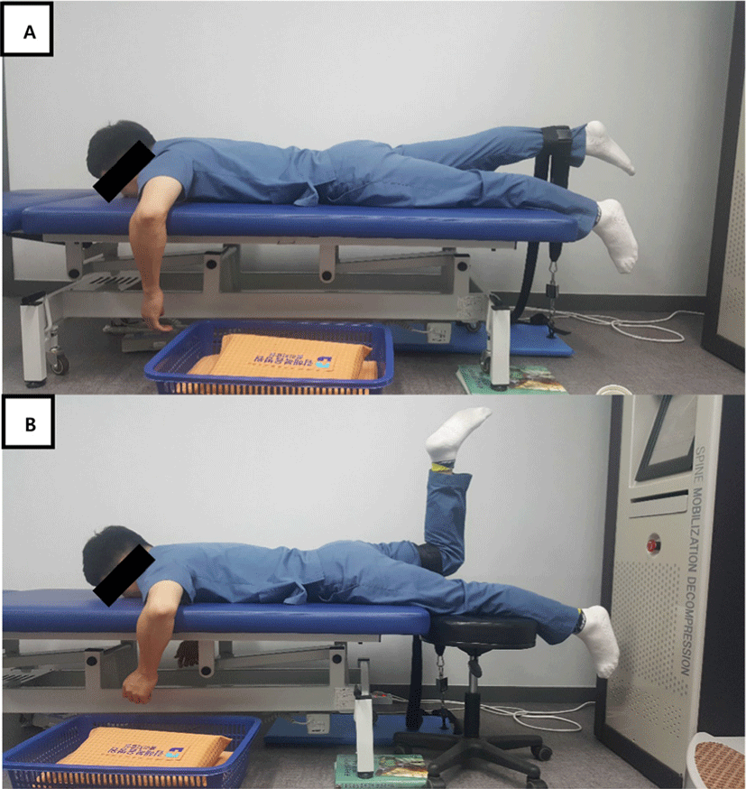
Force of hip extensor was measured using fixed custom-designed instrumentation assessing the isometric PHE-KE and PHE-KF forces. A load cell (RSBA-50L; Radian, Seoul, Korea) linked to a digital indicator displaying the force was connected to a board (a 702 × 300 × 18-mm plate; Figure 1). The detectable force range was 0–50 kg at a resolution of 0.1 kg and the precision was ±0.3 kg. The sampling frequency was 100 Hz. In our previous study, this instrument exhibited excellent intra- and inter-rater correlations (intraclass correlation coefficient 0.97) when used to measure isometric strength.27,34 The maximal force generated during isometric PHE-KE and PHE-KF after application of force to the load cell was automatically recorded.
EMG data were recorded from the contralateral LD and ES during isometric PHE-KE and PHE-KF using a surface EMG system (Trigno Wireless; Delsys, Boston, MA, USA). The signals were amplified and band-pass filtered (20–450 Hz) prior to digital recording at 2,000 Hz, and root mean square (RMS) values then calculated. We used two maneuvers to normalize EMG activity: maximal and submaximal voluntary isometric contractions.35 Pre-test, we confirmed that, under both conditions, the values were equal. However, when the ES was subjected to maximal isometric contraction, SJIP individuals developed acute pain. Therefore, the submaximal method was judged appropriate for ES evaluation. Trunk muscle normalization exhibited excellent within-day reliability in both healthy controls and SJIP individuals.22
To minimize skin impedance before EMG electrodes were attached, hair was removed and the skin cleaned with alcohol-saturated cotton. EMG data were collected bilaterally from the LD (4 cm below the inferior tip of the scapula at half the distance between the spine and lateral edge of the torso) and the ES (2 cm lateral to the spinous process at the L1 level and parallel to the spine).36
All muscles were tested in the PHE position. The LD maximal isometric contractions were measured when manual resistance was applied.17 Each LD was tested with the subject’s arms at the sides, and the shoulders internally rotated to create a palm-up position. Resistance was then applied to the forearm. During submaximal voluntary ES isometric contraction, the subject lifted both knees 5 cm off the examination table, flexed them at 90°, and held that position for 5 s.37 Each maximal and submaximal isometric contraction maneuver was performed twice for 5 s each time, and the muscle activity of average during the middle 3 s of both trials was used for normalization. The EMG results were normalized to the maximal and submaximal EMG RMS values calculated from EMG signals obtained during voluntary isometric contraction of each muscle.
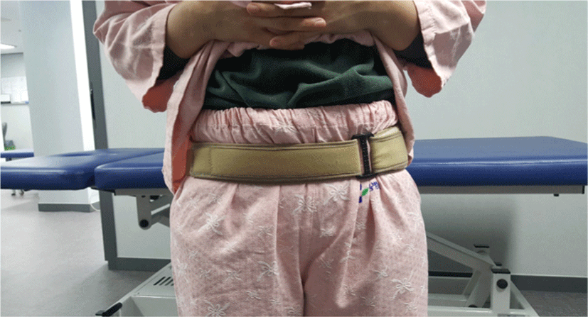
A PCBs (SI-LOC; OPTP, Minneapolis, USA) was worn below the anterosuperior iliac spine (ASIS)25 and fastened firmly to a belt (Figure 2). The device was constructed of non-elastic material with Velcro ends, and was 5 cm wide at the front and back, and 8 cm wide at the sides. The tightness was adjusted by a physiotherapist experienced with SIJP patients; no patient complained of pain or discomfort.
The maximal isometric PHE-KE and PHE-KF were measured with and without the PCBs (in a random order). A non-elastic belt connected to the load cell was placed on the Achilles tendon during PHE-KE, and behind the knee during PHE-KF (Figure 1). The opposite end of the belt was fixed vertically to the ground to maintain a consistent belt length during PHE-KE and PHE-KF, facilitating isometric contraction when the affected hip joint extended from neutral to about 10°. Also, to restrict unwanted trunk movement, individuals lay prone with 90° shoulder abduction and 90° elbow flexion. Isometric PHE-KE and PHE-KF were performed for 5 s three times, with a 1-min break between measurements.
The three mean muscle forces measured during isometric PHE-KE and PHE-KF were analyzed, as were the means of three measurements of contralateral LD and ES activities. Data were analyzed using SPSS software (ver. 18.0; SPSS Inc., Chicago, IL, USA) and are expressed as means±SD. Two-way repeated analysis of variance (ANOVA) was used for within-group comparisons, with and without the PCBs. If significant interactions were evident between PHE-KE and PHE-KF, the post-hoc paired t-test was performed. A p-value <0.05 was considered to reflect statistical significance.
RESULTS
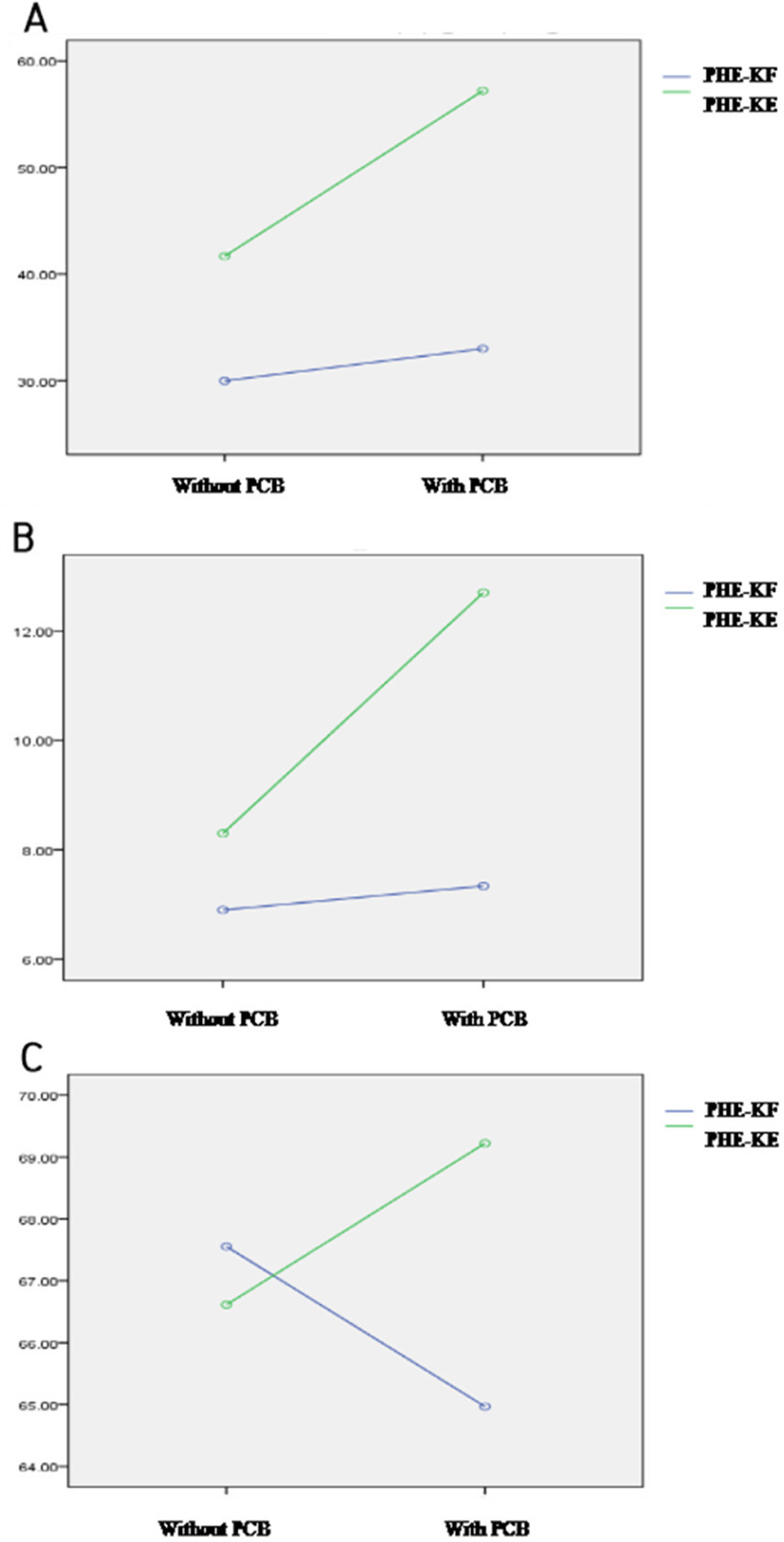
Without and with the PCBs, the PHE-KE force (6.83±3.17 vs. 8.27±3.60 kg; difference, 1.44 kg; 99% CI: 1.11–1.76 kg; p<0.001) and PHE-KF force (7.09±3.85 vs. 12.50±4.38 kg; difference, 5.40 kg; 99% CI: 5.00–5.81 kg; p<0.001) were significantly higher when the PCBs was worn (Table 2, Figure 3). The paired t-test indicated that the force was 3.34 kg higher (99% CI: 2.63–4.91 kg) during PHE-KF than PHE-KE (p<0.001) (Table 2). The post-hoc paired t-test showed that PHE-KE and PHE-KF forces without a PCBs did not differ significantly (p>0.05), but did when the PCBs was worn (p<0.05).
When the PCBs was not worn during PHE-KE, the EMG data varied (28.98±3.12 vs. 39.94±5.03 mV; difference, 11.00 mV; 99% CI: 5.30–16.71 mV; p<0.05), as was also true during PHE-KF (31.66±3.97 vs. 53.68±6.92; difference, 22.02 mV; 99% CI: 13.23–30.80 mV; p<0.05); the muscle activity of the contralateral LD also significantly increased (Table 3, Figure 3). The paired t-test indicated that the mean difference of 16.51 mV (99% CI: 5.41–27.61 mV) was significantly greater during PHE-KF than PHE-KE (p<0.05) (Table 3). However, the muscle activity of the contralateral ES did not differ significantly (p>0.05; Table 4, Figure 3).
DISCUSSION
We explored whether a PCBs passively stabilized the SIJ by improving the strength of hip extensor and LD and ES activities during isometric PHE-KE and PHE-KF. The PCBs significantly improved strength of hip extensor (p<0.001), and also significantly increased activity of LD (p<0.05) but not activity of ES (p>0.05). To stabilize the SIJ, the pelvis and hip muscles contract, forcing closure mediated by a compressive force between the ilium and sacrum.19,38, 39 This represents a particularly important role of the muscles and the fascia; the relevant muscles are the proximal GM, the BF, and the ES; the thoracolumbar fascia is primarily involved.5 Also, appropriate force closure requires SIJ stability.21,22,29 SIJ instability decreased when a PCBs was worn25,26 the PCBs complemented the force closure caused by muscular contraction, by increasing SIJ passive stability (form closure)21 thereby alleviating SIJP. In addition, PCBs aided muscle contraction by increasing intramuscular pressure (IMP), in turn affecting muscle strength and activity.40,41
Yoon et al.24 reported that PCBs application during the active straight leg raise (ASLR) task significantly increased hip flexor muscle strength by enhancing passive stability of the lumbopelvic region, thereby suppressing muscular action form triggering unnecessary compensation, and thereby ultimately promoting hip flexion. The data of Mens et al.,23 Park et al.,39 and Kang et al.27 support this argument. However, in previous studies, hip flexor and adductor strength were assessed by the extent of PCBs wear; strength of hip extensor was not explored. Recently, Arab et al.28 suggested that the GM was weak in those with SIJP, but used only a pressure meter to collect data; real muscle strength was not measured. Pressure meter data are subjective, reflecting the pressure imposed by the examiner. Kim et al.19 showed that muscle activity of hip extensor changed during PHE. Muscle strength was measured via manual strength measurement using an HHD; this is common in clinical practice. However, as strength is assessed as the manual resistance to the examiner, if the patient is stronger than the examiner, the data will be erroneous.42 Thus, we measured objective strength of hip extensor during isometric PHE-KE and PHE-KF in patients wearing or not wearing a non-elastic belt.
The results of previous studies using HHDs, and our results using a non-elastic belt, indicate that PCBs-induced SIJ passive stability promoted action of hip extensor and increased extensor strength. In addition, isometric PHE-KF increased the GM strength more so than isometric PHE-KE increased the hamstring strength; the PCBs reduced reaction time of GM and activity of BF more so than that of the hamstring.21,29 The PCBs contacted the GM and increased the IMP, significantly enhancing muscle strength compared to that of the hamstring.40,41 Our results are clinically important because the proximal GM stabilizes the SIJ5 and the BF; global muscle strengthening can trigger compensatory LBP.29
The activity of the contralateral LD increased significantly when a PCBs was worn during isometric PHE-KE and PHE-KF (p<0.05). However, the activity of the contralateral ES did not change (p>0.05). A PCBs significantly reduced muscle activity of trunk during PHE.19 Jung et al.21 reported that when the PCBs was worn during one leg standing, the reaction time of the GM and the activity of BF decreased, but the BF reaction time increased. However, we did not schedule a PHE task; rather, we measured maximal muscle strength during the PHE-KE and PHE-KF postures. SIJ stability was increased by the PCBs, as was maximal muscle strength. Thus, PCBs-promoted load-transfer via the SIJ increased the maximal strength and activity of the contralateral LD, and also the IMP, through contact with the contralateral LD; this significantly increased muscle activity. However, activity of ES did not change significantly; the ES was not in contact with the PCBs.
Our study had certain limitations. First, we did not divide subjects into acute and chronic SIJP groups, rendering the SDs relatively high. In future studies, patients should be distinguished on this basis. The second limitation was that we measured only strength of hip extensor and muscle activity of trunk during PHE-KE and PHE-KF. It would be useful to additionally assess the strength of the isolated lateral hamstring, and that of external rotation during hip extension. Finally, we enrolled only 15 subjects; it is thus difficult to generalize our results.
CONCLUSION
We found that PCBs use during PHE passively stabilized the SIJ, promoting muscle activity of LD and increasing strength of hip extensor. Thus, the PCBs improved load transfer by the SIJ. Also, the PCBs increased muscle strength and activity by compressing muscles. We recommend using of PCBs for patients with SIJP increased the strength of hip extensor and muscle activity of contralateral latissimus dorsi.