


INTRODUCTION
The anconeus and triceps brachii are primary extensors of the elbow.1 The anconeus extends from the olecranon to the dorsolateral part of the lateral epicondyle.2 The main action of the anconeus is accessory extension of the elbow.3 This muscle also acts as a stabilizer for the elbow joint during supination and pronation.4 The triceps brachii is a three-headed muscle (long, lateral, and medial).5 The long head (LoT) is attached to the olecranon from the infraglenoid tubercle of the scapula.1 Therefore, the LoT serves to extend and adduct the shoulder and extend the elbow.6,7 The medial and lateral heads (LT) originate from the posterolateral aspect of the humerus, and their main function is elbow extension with the LoT.1,7
In clinical, the elbow extensors are strengthened to increase the ability of spinal cord injury patients in a push wheelchair to transfer from one place to another and to reach objects during activities of daily living.8 Athletic trainers’ use several exercises to strengthen the elbow flexors and extensors for elbow balance. Strengthening the triceps is especially important in cross-country sports to improve high propulsion force.9
However, activities that increase muscle strength during elbow extension by shoulder posture remain to be investigated. Two positions are generally adopted to measure the strength of the elbow extensors: supine and prone.5 Therefore, the purpose of this study was to compare the electromyography (EMG) activity of the anconeus, LT, and LoT and the force of the elbow extensors during elbow extension in the supine and prone positions.
METHODS
This study included 21 healthy subjects (19 male, 2 female; mean age: 23.33±1.83 years; height: 174.5±6.90 cm; weight: 75.38±13.01 kg). Exclusion criteria were neuromuscular disorder of the shoulders and upper extremities, limited motion of shoulder and elbow joints, and fracture of the upper extremity (especially the elbow joint). All subjects participated voluntarily. Before the study, the investigator explained the experimental procedures to the subjects.
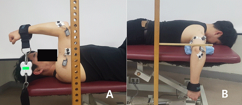
A surface EMG system (TeleMyo DTS, Noraxon, Scottsdale, AZ, USA) was used to measure the activity of the anconeus, LT, and LoT. The sampling rate was 1,024 Hz. Filtered movement artifacts were eliminated using a digital band-pass filter (Lancosh FIR) at a frequency range of 20–450 Hz. Root mean square values were used to process EMG signals with a moving window of 50 ms. EMG signals were recorded for 5 s, and seconds 2–4 were used for data analysis. Two electrodes were placed over the middle of each muscle belly, parallel to the muscle fibers. The electrode sites were shaved, and rubbing alcohol was used to reduce skin impedance. Electrodes were placed on the anconeus (parallel to and below the olecranon on the radial side), LT (lateral, approximately 50% of the distance between the acromion and olecranon or elbow joint), and LoT (medial, approximately 50% of the distance between the acromion and olecranon).10 EMG data were measured in the supine and prone positions. Maximum voluntary isometric contraction (MVIC) was held for 5 s in each position. This procedure was repeated three times in each position, with a 1-minute rest period between sessions. The force of the elbow extensors was simultaneously measured using a Smart KEMA strength measurement system (Factorial Inc., Seoul, Korea); force was measured from the isometric strength of the elbow extensors and transferred to a tablet (Galaxy Tab A6 10.1, Samsung, Inc., Seoul, Korea) via a Bluetooth connection and analyzed using the Smart KEMA application software (Factorial, Inc.).
All subjects were measured in the supine and prone positions randomly. Subjects wore a Smart KEMA force measurement belt on the distal part of the forearm to measure elbow extensor strength. In the supine position, measurements were conducted at maximal isometric elbow extension in 90° shoulder flexion, 90° elbow flexion, and forearm supination (Figure 1A). In the prone position, measurements were conducted at maximal isometric elbow extension in 90° shoulder abduction, 90° elbow flexion, and forearm supination. To limit shoulder movement, we placed a target bar at the elbow joint (Figure 1B).
The data are expressed as mean ± standard deviation. A paired t-test was used to assess the significance of differences in EMG activity and muscle force in each position. Statistical analyses were performed using the SPSS version 21.0 software for Windows (SPSS, Inc., Chicago, IL, USA). Significance was assessed at a level of p<0.05.
RESULTS
The EMG activity of the anconeus and LT was significantly greater in the supine than in the prone position (p<0.05) (Table 1). The EMG activity of LoT in the prone position exhibited greater than in the supine. The force of elbow extensor was significant increase in the prone position (p<0.05) (Table 2).
| In supine | In prone | t | p-value | |
|---|---|---|---|---|
| Anconeus | 74.07±20.26 | 57.40±23.00 | -3.224 | 0.004* |
| LoT | 77.72±19.29 | 92.97±30.45 | -2.143 | 0.005* |
| LT | 77.37±19.91 | 58.76±27.41 | -3.780 | 0.001* |
| In supine (kg) | In prone (kg) | t | p-value | |
|---|---|---|---|---|
| Mean±SD | 12.65±2.71 | 15.94±3.90 | 5.502 | 0.001* |
DISCUSSION
We compared the EMG activity of the anconeus, LT, and LoT and the strength of the elbow extensors in two postures. The anconeus and LT exhibited greater muscle activity in the supine position, whereas the EMG activity of the LoT was greater in the prone position.
The anconeus acts as a stabilizer for the humeroulnar joint.11,12 The muscle fibers of the anconeus combine with those of the LT, some of which are arranged in the same direction.12 Continuity has also been detected between the fascia of the anconeus and that of the LT.13 The EMG patterns observed in this study are consistent with these previous reports.
A previous study found that the LoT, which is connected at two joints, was more effective in the supine position.1 Because the LoT is shortened at both the shoulder and elbow joints during horizontal abduction in the prone position, whereas during shoulder flexion in the supine position, it is shortened at the shoulder and lengthened at the elbow joint.
However, unlike the slight extension of the elbow observed in the previous study, elbow flexion was 90° in this study. This difference in the position of the elbow joint may explain the differential results between this and the previous study.5
When we measured the force and EMG activity of the elbow extensors, the EMG activity of the anconeus and LT were significantly greater in the supine than in the prone position, whereas the muscle strength was greater in the prone position. In a comparison of triceps muscle fibers, Neumann (2002) reported that the LoT had a larger volume and physiological cross-sectional area at the elbow than did the brachialis1. The greater muscle strength of the LoT in the present study may have resulted from the measurement posture and the cross-sectional area of the LoT noted in that study.
It is important that the strength of the elbow extensors and EMG activity of LoT in the prone position was higher than that in the supine position. We recommend the supine position rather than prone to increase the muscle activity of the LT or the anconeus, which serves as an elbow stabilizer.
This study has several limitations. First, the elbow extensors were measured only in the supine and prone positions, although many strategies for strengthening them use other postures. The most common method to determine elbow extensor strength is to measure it in these two postures. Further study is needed to examine muscle strength and EMG activity in other postures. We also compared EMG values when the maximum force was applied. Future research should also measure the activity of each muscle at varying degrees of force.
CONCLUSION
Our study identified the strength and EMG activity of the elbow extensors in two positions. To increase the muscle strength and EMG activity of the LoT, elbow extension should be performed in the prone position; we also recommend that the EMG activity of the anconeus or LT be increased in the supine position.