
 PDF Download
 PubReader
 Export Citation
 Email the Author
 Share Facebook
 Share Twitter
 Cited by 0 Article
 Metrics (View:893)







INTRODUCTION
Osteoarthritis (OA) is a chronic, progressive, and degenerative joint disease with a high global prevalence.1 It leads to joint pain, restricted mobility, and ultimately a decline in quality of life.2 With the accelerating trend of population aging, the increasing prevalence of OA has emerged as a major public health concern, imposing not only individual suffering but also substantial socioeconomic burdens.3
The diagnosis and assessment of OA are primarily based on radiographic examinations, which identify structural changes such as joint space narrowing and osteophyte formation, as well as on analyses of lower limb mechanical alignment indices, including the hip-knee-ankle (HKA) angle.4 However, radiographic evaluations are associated with certain limitations, such as additional costs, the need for patients to visit medical facilities, and potential exposure to ionizing radiation.5 Moreover, radiographic findings do not always correspond precisely with the severity of clinical symptoms experienced by patients.6 Therefore, there is an urgent need to develop objective, noninvasive indicators capable of accurately and conveniently assessing the status and symptom severity of OA, while addressing these limitations.
Recent studies highlight the popliteal crease obliquity angle (PCOA) as a promising noninvasive indicator for OA assessment. PCOA quantifies the obliquity of the knee’s popliteal crease and indirectly reflects lower limb coronal alignment.7 It significantly correlates with the HKA angle, suggesting its relation to knee joint mechanical loading and potential utility in predicting OA pathophysiology.8 Self-reported questionnaires represent another critical evaluation approach, such as early osteoarthritis questionnaire assessment (EOQA) survey, which is widely used to comprehensively assess subjective symptom domains of osteoarthritis, including pain, stiffness, and functional limitation.9
While PCOA and EOQA have both been utilized as independent tools to assess knee OA, research examining the specific association between these measures remains insufficient, particularly in preclinical populations. Identifying a meaningful relationship between these assessment tools through this study could provide valuable clinical implications for the preliminary screening of early-stage OA, potentially enhancing the ability to identify individuals at risk and guiding early intervention strategies. The objective of this study is to investigate the association between PCOA and the clinical features of the EOQA in individuals with preclinical knee OA. By analyzing this relationship, this study evaluates whether PCOA can serve as a potential non-invasive clinical indicator for early screening in primary care and occupational health settings, providing a practical assessment framework that complements existing clinical protocols.
METHOD
A total of 43 manufacturing workers (86 legs) were recruited from a cosmetic production factory. In this study, each limb was treated as an independent unit of analysis. Although potential correlation between bilateral limbs within the same participant has been discussed in musculoskeletal research, this assumption is considered acceptable when the measured variable reflects local anatomical or mechanical characteristics that are not directly influenced by the contralateral side. This approach allows for a more detailed and objective evaluation of limb-specific deviations. Participants were classified into either the experimental or control group according to their responses to the first two items of the EOQA. While the EOQA comprises 11 items in total, this study focused on the first two items, which are categorized as clinical features (e.g., joint stiffness and persistent pain). The remaining nine items are characterized as patient-reported outcomes related to daily activities and quality of life. The decision to use only the clinical feature items for group classification was based on the methodology validated in a previous study, which confirmed that these specific items provide a more direct reflection of early-stage musculoskeletal changes compared to the broader subjective measures. This approach ensures clinical relevance and maintains consistency with established screening protocols for preclinical knee OA.9-10 The experimental (EOQA) group included 42 legs from participants who responded “frequently” or “rarely,” whereas the control (Non-EOQA) group comprised 44 legs from those who responded “never.”9 To ensure a homogeneous study population and minimize potential confounding factors, specific inclusion and exclusion criteria were applied. Individuals were excluded if they (1) had sustained a lower extremity injury within the previous six months, (2) had a history of hip surgery, (3) had been diagnosed with rheumatoid arthritis or osteoarthritis, or (4) had any neurological disorders that could affect lower limb function. No significant baseline differences between groups, confirming demographic comparability (Table 1). The study protocol was reviewed and approved by the Institutional Review Board of Sangji University (IRB No. 1040782-230814-HR-09-117). All participants received detailed information about the study procedures and provided written informed consent prior to participation.
The EOQA is a recently developed tool designed specifically to assess and monitor early-stage knee osteoarthritis. It comprises 11 items divided into two main domains: Clinical Features, which includes 2 questions focusing on objective symptoms such as pain during prolonged walking and episodes of knee instability, and Patient-Reported Outcomes, which consists of 9 questions exploring subjective experiences and functional limitations related to early knee OA. The questionnaire was developed through a rigorous process of item generation, reduction, and patient feedback to ensure clarity, relevance, and ease of use. Its purpose is to sensitively detect subtle symptomatic and functional changes characteristic of the early disease stage, facilitating timely diagnosis and appropriate intervention.9
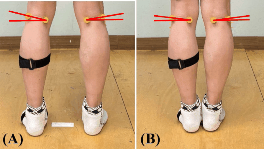
The PCOA was measured with participants standing barefoot in two positions: with feet shoulder-width apart and with feet together. Photographs (2,556×1,179 pixels) were taken from behind at knee height, 1 meter away, using a smartphone (iPhone 15; Apple Inc., USA) under consistent indoor fluorescent lighting. Yellow spherical markers were placed at the medial and lateral ends of the knee crease to improve measurement accuracy. The PCOA was defined as the angle between the line connecting these markers along the knee crease and a horizontal reference line. A single trained researcher performed three independent measurements per side, averaging them for the final analysis. Angle measurements were conducted using image analysis software (Kinovea® version 0.8.15, Bordeaux, France). Lee et al. reported excellent reliability for this method, with an intraclass correlation coefficient (ICC) of 0.991 (95% CI: 0.987–0.994) for inter-observer and 0.957 (95% CI: 0.906–0.977) for intra-observer variability7 (Figure 1).
Data analysis was performed using SPSS version 27.0. The Shapiro–Wilk test showed that some variables were not normally distributed. Therefore, we used the Mann–Whitney U test to compare PCOA values between the EOQA and Non-EOQA groups under two different conditions: shoulder-width apart and feet together. We also calculated Cohen’s d to determine the size of the differences, ranging from small to very large. Statistical significance was set at p<0.05. Effect sizes were calculated as Cohen’s d to quantify the magnitude of group differences and were interpreted as small (0.2), medium (0.5), large (0.8), and very large (≥2.0). Statistical significance was set at p<0.05. To evaluate the screening performance of the PCOA for preclinical knee OA, a Receiver Operating Characteristic (ROC) curve analysis was performed. The Area Under the Curve (AUC) was calculated to determine the overall diagnostic accuracy. The optimal cutoff value was identified using Youden’s Index (J=sensitivity+specificity–1). To ensure the precision of the estimated effect sizes and diagnostic metrics, 95% confidence intervals (CIs) were calculated for all variables, including AUC, sensitivity, and specificity, using a bootstrapping procedure with 1,000 iterations.
RESULTS
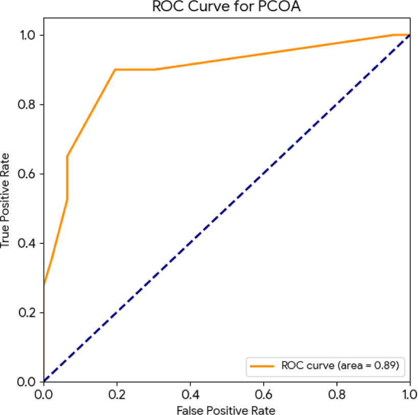
The EOQA classification showed a strong ability to distinguish PCOA values in both standing positions. In the shoulder-width stance, the EOQA group had significantly higher PCOA angles than the Non-EOQA group, with a mean difference of 8.71° and a very large effect size (d=2.325). A similar pattern was found in the feet-together stance, where the mean difference was 5.22° with a large effect size (d=1.818). For both positions, the differences were statistically highly significant (p<0.001), confirming a strong link between EOQA status and PCOA regardless of other factors (Table 2). The ROC curve analysis revealed that PCOA is a highly effective clinical indicator for distinguishing between the EOQA (Preclinical OA) group and the Non-EOQA group (Figure 2). The AUC was 0.888 (95% CI: 0.815–0.951), indicating excellent discriminatory power. At the optimal threshold of 7.0°, PCOA demonstrated a sensitivity of 0.900 (95% CI: 0.800–0.976) and a specificity of 0.804 (95% CI: 0.688–0.911) (Table 3).
DISCUSSION
This study identifies PCOA as a highly sensitive, non-invasive potential clinical indicator that effectively distinguishes between EOQA and Non-EOQA limbs in manufacturing workers. Specifically, EOQA limbs exhibited significantly higher PCOA values compared to the negative group in both measured positions. In the shoulder-width stance, the difference was most pronounced (16.12° vs. 7.41°; d=2.325), while the feet-together stance also showed a substantial difference (8.90° vs. 3.68°; d=1.818). To validate the screening performance and address the need for objective metrics, a ROC curve analysis was conducted, yielding an excellent Area Under the Curve (AUC) of 0.888 (95% CI: 0.815–0.951). At the optimal threshold of 7.0°, the PCOA demonstrated a sensitivity of 0.900 (95% CI: 0.800–0.976) and a specificity of 0.804 (95% CI: 0.688–0.911). These results, supported by rigorous 95% CIs for all major effect sizes, confirm that PCOA is a powerful tool for identifying early signs of OA while ensuring statistical precision.
The superiority of the shoulder-width stance is evident through enhanced diagnostic discrimination (d=2.325 vs. 1.818), greater absolute separation between groups (8.71° vs. 5.22°), and better practical implementation. Biomechanically, this stance mimics natural weight-bearing conditions found in daily activities, such as gait initiation or turning. This allows for the maximal expression of underlying varus alignment without the forced stability induced by standing with feet together. In contrast, the feet-together stance increases muscle tension around the hip and causes changes in hip-knee-ankle (HKA) alignment, which collectively attenuate the expression of the true PCOA value by approximately 45%. Furthermore, the larger angles recorded in the shoulder-width stance help minimize the relative impact of smartphone photography errors (±0.5–1°). Finally, this stance eliminates balance demands, enhancing patient safety and ensuring more consistent clinical standardization.
The observed increase in PCOA among EOQA limbs likely reflects early soft tissue changes caused by repetitive stress on the inner knee during weight-bearing activities.11 The classification of these limbs was based on the clinical feature items of the EOQA, following the methodology validated in our previous study. By focusing on these core clinical symptoms (Items 1 and 2) rather than broader patient-reported outcomes (Items 3 to 11), we ensured that the PCOA measurements were correlated with objective physical impairments and clinical objectivity. By capturing these cumulative biomechanical adaptations before joint space narrowing appears on X-rays, PCOA serves as a robust pre-clinical indicator.12 Identifying this critical therapeutic window is essential, as it allows for targeted treatments during the reversible stage, potentially preventing the progression to permanent degenerative OA.12 These findings address the limitations of conventional X-rays—such as high costs, radiation exposure, and poor correlation with patient symptoms—by providing an immediate and accessible evaluation method via smartphone photography. Furthermore, the accuracy of this smartphone-based approach, optimized through standardized stance protocols, ensures reliability in primary care and occupational health settings. Integrating PCOA with EOQA results establishes a comprehensive screening protocol that shifts clinical practice from reactive treatment to proactive prevention.
The findings of this study should be interpreted with several limitations. First, the cross-sectional design means we cannot yet prove a direct cause-and-effect relationship between EOQA results and the progression of PCOA. Second, because all participants were manufacturing workers, the results may not apply to the general population or other occupations. Additionally, this study focused only on frontal plane measurements (front view), which might overlook biomechanical factors from the side (sagittal) or rotational perspectives. Regarding statistical independence, although both limbs were analyzed, we followed clinical biomechanics conventions where local musculoskeletal deviations are treated as independent units of analysis. Therefore, future long-term studies involving more diverse groups and different health conditions are needed to fully confirm PCOA’s clinical value as a screening tool. Third, this study did not include radiographic confirmation of knee osteoarthritis (e.g., Kellgren–Lawrence grading); therefore, the findings should be interpreted as preliminary evidence rather than diagnostic validation. The variables examined in this study may serve as potential clinical indicators for early degenerative changes but cannot be regarded as definitive biomarkers or diagnostic tools without imaging-based verification. Future studies incorporating radiographic or other objective diagnostic criteria are warranted to validate the clinical applicability of these measures.
CONCLUSIONS
Our findings indicate that smartphone-based PCOA measurement, particularly when a 7.0° threshold is applied, serves as a robust and potential clinical indicator for identifying individuals at risk of preclinical knee OA. The integration of objective PCOA with clinical feature-based EOQA assessments offers a streamlined, radiation-free biomechanical evaluation tool that is highly applicable in primary care and occupational health settings. The excellent diagnostic performance, evidenced by an AUC of 0.888 and a sensitivity of 0.900, underscores its effectiveness for early screening. While PCOA shows great promise as an accessible assessment tool, further longitudinal research incorporating radiographic validation remains necessary to confirm its long-term predictive value and diagnostic accuracy.


