


INTRODUCTION
Taekwondo was introduced as a demonstration sport at the 1988 Seoul Olympics and has been included as an official sport since the 2000 Sydney Olympics, continuing to the present.1 Among its disciplines, Taekwondo poomsae was recognized for its professionalism when it was adopted as an official event at the 2018 Jakarta–Palembang Asian Games.2 Taekwondo poomsae is part of the Taekwondo technical system and allows practitioners to independently practice offensive and defensive techniques against an imagined opponent. Each poomsae consists of movements and forms corresponding to its intended meaning.3
Taekwondo movements can be classified into stances, blocks, strikes, thrusts, punches, kicks, and evasive actions, depending on their type.4 Beginners in Taekwondo are typically introduced to hand techniques first because these movements can be executed quickly, accurately, and with fine control. These characteristics make hand techniques highly effective defensive skills that allow faster responses than foot techniques. Moreover, the proportion of hand techniques in poomsae performed by color belt practitioners was 81.9%, and 86% in poomsae performed by black belt practitioners, demonstrating a markedly higher ratio compared with foot techniques.5
The trunk muscles play a crucial role in performing functional body movements.6 A study by Nuhmani involving 61 male collegiate athletes reported that individuals with greater core stability were able to throw a medicine ball farther.7 In addition, Kim and Woo found that greater rotational angular velocity and impact force were generated when a hook punch was performed with trunk rotation.8 Moreover, stronger lumbar extensor muscles were associated with faster Taekwondo trunk strikes.9 Collectively, these findings suggest that greater core stability and trunk muscle activation enable the upper limb muscles to exert force more efficiently. This evidence indicates that the muscles responsible for abdominal and trunk stability play an important role in enhancing athletic performance.7
Although hand techniques account for a high proportion of Taekwondo poomsae, previous studies have focused primarily on kicking techniques, and research on hand movements such as knife hand strikes or blocks remains limited.5 In particular, a review of Taekwondo poomsae research trends from December 1992 to June 2021 revealed that only 18 studies addressed biomechanics, highlighting the limited research on hand techniques.10
Therefore, this study aimed to examine how abdominal muscle activation influenced trunk muscle activity and upper-limb kinematic characteristics during the knife hand block, with particular focus on the transfer of kinetic energy along the upper kinetic chain in Taekwondo poomsae athletes. In this study, we hypothesized that the presence or absence of abdominal muscle activation would result in differences in trunk muscle activity and kinematic characteristics during performance of the knife hand block. Specifically, we hypothesized that activation of the abdominal muscles would increase the muscle activity of the oblique muscles and erector spinae, thereby enhancing the angular velocity and angular acceleration of the knife hand block.
METHOD
The experiment was conducted with 24 male Taekwondo undergraduate students in their twenties who were enrolled at University B in Cheonan, Chungcheongnam-do. The participants’ mean age was 20.0 ± 1.16 years, mean height was 173.7 ± 4.83 cm, and mean body weight was 68.77 ± 8.63 kg. Participants were instructed to perform the knife-hand block using their preferred hand, and the hand used was defined as the dominant hand. The dominant hand was left in 79.2% of participants and right in 20.8%. Their mean Taekwondo experience was 4.75 ± 2.11 years. The inclusion criteria were as follows: Taekwondo athletes who were registered poomsae players with the Korea Taekwondo Association and had participated in official competitions; individuals with more than three years of Taekwondo experience; individuals without functional limitations during the knife hand block motion; and those who voluntarily agreed to participate in the study.11 The exclusion criteria included individuals who had sustained musculoskeletal injuries within the previous six months, were taking medication, or had experienced significant body weight changes during the previous six months.12 This study was approved by the Institutional Review Board of Baekseok University (IRB No. BUIRB-202505-HR-031).
To ensure accurate motion analysis, a 3D motion capture system (OptiTrack, NaturalPoint, Inc., Oregon, USA) with Motive software was used. A total of eight cameras were employed, each set to record at 100 frames per second. The Conventional Upper Body marker set (27 markers), which is the basic marker set provided by Motive, was used.13 Additionally, two supplementary markers were attached to obtain more accurate measurements.14 These additional markers were placed to ensure accurate kinematic data collection and to prevent marker occlusion or misidentification during the complex knife hand block motion. Markers were placed on the upper body at the vertex, temporal bone, occipital bone, sternum, xiphoid process, C7, T10, medial border of the scapula, acromion, lateral humerus, elbow, forearm, radial and ulnar styloid processes, and second metacarpal. On the lower body, markers were attached to the anterior superior iliac spine, posterior superior iliac spine (PSIS), and coccyx. To facilitate marker identification, participants were shirtless and wore 3–5-inch cycling shorts during the experiment. The collected kinematic data were processed using Visual3D software (C-Motion, Inc., Maryland, USA). The axes of the motion analysis system were defined as follows: for the elbow, the x-axis represented flexion/extension, the y-axis represented deviation, and the z-axis represented supination/pronation; for the wrist, the x-axis represented flexion/extension, the y-axis represented ulnar/radial deviation, and the z-axis represented rotation. Based on these axis definitions, angular velocity and angular acceleration were calculated for each axis during performance of the knife hand block. These movements are characteristic motions that occur during the knife hand block, making this analysis appropriate for evaluating the functional aspects of the Taekwondo technique. The definitions of each variable are as follows. Angular velocity refers to the rate of change of joint angle over time and was extracted using Visual3D’s automatic calculation of joint angle changes. Angular acceleration refers to the rate of change of angular velocity over time and was obtained through Visual3D’s automatic calculation of changes in angular velocity.
The participants performed the experiment with their upper body uncovered and wore cycling shorts on the lower body. Surface electromyography (sEMG) data were collected using a Trigno EMG sensor (Delsys Inc., Boston, MA, USA). The EMG signals were sampled at 1,000 Hz, and a 20–450 Hz band-pass filter was applied to remove noise and artifacts. To obtain smoothed muscle activity values, root mean square processing was applied. The EMG amplitudes during the knife hand block were normalized to the maximal voluntary isometric contraction (MVIC) values and expressed as %MVIC.15 To minimize skin impedance and ensure high-quality signal acquisition, body hair was removed, and the electrode placement sites were cleaned with alcohol swabs prior to electrode attachment. Electrodes were attached to a total of nine channels: the bilateral external oblique (EO), bilateral internal oblique (IO), bilateral erector spinae (ES), dominant-side serratus anterior (SA), dominant-side pectoralis major (PM), and dominant-side middle deltoid (MD). The electrode for the EO was attached 3 cm below the SA.16 The electrode for the IO was attached 3 cm above the iliac crest and directed toward the umbilicus.16
The MVIC of the EO was measured in a sit-up position while the participant drew the navel inward to stabilize the abdomen, and resistance was applied to the scapula during trunk rotation to the opposite side. The MVIC of the IO was measured in the same position as that used for the EO, except that resistance was applied to the scapula during trunk rotation to the same side.17 The ES electrode was attached at the L3 level, approximately 5 cm above the PSIS, after palpation.9 For ES MVIC, participants lay prone on a bench with their knees secured by a strap and performed trunk extension against resistance.18 The SA electrode was placed along the fifth rib.19 For SA MVIC, participants sat with the shoulder flexed to 125° and protracted the scapula. The examiner stabilized the inferior angle of the scapula with one hand and applied posterior resistance to the upper arm with the other hand while instructing the participant to hold the position.20 The PM electrode was attached 2 cm below the clavicle, near the anterior axillary fold, after palpation of the clavicle.5 PM MVIC was assessed in a seated position with the shoulder and elbow flexed to 90°, while horizontal adduction was performed against manual resistance.21 The MD electrode was placed 3 cm below the acromion after palpation.19 For MD MVIC, participants sat with the arm abducted to 90° and the palm facing the floor, while the examiner applied downward manual resistance.22 After electrode placement on all muscles, MVIC was measured three times, and muscle activity for each phase was subsequently analyzed.
Participants performed the following intervention to facilitate abdominal muscle activation. They first assumed a quadruped position and aligned the trunk in a neutral posture, after which they were instructed to perform diaphragmatic breathing.23 During diaphragmatic breathing, participants were instructed to expand the anterior and posterior aspects of the abdomen evenly and to allow the abdomen to expand in a cylindrical manner.24 Verbal cues were provided to ensure that participants maintained this breathing pattern while performing the movement. In addition, to objectively verify performance, the examiner directly palpated the abdomen. Appropriate abdominal muscle activation was considered to have occurred when abdominal tension increased without excessive rib elevation or compensatory trunk movements.25
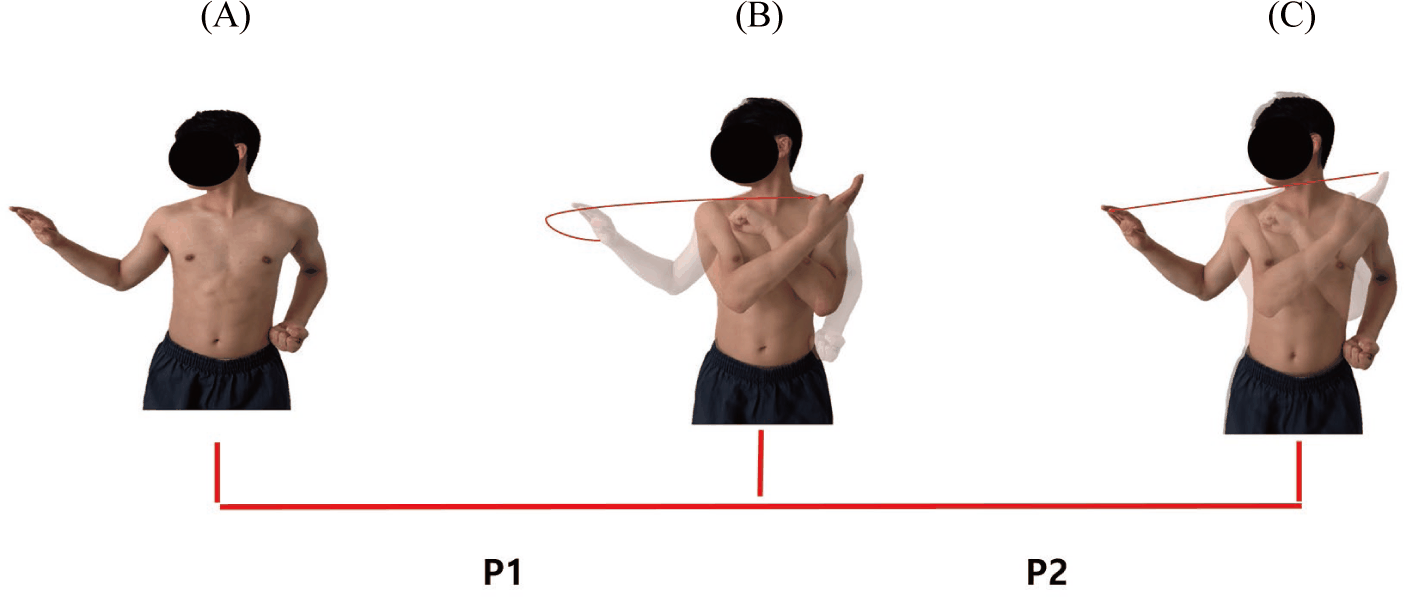
Prior to data collection, participants received a detailed explanation of abdominal muscle activation and the knife hand block motion. Because all participants were experienced Taekwondo athletes with sufficient proficiency in performing the knife hand block, no additional practice time was provided. The knife hand block movement was divided into three positions—Start, Mid, and End—and two phases (Figure 1).5 The Start and End positions shared the same posture but represented different temporal events marking the initiation and completion of the movement, respectively. The Mid position was defined as the moment when the dominant hand completely passed the fingertip of the nondominant hand. The interval between the Start and Mid positions was defined as Phase 1 (P1), and the interval between the Mid and End positions was defined as Phase 2 (P2).
Following the explanation, surface electrodes were attached to the corresponding muscles of each participant, and MVIC was measured three times. After a 5-minute rest period, participants performed the knife hand block three times without abdominal muscle activation. Following another 5-minute rest period, participants were verbally instructed to activate their abdominal muscles. Activation was confirmed through direct palpation by the examiner before proceeding. Upon confirmation, the knife hand block was performed three times. The experimental order was fixed such that the nonactivation condition was performed first, followed by the abdominal activation condition, because muscle activation and fatigue could potentially influence the performance of subsequent movements.26
All statistical analyses were performed using SPSS version 20.0 (SPSS Inc., Chicago, IL, USA). The mean and standard deviation were calculated for each variable. To assess data normality, the Shapiro–Wilk test was conducted. Upper-limb kinematic variables were designated as the primary outcomes, and muscle activity variables were designated as the secondary outcomes. Paired t tests were conducted for all variables, with the level of statistical significance set at p<0.05. Effect sizes were calculated using Cohen’s dz, and 95% confidence intervals were reported for all paired comparisons.
RESULT
The results of the paired t test comparing muscle activity, angular velocity, and angular acceleration of the elbow and wrist during the knife hand block between conditions with and without abdominal muscle activation in P1 are presented below.
Muscle activity in P1 and P2 is presented in Table 1. In P1, the dominant internal oblique (D-IO) showed a significant increase when the abdominal muscles were activated compared with the nonactivation condition (p<0.05; Table 1). The dominant erector spinae (D-ES) also demonstrated a significant difference between conditions (p<0.05; Table 1). In contrast, no significant differences were observed in the dominant external oblique (D-EO), nondominant external oblique (ND-EO), nondominant internal oblique (ND-IO), nondominant erector spinae (ND-ES), MD, PM, SA (p>0.05). In P2, D-IO showed a significant increase during abdominal activation compared with the nonactivation condition (p<0.05; Table 1). However, unlike in P1, no significant difference was observed in D-ES. Similar to P1, no significant differences were observed in MD, PM, or SA (p>0.05).
ANA, abdominal nonactivation; AA, abdominal activation; ES, effect size; CI, confidence interval; P1, phase 1; P2, phase 2; MD, middle deltoid; PM: pectoralis major; SA, serratus anterior; D-EO, dominant external oblique; D-IO, dominant internal oblique; ND-EO, nondominant external oblique; ND-IO, nondominant internal oblique; D-ES, dominant erector spinae; ND-ES, nondominant erector spinae.
The angular velocity of the elbow in P1 and P2 is presented in Table 2. A significant difference was observed in the z-axis during P2, with higher values under the abdominal activation condition than under the nonactivation condition (p<0.05; Table 2). However, no significant differences were observed in the x-, y-, or z-axes during P1 or in the x- or y-axes during P2. In addition, the angular velocity of the wrist showed no significant differences in the x-, y-, or z-axes in either P1 or P2.
The angular acceleration of the elbow in P1 and P2 is presented in Table 3. A significant difference was observed in the x-axis during P2, with higher values under the abdominal activation condition than under the nonactivation condition (p<0.05; Table 3). A significant difference was also observed in the z-axis during P2 (p<0.05; Table 3). However, no significant differences were observed in the x-, y-, or z-axes during P1 or in the y-axis during P2. In addition, the angular acceleration of the wrist showed no significant differences in the x-, y-, or z-axes in either P1 or P2.
DISCUSSION
This study was conducted to compare and analyze kinematic changes during performance of a knife hand block according to the presence or absence of abdominal muscle activation. With abdominal muscle activation, muscle activity of the D-IO and D-ES significantly increased, and significant differences were observed in elbow velocity and acceleration during P2. These findings suggest that activation of the trunk muscles may have enhanced movement performance by improving the transfer of kinetic energy from proximal to distal segments. A review of previously published Taekwondo-related studies indicates that the present study has several distinguishing features. Park et al. analyzed muscle activation and joint angles according to the type of trunk inward block in poomsae, focusing on activation of the shoulder joint and upper-limb muscles.5 However, their study did not examine detailed kinematic variables such as velocity and acceleration. In addition, Kang et al. analyzed trunk thrust performed in the Juchum-seogi stance and discussed trunk rotation and upper-limb movement; nevertheless, axis-specific comparisons of velocity and acceleration were not conducted.4 Although several Taekwondo studies have investigated physiological variables such as muscle activation and angular measures, research comparing axis-specific velocity and acceleration remains limited. Therefore, the present study differs from previous research in that it expands upon earlier findings by analyzing phase-specific and axis-specific kinematic variables in conjunction with muscle activation.
In the present study, large standard deviations were observed in several EMG outcomes, indicating interindividual variability among participants. This variability may be attributed to differences in movement strategies even among skilled athletes; some participants may have performed the knife hand block using a trunk-dominant strategy, whereas others may have relied more on an upper-limb–dominant approach. Such differences may be associated with variations in muscle activation timing and neuromuscular control, which may have contributed to inconsistencies in muscle activation levels across individuals. Previous research has reported that muscle activation patterns can vary within skilled athlete populations because of differences in coordination strategies and neuromuscular control characteristics.27 Therefore, the large standard deviations observed in the present study should be interpreted not merely as measurement error but as reflecting differences in motor control strategies during complex poomsae movements.
According to Ng et al., as the intensity of axial trunk rotation increases, muscle activity of all trunk muscles increases, with the IO demonstrating particularly increased activation in response to higher loads.28 Furthermore, Teyhen et al. reported that the muscle thickness of the transversus abdominis and IO increased during trunk-strengthening exercises compared with a resting state.29 Regarding trunk extension, Park and Park observed high levels of muscle activity in the ES when extension was performed in conjunction with trunk rotation.30 In terms of energy transfer, a review by Chu et al. indicated that more than 51%–55% of the kinetic energy delivered to the hands was generated from the core and lower extremities.31 Similarly, Palmer and McCabe found that increased power during upper-limb rotation led to higher trunk and upper-limb rotational velocities, which in turn enhanced bat swing speed in softball players.32 Synthesizing these prior findings suggests that the IO and ES play critical roles in trunk rotation and extension. Moreover, trunk rotation serves as a vital mechanism for transferring kinetic energy to distal segments. When applying these principles to the knife hand block in Taekwondo—a technique performed with simultaneous trunk rotation—the significant increase in IO and ES muscle activity observed in this study suggests that rotational energy generated in the trunk was more efficiently transferred to the elbow. This efficient energy transfer likely contributed to the observed increases in elbow angular velocity and angular acceleration.
In P2, the abdominal muscle activation condition showed a significant difference in elbow x-axis (flexion/extension) acceleration compared with the nonactivation condition. This change is unlikely to represent an isolated alteration at the elbow joint; rather, it may be interpreted as a kinetic chain effect, in which activation of the abdominal muscles increases trunk rotational velocity, thereby influencing the transmission of movement from proximal to distal segments. According to Hirashima et al., velocity is generated in proximal segments, such as the trunk and shoulder, and this generated velocity is subsequently utilized to produce greater velocity in distal segments, including the elbow and wrist. In particular, during an overarm throwing motion, elbow extension angular velocity reached its peak immediately prior to ball release, accompanied by a substantial increase in distal segment acceleration.33 In this context, the increased elbow x-axis acceleration observed under the abdominal activation condition in the present study may be interpreted similarly. Specifically, the significant increase in elbow extension acceleration during P2—the primary phase of the knife hand block—resembles the previously reported phenomenon in which elbow extension angular velocity reached its peak immediately before ball release, leading to increased distal acceleration. Therefore, the results of this study suggest that abdominal muscle activation may enhance trunk rotational velocity, which in turn contributes to increased extension acceleration of the distal segment, namely, the elbow.
In P2, the abdominal activation condition showed significant differences in elbow z-axis (supination/pronation) velocity and acceleration compared with the nonactivation condition. This finding suggests that rotational motion generated in proximal segments may have been transmitted to distal segments. According to the review by Marshall and Elliott, rotational movements such as supination and pronation play an important role in transferring rotational energy generated in proximal segments to distal segments.34 In the present study, z-axis velocity also increased with abdominal muscle activation, suggesting that trunk kinetic energy may have been enhanced. Accordingly, rotational motion generated in the trunk may have been transmitted along the upper limb, resulting in a kinetic chain effect.
In contrast, no significant changes were observed at the wrist. According to Van Ginneken et al., conscious control during the motor learning process tends to freeze mechanical degrees of freedom. This finding can be interpreted to mean that conscious control during task execution reduces variability and promotes more stable performance.35 In Taekwondo, the knife hand block is a high-speed movement that must be performed both quickly and accurately. Therefore, participants in this study may have employed conscious control to achieve stable and precise performance during the knife hand block, which may have increased wrist stiffness and consequently limited wrist motion.
Several limitations should be acknowledged in this study. First, the participants consisted exclusively of university athletes in their twenties. Therefore, generalization of the present findings is limited because neuromuscular control strategies and muscle activation patterns may vary according to sex and age.36 In addition, because the participants were trained athletes, the observed results may differ in novice populations; thus, caution is required when generalizing these findings to untrained individuals. Second, this study employed a pre–post design to examine the immediate effects of abdominal muscle activation. However, this short-term comparison does not adequately reflect the mid- to long-term adaptive effects that may result from abdominal stabilization or core strengthening training. Therefore, future studies should adopt a research design that implements a more systematic abdominal stabilization or core strengthening program over a defined period and analyzes changes before and after the intervention. Third, the elbow kinematic results may be associated with shoulder and trunk movements; however, these movements were not directly measured. Therefore, the interpretations should be made with caution. Future studies should directly measure shoulder and trunk movements to clarify their contributions to distal segment kinematics.
CONCLUSION
In this study, abdominal muscle activation significantly increased D-IO and D-ES muscle activity and produced significant differences in elbow z-axis velocity and acceleration, as well as elbow x-axis acceleration during P2. These findings suggest that trunk muscle activation enhances kinetic energy transfer from proximal to distal segments and that incorporating abdominal muscle activation into Taekwondo poomsae training may be an effective strategy for optimizing knife hand block performance. These findings may also have implications for rehabilitation settings, in which trunk muscle activation strategies could be incorporated to enhance upper-limb movement efficiency. Future studies should implement long-term trunk strengthening interventions and examine whether these effects differ according to skill level and across diverse participant populations.