


INTRODUCTION
In modern society, where time spent using information and communication devices such as smartphones and computers has increased sharply, musculoskeletal dysfunction involving the cervical spine, thoracic spine, and scapula has been rising rapidly.1 In particular, prolonged sitting posture induces early fatigue of trunk muscles,2 leading individuals to habitually adopt a relaxed slouched posture. This slouched posture can cause anterior translation of the humeral head and increased scapular anterior tilt, resulting in rounded shoulder posture (RSP).3,4 Rounded shoulder posture is a key component of upper crossed syndrome and has been reported to accompany various secondary problems such as pain, decreased respiration, and functional decline.5
Choi et al.6 reported that individuals with rounded shoulder posture have significantly shorter pectoralis minor length than those with normal posture. Shortening of the pectoralis minor increases scapular protraction and anterior tilt, decreases the pectoralis minor index, and is known as a major factor contributing to forward shoulder posture.7 Based on these prior findings, interventions for individuals with rounded shoulder posture have relatively focused on stretching anterior soft tissues, particularly the pectoralis minor.8
However, regression analysis by Lee et al.9 indicated that approximately 93% of the variance in forward shoulder posture was associated not only with pectoralis minor length but also with multiple factors such as posterior shoulder tightness, thoracic kyphosis, and serratus anterior weakness. Among these, posterior shoulder tightness was presented as a variable with moderate or greater predictive power independent of pectoralis minor length. Posterior shoulder tightness is a common condition observed in both athletes and the general population,10 and it is defined as a limitation in extensibility across posterior shoulder structures, including contractile tissues such as muscles, non-contractile tissues such as the capsule and ligaments, and even bony factors such as humeral torsion.11
Posterior shoulder stretching has been proposed as a strategy to improve posterior shoulder tightness and restricted shoulder internal rotation. In a meta-analysis by Guo et al.,12 posterior shoulder stretching was reported to reduce subacromial space pressure, alleviate rotator cuff compression, significantly improve shoulder function, and decrease pain. In addition, Rosa et al.13 examined correlations between pectoralis minor shortening, posterior shoulder tightness, and the Shoulder Pain and Disability Index in patients with shoulder pain, and reported that disability scores were significantly associated with the degree of posterior shoulder tightness but were not significantly related to pectoralis minor length.
Although previous studies have shown that both pectoralis minor stretching and posterior shoulder stretching have significant effects on shoulder alignment and improvements in pain and function, no study has directly compared the effects of these two interventions in adults with rounded shoulder posture. Therefore, this study aimed to compare the effects of posterior shoulder stretching and pectoralis minor stretching, each combined with scapular posterior tilting exercises, in adults with rounded shoulder posture. We hypothesized that both interventions would improve shoulder alignment, muscle tone, and shoulder-stabilizing muscle activity.
METHODS
Twenty-six adults with rounded shoulder posture living in Andong-si were recruited according to predefined inclusion and exclusion criteria. The required sample size was calculated using G*Power 3.1.9.4. Based on a two-way repeated-measures ANOVA (within–between interaction), with α=0.05, power (1–β)=0.95, and an effect size of f=0.40, the minimum required sample size was 24 participants. To account for potential dropout, 26 partici-pants were enrolled. The assumed effect size (f=0.40) corresponds to a large effect according to Cohen’s criteria and was selected with reference to prior rounded-shoulder intervention research reporting moderate-to-large pre–post improvements in shoulder alignment measures, including acromion–table distance, following a 4-week exercise regimen.14,15
The inclusion criterion was a rounded shoulder posture distance ≥ 2.5 cm measured in the supine position.16 Exclusion criteria were: presence of shoulder pain or neurological impairment; orthopedic or neurological history involving the cervical spine and upper extremity; and dermatological history involving the cervical spine and upper extremity.17 Prior to participation, all participants received sufficient explanation of the purpose and procedures of the study and provided written informed consent. The study was conducted after approval by the Institutional Review Board of Daegu University (1040621-202503-HR-018) (Table 1).
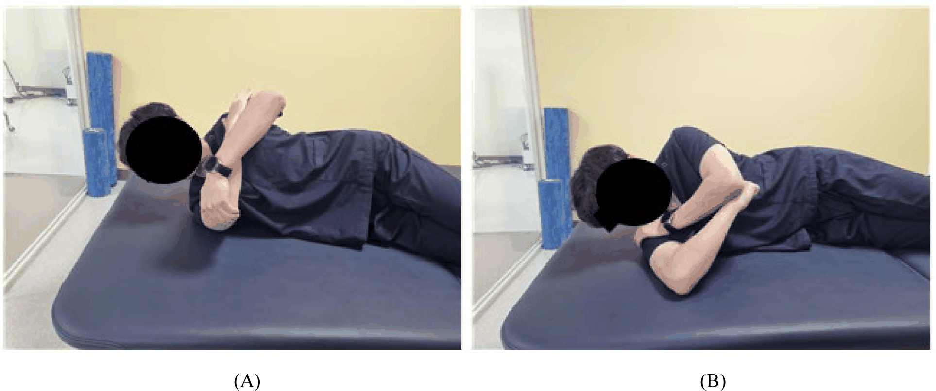
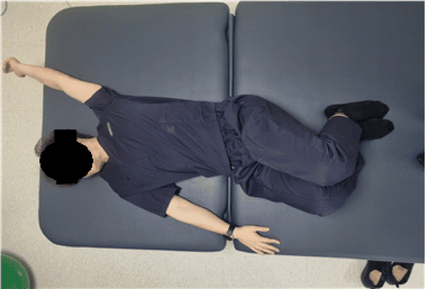
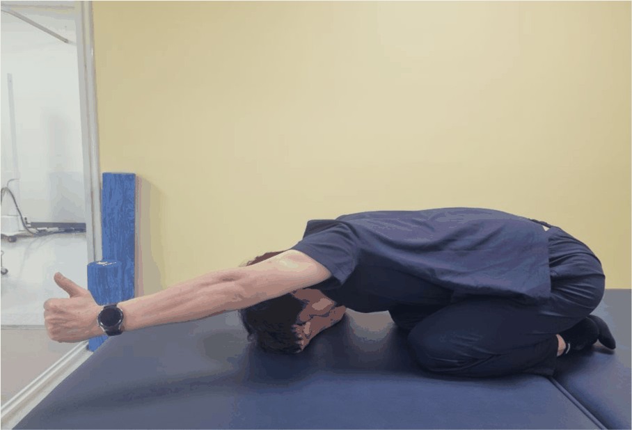
This study was a randomized comparative trial with a two-way repeated-measures design, with group (experi mental vs.comparison) and time (pre- vs. post-intervention) as factors. Participants were allocated to an experimental group that performed posterior shoulder stretching plus scapular posterior tilting exercises (n=13; Figures 1 and3) or a comparison group that performed pectoralis minor self-stretching plus scapular posterior tilting exercises (n=13; Figures 2 and 3).
For random allocation, cards numbered 1–26 were placed in a sealed box, and each participant drew one card. Odd numbers were assigned to the comparison group and even numbers to the experimental group. After baseline assessment, both groups received the intervention three times per week for 4 weeks, and post-intervention assessment was conducted immediately after the 4-week program using the same outcome measures.
The experimental group performed two types of posterior shoulder stretching for a total of 10 sets (each set: 30 s hold, 10 s rest; 5 sets per stretch). The comparison group per-formed pectoralis minor self-stretching for 10 sets (30 s hold, 10 s rest). Both groups additionally performed scapular posterior tilting exercises for 3 sets of 15 repetitions. All exercise interventions were supervised directly by the investigator, a physical therapist with more than 5 years of clinical experience. Before the intervention, the purpose and methods of each exercise were explained in detail, demonstrated, and participants completed three practice trials. If a participant could not attend due to unavoidable circumstances, they were instructed to perform the same exercises at home using pre-provided video materials.
All analyses were conducted as per-protocol analyses including participants who completed at least 10 of 12 total sessions over 4 weeks; no dropouts occurred.
All outcome measures were assessed pre-intervention and immediately after the 4-week intervention.
Rounded shoulder posture distance was measured in the supine position with the arms relaxed at the sides. Using a digital caliper, the vertical distance between the posterior aspect of the dominant-side acromion and the table surface was measured. Measurements were repeated three times and averaged. The final value was calculated after correcting for the 1 cm thickness of the caliper base.18
Pectoralis minor length was measured in standing using a digital caliper as the distance between the inferior-medial aspect of the coracoid process on the dominant side and the junction of the fourth rib with the sternum.7 Each measurement was performed three times and the mean value was used for analysis.
Muscle mechanical properties of the dominant-side pectoralis major and posterior deltoid were assessed using a handheld myotonometer (Myoton®PRO, Myoton AS, Estonia). Assessments were performed in a relaxed sitting posture, and tone (Hz) and stiffness (N/m) were recorded.
For the pectoralis major, the measurement point was defined as the muscle belly at the intersection of a vertical line drawn inferiorly from the midpoint of the clavicle and a horizontal line drawn from the axilla.19 For the posterior deltoid, the muscle belly was defined as a point two finger-widths inferior to the posterior aspect of the acromion.20 Each site was measured three times and the mean value was used for analysis.
Surface EMG was recorded to evaluate muscle activity of the dominant-side upper trapezius and posterior deltoid using a TeleMyoDTS system (Noraxon Inc., AZ, USA). EMG signals were recorded during shoulder flexion at 60°, 90°, and 120°. Before electrode placement, the skin was prepared by shaving if necessary and cleaning with alcohol, and bipolar surface electrodes were applied according to standard guidelines.21
For the upper trapezius, electrodes were placed at the midpoint between the C7 spinous process and the lateral tip of the acromion with the arm relaxed at the side. For the posterior deltoid, electrodes were placed parallel to the muscle fiber direction along the lateral border region of the scapula, oriented obliquely from the posterior acromion toward the upper limb.
Maximum voluntary isometric contraction (MVIC) was obtained for normalization.
Upper trapezius MVIC: Participants were seated and maintained an elevated shoulder girdle (scapular elevation). The examiner applied downward resistance over the acro-mion/clavicular region.
Posterior deltoid MVIC: Participants were positioned prone and abducted the shoulder horizontally with slight external rotation, while the examiner applied downward resistance over the humerus.8
After MVIC testing, participants actively raised the arm to each target angle (60°, 90°, 120°) while maintaining neutral trunk alignment. EMG signals were recorded during a 5-second isometric hold at each angle and were normalized to MVIC to calculate %MVIC. EMG signals were sampled at 1000 Hz, band-pass filtered at 20–400 Hz, rectified, and smoothed using a 50 ms time constant. Root mean square (RMS) values were computed from the middle 3 seconds of each trial and used for analysis.
All data were analyzed using SPSS version 29.0 for Windows (IBM Corp., Armonk, NY, USA). Between-group differences in general characteristics and baseline values were examined using independent-samples t-tests. Normal-ity was assessed using the Shapiro–Wilk test. To evaluate the effects of the 4-week intervention, a two-way repeated-measures ANOVA with group (experimental vs. comparison) and time (pre vs. post) as factors was conducted to test the main effects of time and group × time interaction effects. Rounded shoulder posture distance and pectoralis minor length were defined as the primary outcomes.
When a significant group × time interaction was identified, between-group comparisons of change scores (post–pre) were conducted using independent-samples t-tests as post-hoc analyses. Because two post-hoc between-group tests were performed (posterior deltoid tone and posterior deltoid EMG activity at 60° shoulder flexion), Bonferroni correction was applied (adjusted significance level: p<0.025) to control the family-wise type I error rate. Within-group pre–post differences were assessed using paired-samples t-tests. The overall significance level was set at α=0.05. Effect sizes were calculated as Cohen’s dz for pre–post changes.
RESULTS
In both groups, rounded shoulder posture distance de-creased and pectoralis minor length increased, showing significant within-group improvements. However, the group × time interaction effect was not significant (Table 2).
RSP, rounded shoulder posture; PML, pectoralis minor length; PM, pectoralis major muscle; PD, posterior deltoid muscle; F, muscle tone; S, muscle stiffness; EG, posterior shoulder stretching with scapular posterior tilting exercise group; CG, pectoralis minor stretching with scapular posterior tilting exercise group; M±SD, mean ± standard deviation.
Within-group effect sizes (Cohen’s dz) were large in both groups (experimental group: 2.20 for RSP distance and 2.40 for pectoralis minor length; comparison group: 1.86 and 2.27, respectively) (Table 3).
RSP, rounded shoulder posture; PML, pectoralis minor length; PM, pectoralis major muscle; PD, posterior deltoid muscle; F, muscle tone; S, muscle stiffness; EG, posterior shoulder stretching with scapular posterior tilting exercise group; CG, pectoralis minor stretching with scapular posterior tilting exercise group; M±SD: mean ± standard deviation.
Post-hoc power analyses for between-group differences in change scores indicated very low achieved power for RSP distance (1−β=0.05) and pectoralis minor length (1−β=0.19).
Consistent with the non-significant interaction, the between-group differences in change were small: the difference in change (EG−CG) was 0.03 cm (95% CI −0.33 to 0.39) for RSP distance and −0.32 cm (95% CI −0.93 to 0.29) for pectoralis minor length.
Tone and stiffness of the pectoralis major significantly decreased over time in both groups. For posterior deltoid tone, both the main effect of time and the group × time interaction were significant; post hoc analysis showed that the reduction in the experimental group was significantly greater than that in the comparison group (Tables 2 and 5). The between-group difference in change (EG−CG) was −0.76 Hz (95% CI −1.08 to −0.44).
For posterior deltoid stiffness, the main effects of time and group were significant, but the interaction was not; within-group analysis showed a significant decrease in the experimental group, whereas the comparison group did not show a significant change.
For the pectoralis major, post-intervention effect sizes in the experimental group were 0.79 (tone) and 0.64 (stiffness), indicating moderate effects, while the comparison group showed effect sizes of 1.27 (tone) and 0.84 (stiffness), indicating large effects (Table 3).
For the posterior deltoid, post-intervention effect sizes in the experimental group were 2.18 (tone) and 1.54 (stiffness), indicating large effects, whereas the comparison group showed 0.93 (tone; large effect) and 0.49 (stiffness; small effect) (Table 3).
Upper trapezius activity showed a significant time effect only at 120° shoulder flexion.
Posterior deltoid activity showed a significant group × time interaction at 60° shoulder flexion; change-score comparisons indicated that the increase in the comparison group was significantly greater than that in the experimental group (Tables 4 and 5). The between-group difference in change (EG−CG) was –6.55 %MVIC (95% CI –1.26 to –11.84).
DISCUSSION
This study compared the effects of posterior shoulder stretching versus pectoralis minor stretching, each combined with scapular posterior tilting exercises, in adults with rounded shoulder posture. Both interventions were effective in improving rounded shoulder alignment and reducing tone in the pectoralis major and posterior deltoid. Regarding muscle activity, upper trapezius activity tended to slightly decrease across most angles, whereas posterior deltoid activity significantly increased only in the comparison group at 60° shoulder flexion, showing a between-group difference in the pattern of change. The different change pattern in posterior deltoid tone suggests a possible additional benefit of stretching that directly targets posterior soft tissues. However, because clinically agreed minimal clinically important differences (MCIDs) for RSP distance, pectoralis minor length, and Myoton outcomes in rounded shoulder posture have not been sufficiently established, clinical interpretation in this study was supported primarily by change scores and effect sizes.22,23
For shoulder alignment, both groups showed significant improvements in rounded shoulder posture distance and pectoralis minor length. Posterior shoulder stretching may have reduced excessive passive tension by lengthening posterior soft tissues such as the posterior deltoid and posterior capsule, thereby decreasing superior and anterior translation of the humeral head and creating a mechanical environment more favorable for scapular posterior tilt and upward rotation. This interpretation is consistent with a previous study reporting significant improvements in scapular alignment after applying posterior shoulder stretching in adults characterized by scapular anterior tilt, a typical feature of rounded shoulder posture.24 These findings suggest that programs including posterior shoulder stretching may serve as a useful intervention option for correcting rounded shoulder alignment.
F, muscle tone; EG, posterior shoulder stretching with scapular posterior tilting exercise group; CG, pectoralis minor stretching with scapular posterior tilting exercise group; diff in change, between-group difference in change, computed as (EG post−pre) − (CG post−pre); 95% CI, calculated using an independent-samples t distribution (df=24; n=13 per group).
In addition, the comparison group’s increases in pectoralis minor length and reductions in rounded shoulder posture distance align with previous work indicating that inadequate scapular positioning contributes to scapular anterior tilt and anterior/superior translation of the humeral head,25 and further support that lengthening of the pectoralis minor is a key mechanism contributing to improved rounded shoulder alignment.26
For muscle tone, decreases in tone and stiffness of the pectoralis major and posterior deltoid in both groups indicate that stretching combined with scapular stabilization exercises can effectively alleviate excessive tension and reduce soft tissue stiffness around the shoulder. The between-group difference observed in posterior deltoid tone suggests that anterior-soft-tissue–focused stretching may have limited effects on reducing posterior muscle tone, highlighting the need to consider interventions that directly target posterior soft tissues. Guduru et al.20 reported that posterior deltoid stiffness is 10–12% higher in individuals with rounded shoulder posture than in those with normal posture; thus, the present findings provide clinical support by demonstrating that such increased stiffness can be significantly reduced through posterior shoulder stretching.
For EMG outcomes, upper trapezius activity tended to decrease slightly across most angles. Pectoralis major shortening and posterior shoulder tightness are known to reduce scapular upward rotation and induce compensatory overactivity of the upper trapezius,27,28 and decreased upper trapezius activity has been reported following posterior stretching and joint mobilization in individuals with shoulder impingement syndrome and posterior shoulder tightness.29 Considering these findings, the decreasing tendency in upper trapezius activity observed in this study may reflect partial reduction in compensatory overactivation due to improved anterior–posterior soft tissue balance and scapular alignment.
Posterior deltoid activity significantly increased only in the comparison group at 60° shoulder flexion, with a significant between-group difference in the pattern of change. Given reports that coactivation of the rotator cuff and deltoid activation patterns contributing to stabilization in the early range of elevation may differ in symptomatic populations,30 these findings suggest the possibility of altered motor recruitment strategies or compensatory increases in activation due to increased stabilization demands. Moreover, because Myoton®-measured stiffness has been reported to increase with higher levels of voluntary muscle contraction,31 the observed changes in posterior deltoid activity at 60° may reflect both passive mechanical properties of posterior soft tissues and changes in neuromuscular control strategies. Overall, these results support the need to individualize intervention strategies according to the distribution of anterior and posterior soft tissue tension.
Although both groups showed statistically significant within-group improvements in RSP distance and pectoralis minor length, the between-group differences in change scores were small. The post-hoc powers for detecting these between-group effects were very low (1−β=0.05 for RSP distance and 0.19 for pectoralis minor length), indicating that the present study was underpowered to detect small between-group differences. Therefore, the non-significant group × time interactions should not be overinterpreted as evidence of true equivalence between the two interventions. Beyond these statistical considerations, the clinical meaningfulness of the observed changes also warrants cautious interpretation. Although reductions in rounded shoulder posture distance, increases in pectoralis minor length, and decreases in tone and stiffness of the pectoralis major and posterior deltoid were statistically significant in both groups, it is difficult to conclude that these changes represented clearly meaningful clinical improvements for every individual participant, although the moderate-to-large group-level effect sizes suggest that clinically relevant benefits were likely achieved in at least a subset of participants. In particular, for certain posterior deltoid tone and muscle activity indices, the standard deviation exceeded the mean change, suggesting substantial inter-individual heterogeneity in response—some participants may have experienced meaningful improvements, whereas others may have shown relatively small changes. In this study, effect sizes were moderate-to-large for rounded shoulder posture distance and pectoralis minor length, mostly small-to-moderate for Myoton outcomes, and generally small for EMG (%MVIC) outcomes. These findings suggest that the interventions can induce a certain degree of short-term improvement in alignment and muscle mechanical properties, but may be limited in ensuring long-term structural changes or clearly distinct functional improvements. Future research should apply combined programs including resistive shoulder stabilization exercises and neuromuscular control training using unstable surfaces, and should systematically investigate causal relationships among muscle tone, muscle activity of various peri-scapular muscles, and functional outcomes.
This study has several limitations. First, the sample size was small, limiting generalizability to all individuals with rounded shoulder posture. Second, the intervention duration was relatively short (4 weeks), and maintenance of effects or long-term impacts could not be determined. Third, posterior shoulder tightness was assessed only via the posterior deltoid and did not include other posterior stabilizing muscles. Fourth, daily habits such as working posture, smartphone use, and leisure activities were not controlled, and the influence of these factors cannot be excluded. Fifth, outcome assessments were performed by the treating therapist, and assessor blinding was not possible, which may have introduced potential measurement bias.
CONCLUSIONS
This study compared the effects of posterior shoulder stretching plus scapular posterior tilting exercises versus pectoralis minor stretching plus scapular posterior tilting exercises in adults with rounded shoulder posture, focusing on rounded shoulder alignment, muscle tone, and muscle activity. Both interventions significantly improved rounded shoulder alignment through decreases in rounded shoulder posture distance and increases in pectoralis minor length, and both reduced tone and stiffness of the pectoralis major and posterior deltoid overall. In contrast, EMG outcomes showed only limited changes in the upper trapezius, and a significant group × time interaction was observed for posterior deltoid activity at 60° shoulder flexion, with a significant increase in the comparison group. These findings suggest that, when correcting rounded shoulder posture, clinicians should evaluate shortening and hypertonicity patterns of both anterior and posterior soft tissues to select appropriate stretching targets, and should incorporate a variety of strengthening programs for shoulder-stabilizing muscles as an effective treatment approach.