Research Report

Effects of a 4-Week Trunk Stabilization Exercise Program on Lower-Limb Muscle Activation during Gait and Functional Disability in University Students with Low Back Pain

Kihyeon Park1, Tae-ho Kim1,*
Author Information & Copyright
1Department of Physical Therapy, College of Rehabilitation Science, Daegu University, Gyeongsan, Republic of Korea
*ptpkh2@gmail.com, Tae-ho Kim, Department of Physical Therapy, College of Rehabilitation Science, Daegu University, Gyeongsan, Republic of Korea

© Copyright 2026, Academy of KEMA. This is an Open-Access article distributed under the terms of the Creative Commons Attribution Non-Commercial License (http://creativecommons. org/licenses/by-nc/4.0) which permits unrestricted non-commercial use, distribution, and reproduction in any medium, provided the original work is properly cited.

Received: Nov 11, 2025 ; Revised: Dec 24, 2025 ; Accepted: Dec 26, 2025

Published Online: Jun 30, 2026

ABSTRACT

Background

Prolonged sitting, low physical activity, and trunk muscle weakness in university students increase the risk of low back pain (LBP), which can alter gait and lower-limb muscle activation.

Purpose

To investigate the effects of a 4-week trunk stabilization exercise program on spatiotemporal gait parameters, lower-limb muscle activation, and functional disability in university students with LBP.

Study design

Single-group pre–post intervention study.

Methods

Twenty-seven university students with LBP performed 16 trunk stabilization exercises once daily for 40 minutes, 5 days per week, for 4 weeks. Gait was assessed using a pressure-sensing walkway to obtain stance phase, single-limb stance, and stride length. Surface electromyography recorded activation of six lower-limb muscles during gait and was normalized to maximum voluntary isometric contraction. Functional disability was evaluated using the Korean version of the Oswestry Disability Index (K-ODI). Pre–post differences were analyzed using repeated measures ANOVA.

Results

Stance phase duration significantly decreased, whereas single-limb stance and stride length significantly increased (p<.01). Soleus and biceps femoris activity increased (p<.01), while rectus femoris, tibialis anterior, and popliteus activity decreased (p<.05); gastrocnemius showed a non-significant increase. K-ODI scores decreased from 29.40±5.60 to 23.62±5.01 (p<.01).

Conclusions

A 4-week trunk stabilization program improved gait efficiency, reorganized lower-limb neuromuscular activation, and reduced functional disability in young adults with LBP. Trunk stabilization may be used as a practical, non-invasive intervention in university populations.

Keywords: Electromyography; Gait; Low back pain; Oswestry disability index; Trunk stabilization

Key Points

Question Does a 4-week trunk stabilization exercise program improve gait parameters, lower-limb muscle activation, and functional disability in university students with low back pain (LBP)?

Findings After 4 weeks, stance phase decreased and single-limb stance and stride length increased; soleus and biceps femoris activation increased, while rectus femoris and tibialis anterior overactivation decreased, and K-ODI scores improved.

Meaning Trunk stabilization can normalize lower-limb neuromuscular control and enhance functional capacity in young adults with lifestyle-related LBP and may be included in rehabilitation or preventive exercise programs for this population.


INTRODUCTION

The university period is characterized by heavy academic demands, irregular daily routines, and prolonged sedentary behaviors, all of which increase the risk of musculoskeletal problems. Among these, low back pain (LBP) is common, and up to 80% of adults experience LBP at least once in their lifetime.1 Previous studies reported that the prevalence of LBP among university students is high and is closely associated with prolonged sitting, excessive use of digital devices, low physical activity, core muscle weakness, and postural imbalance.2 Spending more than 6 hours per day seated induces repeated compression on the lumbar spine and fatigue in trunk muscles, overloading the intervertebral discs and negatively affecting daily activity and even psychological status.3 When LBP appears early and remains untreated, it may progress to chronic LBP and lead to long-term functional limitation; therefore, prior research emphasized that early intervention in younger adults is important.4

LBP in young adults can reduce trunk stability and neuromuscular control, which is reflected in gait.5 Trunk instability alters the recruitment and coordination of lower-limb muscles and produces inefficient gait strategies.6 Previous research indicated that individuals with LBP often show abnormal or asymmetric activation in the rectus femoris, tibialis anterior, gastrocnemius, soleus, biceps femoris, and popliteus during gait.7 These neuromuscular deficits typically manifest as slower walking speed, shorter stride length, stance-time asymmetry, and increased energy expenditure.8 In some cases, excessive co-contraction of the hamstrings or gastrocnemius was observed as a compensatory strategy to stabilize the trunk when trunk control is insufficient.9-11

Trunk stabilization exercise (TSE) is a representative non-surgical, non-pharmacological intervention designed to activate deep stabilizers such as the transversus abdominis and multifidus, thereby enhancing lumbopelvic stability and redistributing mechanical load.12,13 Previous studies demonstrated pain reduction, improved postural control, and reorganization of neuromuscular coordination after stabilization training.14-16 However, a clear research gap exists in the current literature. Most previous studies concerning trunk stabilization have focused on older adults or patients with structural degeneration. Unlike degenerative LBP in older adults, LBP in university students is primarily driven by functional instability and lifestyle factors (e.g., prolonged sitting), yet research integrating gait analysis with electromyography to evaluate rehabilitation outcomes in this specific demographic is scarce. Investigating this population is crucial because their functional impairments are often reversible if addressed early, preventing the transition to chronic pathology.

Establishing an effective intervention for this population is clinically important to prevent chronicity and restore functional movement patterns. Therefore, the purpose of this study was to investigate the effects of a 4-week trunk stabilization exercise program on spatiotemporal gait parameters, lower-limb muscle activation during gait, and functional disability in university students with LBP. We hypothesized that the 4-week intervention would improve gait efficiency, optimize neuromuscular activation patterns, and reduce functional disability scores.

METHODS

Participants

This study was conducted with university students in their 20s enrolled at a university in Busan, Republic of Korea. The required sample size was calculated using G*Power 3.1 based on an effect size of 0.5, α=.05, and power=0.80; the minimum required sample size was 27, and 27 students who completed both pre- and post-intervention assessments were included. Inclusion criteria were: (1) chronic (lasting>3 months) or recurrent LBP within the last year, (2) moderate disability (21–40%) on the Korean version of the Oswestry Disability Index (K-ODI), and (3) a pain intensity score of ≥ 3 on Visual Analog Scale (VAS). Exclusion criteria were marked pain due to limited lumbar flexibility, inflammatory disease, spinal tumor or infection, history of lumbar surgery, or current pharmacological/physical/exercise therapy. All participants were informed about the study and signed written informed consent. The study was approved by the Bioethics Committee of Daegu University (Approval No.: 1040621-202509-HR-073) and conducted in accordance with the Declaration of Helsinki.

Instruments
1) Gait analysis

A pressure-based gait analysis system (Gait Checker, GHW-1100, GHiWell, Korea) was used to obtain spatiotemporal parameters. Prior to data collection, partici-pants performed 3 minutes of practice walking on the walkway to familiarize themselves with the experimental environment. Participants then walked at a self-selected speed across the walkway; three trials were performed and averaged. Stance phase, single-limb stance, and stride length were selected as main variables. Single-limb stance was defined as the period from contralateral toe-off to ipsilateral toe-off.17

2) Surface electromyography (EMG)

An 8-channel wireless surface EMG system (TeleMyo DTS, Noraxon Inc., USA) was used to measure activation of the rectus femoris, tibialis anterior, gastrocnemius, soleus, biceps femoris, and popliteus during gait. Electrode placement followed standardized locations.18 EMG signals were band-pass filtered (20–450 Hz), rectified, and smoothed (50 ms RMS). Each muscle’s activation was normalized to its maximal voluntary isometric contraction (%MVIC), obtained from three trials in standardized manual muscle testing positions. To synchronize EMG with gait events, gait was recorded using a smartphone (60 fps), and participants tapped the floor twice at the start and end of the trial to create synchronization markers.19 Heel-strike and toe-off were identified frame by frame, and mean %MVIC in the stance phase was used for analysis.

3) Functional disability

Functional disability was assessed with the Korean version of the Oswestry Disability Index (K-ODI), which has shown high reliability and validity in Korean populations.20

INTERVENTION

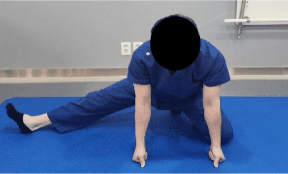
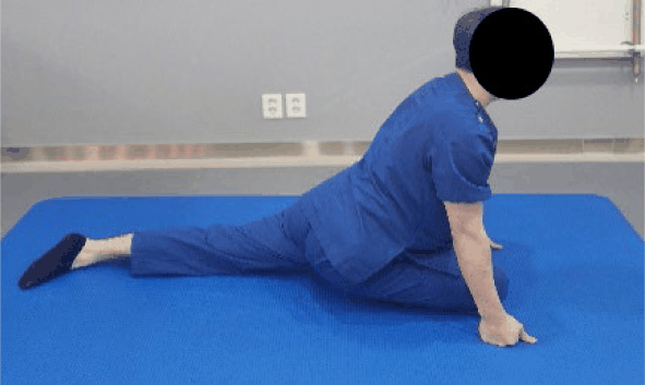
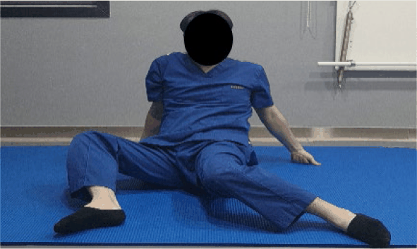
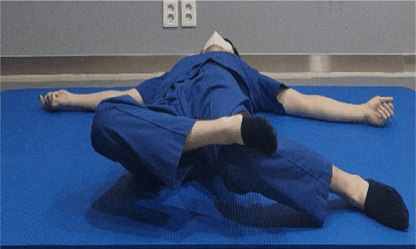
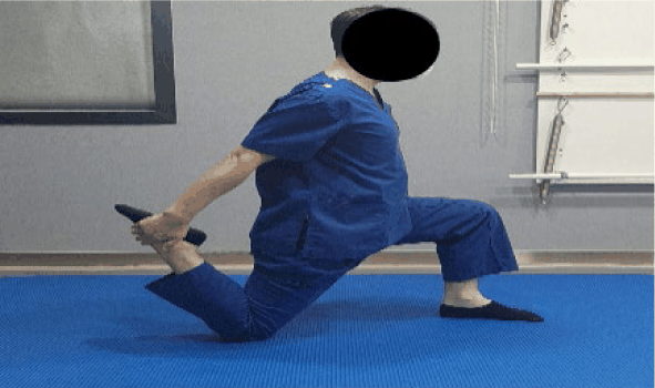
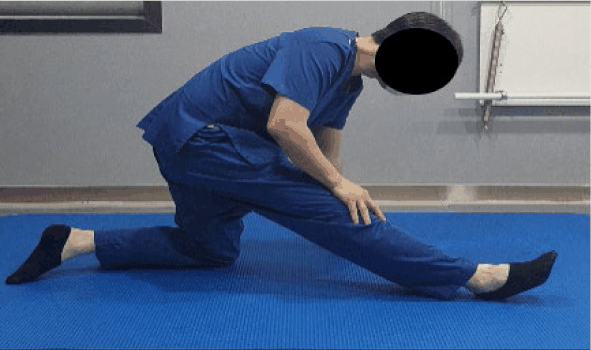
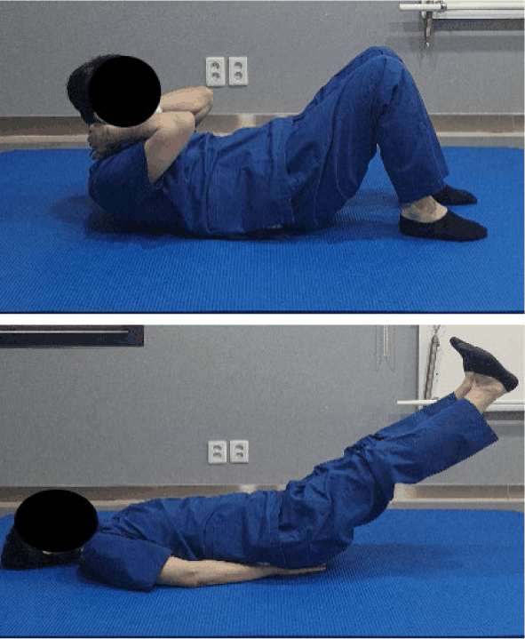
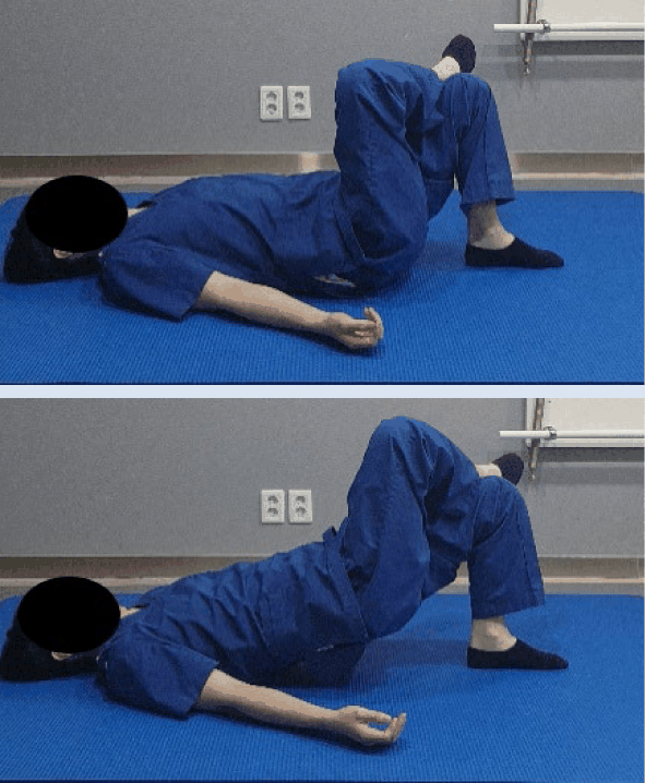
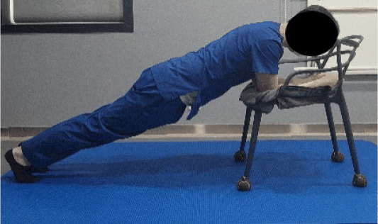
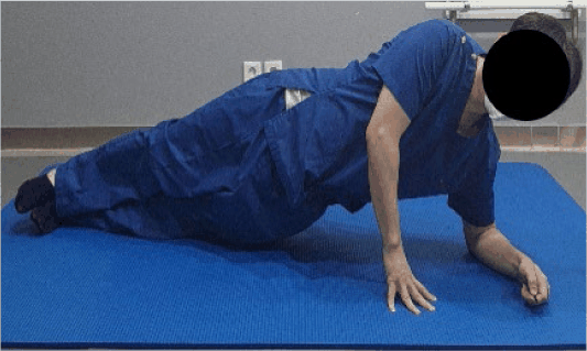
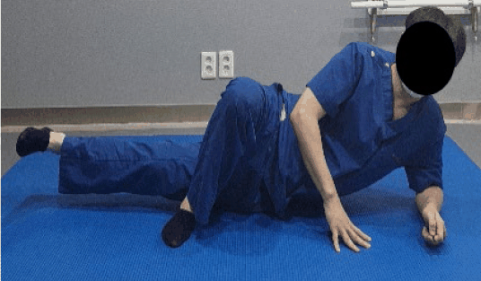
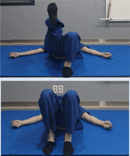
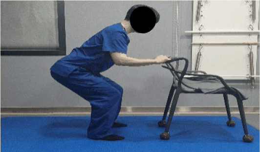
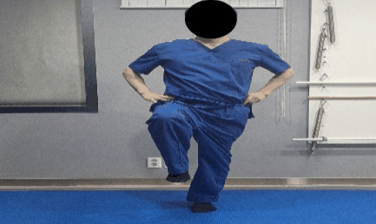
Participants performed a 4-week trunk stabilization exercise program once daily for about 40 minutes, 5 days per week, following an instructional video. The program consisted of 16 flexibility and stabilization exercises organized from supine/frog positions to bridge, plank, side plank, squat, and standing single-leg raises, based on previous studies (Table 1).21,22 A rest period of 30–60 seconds was allowed between exercises, and intensity was adjusted using the Modified Borg Scale to avoid excessive fatigue. Before gait reassessment, treadmill walking was performed until a fatigue level of 4 (“somewhat strong”) was reached.23 To ensure adherence and accuracy, participants were required to record their daily performance, difficulty level, and pain intensity in an exercise log. The researcher monitored their progress once a week via mobile messages and requested short exercise videos if necessary to provide feedback on posture.
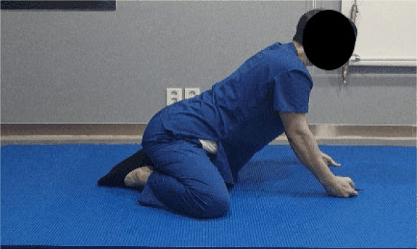
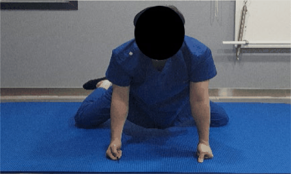
Table 1. Trunk stability exercise program
Posture Description
jkema-10-1-13-g1
(A) Frog position adductor stretch
Position your knees wide apart with feet turned outward. Extend your arms forward and place your elbows lightly on the floor. While maintaining mild abdominal tension, slowly shift your upper body forward and backward to create a gentle stretch in the inner thighs. Ensure the pelvis does not tilt to one side. Perform continuously for 1 minute within a pain-free range.
jkema-10-1-13-g2
(B) Frog position internal rotator strengthening
From the frog position, widen your knees slightly beyond shoulder width. Using the knee as a pivot axis, slowly rotate one foot outward (internally rotating the hip) so the toes point inward. Engage the core to prevent lumbar swaying or pelvic rotation. Perform 2 sets of 15 repetitions at a controlled tempo, focusing on the sensation of internal rotation. Repeat on the opposite side.
jkema-10-1-13-g3
(C) Frog position hamstring stretch
In the frog position, extend one leg out to the side with the knee straight. Slowly push your hips backward to elongate the posterior thigh muscles. Tighten the abdominals to maintain a neutral spine and prevent rounding of the lower back. Hold the stretch for 1 minute without bouncing, then switch sides.
jkema-10-1-13-g4
(D) Piriformis stretch
Position the front leg in a bent posture (similar to a pigeon pose) and extend the opposite leg straight back, allowing the hips to sink naturally toward the floor. Keep the chest upright to feel a stretch deep in the gluteal region. Hold for 1 minute. Adjust the intensity by leaning the torso forward if needed, but reduce the range if pain occurs.
jkema-10-1-13-g5
(E) Hip internal and external rotation
Sit on the floor with legs wide apart and spine upright to prevent posterior pelvic tilt. Pivot one knee inward and outward to explore the hip’s range of motion. Repeat 15 times on each side to gradually increase mobility. Focus on maintaining a stable center to prevent lumbar or pelvic rotation.
jkema-10-1-13-g6
(F) Tensor fasciae latae stretch
Lie supine with knees bent. Place the heel of the opposite foot on the outside of the knee being stretched and gently guide the leg inward toward the floor. You should feel a stretch along the outer thigh and hip. Engage the core to keep the pelvis grounded and relax the shoulders. Hold for 1 minute.
jkema-10-1-13-g7
(G) Lunge position rectus femoris stretch
Assume a lunge position with the front leg at 90 degrees and the back knee on the floor. Grasp the ankle of the rear leg with the same-side hand and gently pull it toward the glutes to stretch the front of the thigh. Slightly push the pelvis forward to lengthen the rectus femoris. Brace the core to prevent lumbar hyperextension. Hold for 1 minute, then switch sides.
jkema-10-1-13-g8
(H) Lunge position hamstring stretch
From a kneeling position, extend the front leg straight and dorsiflex the foot (pull toes toward the body). Slowly push the hips back while leaning the torso forward to stretch the entire posterior thigh. Contract the abdominals to prevent rounding of the back. Hold for 1 minute.
jkema-10-1-13-g9
(I) Upper and lower abdominal crunch
Lie supine with knees bent and feet flat. Place hands behind the head or on the chest. Gently tuck the chin and engage the abdominals to lift the shoulders and scapulae off the floor. Focus on curling the ribcage down rather than lifting the torso high. Pause for 1 second at the top and slowly lower. Perform 15 repetitions.
jkema-10-1-13-g10
(J) Single-leg bridge
Start in a standard bridge position. Lift one leg off the floor, keeping only the supporting leg grounded. Drive through the supporting heel to lift the hips, maintaining a level pelvis. Align the shoulders, hips, and knees. Hold for 1 second at the top and slowly lower. Perform 2 sets of 15 repetitions per leg.
jkema-10-1-13-g11
(K) Plank
Place elbows on a chair (or floor) directly under the shoulders. Extend legs back to form a straight line from head to heels. Brace the abdominals as if tightening a corset to prevent the hips from sagging or hiking up. Keep the neck neutral. Hold for up to 2 minutes.
jkema-10-1-13-g12
(L) Side plank
Lie on your side with the lower elbow under the shoulder. Extend legs straight or stagger the top leg forward for balance. Lift the hips to form a straight line from shoulder to ankle, engaging the obliques and glutes. Prevent the waist from sagging. Hold for 1 minute on each side.
jkema-10-1-13-g13
(M) Side-lying adductor raise
Lie on your side. Extend the bottom leg straight and bend the top leg, placing the foot on the floor in front. Dorsiflex the bottom foot and slowly lift the leg to engage the inner thigh. Hold for 1 second and lower with control. Perform 2 sets of 15 repetitions.
jkema-10-1-13-g14
(N) Bridge with hip flexion
From a bridge position with hips lifted, maintain pelvic stability and slowly lift one knee toward the chest (hip flexion). Keep the core tight to prevent the lower back from arching. Lower the foot and repeat on the opposite side. Perform 2 sets of 15 repetitions per leg.
jkema-10-1-13-g15
(O) Squat
Stand with feet shoulder-width apart, holding a chair for stability if needed. Push hips back and bend knees as if sitting in a chair, ensuring knees do not collapse inward or extend excessively beyond toes. Keep the chest up and back straight. Drive through the heels to return to standing. Perform 2 sets of 15 repetitions.
jkema-10-1-13-g16
(P) Standing single-leg raise
Stand upright, lightly holding a wall or chair. Lift one knee to 90 degrees while engaging the core and glutes to prevent the pelvis from tilting. Lower the leg slowly with control. Perform 2 sets of 15 repetitions per side.
Download Excel Table
Statistical analysis

Data were analyzed using SPSS Statistics 29.0 (IBM Corp., Armonk, NY, USA). Descriptive statistics were used for general characteristics. The normality of the data distribution was confirmed using the Shapiro–Wilk test. To investigate the overall changes in muscle activity and gait parameters, Repeated Measures ANOVAs (Time × Muscle; Time × Variable) were performed. Upon observing significant interaction effects, paired t-tests were conducted as post-hoc analyses to identify specific pre-post differences for each muscle and gait variable. The significance level was set at $\alpha$=.05. Effect sizes were calculated using partial eta squared (ηp2).

RESULTS

General characteristics

General Characteristics A total of 27 participants (17 males, 10 females) completed the study. All 27 participants completed the 4-week program with an attendance rate of 100%. The mean age was 26.24±3.93 years, height 169.90±7.74 cm, and weight 74.71±12.41 kg. The baseline K-ODI score was 29.4±5.6, indicating a moderate level of disability, and the mean duration of LBP was greater than 3 months (Table 2).

Table 2. General characteristics of the participants
Characteristics Values (N=27)
Age (years) 26.24±3.93
Sex (male/female) 17 / 10
Height (cm) 169.90±7.74
Weight (kg) 74.71±12.41
Duration of LBP > 3 months
VAS (score) 6.96±0.85
K-ODI (score) 29.40±5.60

Values are presented as mean ±standard deviation or frequency. K-ODI, Korean version of the Oswestry Disability Index; LBP, Low back pain.

Download Excel Table
Changes in gait parameters

To determine the effects of the intervention on gait, a repeated measures ANOVA was performed. The results revealed a significant interaction effect between time and gait variables (F=362.01, p<.001, ηp2=.933). Post-hoc analysis indicated significant improvements in all measured parameters. Specifically, the stance phase significantly decreased from 63.57±3.31% to 60.96±2.79% (p<.01). The single-limb stance phase significantly increased from 29.96±1.86% to 32.91±2.29% (p<.01). Furthermore, stride length showed a substantial increase from 77.16±22.21 cm to 96.71±7.05 cm (p<.01) (Table 3).

Table 3. Comparison of gait parameters pre- and post-intervention
Variables Pre-test (Mean±SD) Post-test (Mean±SD) t p Effect size (ηp2)
Stance phase (%) 63.57±3.31 60.96±2.79 24.713 <.001* .933
Single-limb stance (%) 29.96±1.86 32.91±2.29 –36.225 <.001* -
Stride length (cm) 77.16±22.21 96.71±7.05 –4.784 <.001* -

* Values are presented as mean±standard deviation.Significant difference between pre- and post-intervention (p<.05).

Partial eta squared (ηp2) for the Time × Gait interaction effect from repeated measures ANOVA.

Download Excel Table
Changes in lower-limb muscle activation

A significant interaction effect between time and muscle type was observed (F=3.23, p=.047, ηp2=.111), indicating that the intervention had differential effects on individual muscles. Post-hoc paired t-tests revealed significant changes in specific muscles. The %MVIC of the soleus increased significantly from 73.40±19.10% to 78.40±24.87% (p<.01), and the biceps femoris increased from 80.00±31.81% to 83.00±31.49% (p<.01). Conversely, significant decreases were observed in the rectus femoris (80.00±21.81% to 77.00±28.23%, p<.05), tibialis anterior (92.00±26.09% to 84.00±25.07%, p<.01), and popliteus (75.00±23.85% to 74.00±21.60%, p<.05). The gastrocnemius showed a slight increase (89.17±19.44% to 92.17±32.92%), but this change was not statistically significant (p>.05) (Table 4).

Table 4. Comparison of lower-limb muscle activation during the stance phase of gait pre- and post-intervention
Muscles (%MVIC) Pre-test (Mean±SD) Post-test (Mean±SD) t p Effect size (ηp2)
Soleus 73.40±19.10 78.40±24.87 –4.508 .001* .111
Gastrocnemius 89.17±19.44 92.17±32.92 –1.157 .258 -
Biceps femoris 80.00±31.81 83.00±31.49 –48.468 .001* -
Popliteus 75.00±23.85 74.00±21.60 2.320 .028* -
Rectus femoris 80.00±21.81 77.00±28.23 2.428 .022* -
Tibialis anterior 92.00±26.09 84.00±25.07 40.773 .001* -

* Values are presented as mean±standard deviation. %MVIC: percentage of maximum voluntary isometric contraction.Significant difference between pre- and post-intervention (p<.05).

Partial eta squared (ηp2) for the Time × Muscle interaction effect from repeated measures ANOVA. Note: EMG data represent the mean activation measured specifically during the stance phase.

Download Excel Table
Changes in functional disability

The K-ODI score decreased significantly from 29.40±5.60 to 23.62±5.01 (p<.01), indicating a meaningful improvement in functional status (Table 5).

Table 5. Comparison of functional disability (K-ODI) pre- and post-intervention
Variables Pre-test (Mean±SD) Post-test (Mean±SD) t p
K-ODI (score) 29.40±5.60 23.62±5.01 7.180 <.001*

* Values are presented as mean±standard deviation. K-ODI: Korean version of the Oswestry Disability Index.Significant difference between pre- and post-intervention (p<.05).

Download Excel Table

DISCUSSION

This study aimed to investigate the effects of a 4-week trunk stabilization exercise program on spatiotemporal gait parameters, lower-limb muscle activation, and functional disability in university students with chronic LBP. The results demonstrated that the intervention significantly improved gait efficiency, reorganized neuromuscular control patterns, and reduced functional disability. Notably, the Repeated Measures ANOVA revealed significant interaction effects for both gait variables and muscle activation, indicating that the trunk stabilization exercise produced differential, function-specific adaptations rather than a generalized increase or decrease in all parameters.

Gait analysis showed that the stance phase significantly decreased, while the single-limb stance and stride length significantly increased following the intervention. Individuals with LBP typically adopt a "defensive gait strategy" characterized by a shortened stride length and prolonged stance phase to minimize trunk motion and pain.17 The increase in stride length and single-limb stance observed in this study suggests a reversal of this protective mechanism. By enhancing lumbopelvic stability, the participants likely gained the confidence to transfer weight onto a single limb more effectively, leading to a more dynamic and efficient gait pattern consistent with previous reports.24

The EMG results provide further insight into the mechanism of these gait improvements. The significant interaction effect found in our analysis supports the hypothesis of "selective neuromuscular reorganization." Specifically, the activation of the soleus and biceps femoris significantly increased, whereas the rectus femoris, tibialis anterior, and popliteus significantly decreased. The increased activity of the soleus and biceps femoris suggests an enhancement of the posterior chain, which is crucial for generating propulsion (push-off) and maintaining pelvic stability during the stance phase.25-27 This aligns with the observed increase in stride length. Conversely, the significant reduction in rectus femoris and tibialis anterior activity indicates a decrease in excessive anterior-chain co-contraction. In LBP populations, excessive co-contraction is often a compensatory strategy to artificially stiffen the spine.28 The reduction of this unnecessary muscle tension suggests that as intrinsic trunk stability improved, the reliance on superficial global muscle guarding diminished, allowing for more economical movement.29

Functional improvements were confirmed by significant reductions in K-ODI scores. These findings align with previous studies reporting that core stabilization reduces mechanical load on the lumbar spine, thereby improving daily function.30,31 The improvement in functional status likely contributed to the normalization of gait and muscle activation patterns.

There are several limitations to this study. First, the single-group pre–post design limits the ability to draw definitive causal inferences; future randomized controlled trials are needed to control for placebo effects and natural history. Second, the relatively high %MVIC values observed in the soleus muscle warrant cautious interpretation. This finding may be attributed to "pain inhibition" or fear-avoidance mechanisms common in LBP populations. According to the pain adaptation theory, individuals with pain often fail to exert true maximum force during MVIC testing due to neural inhibition, leading to an underestimation of the MVIC reference value.32,33 Consequently, normalized gait EMG values may appear numerically inflated. Therefore, interpretations should focus on the relative pre-post changes rather than absolute magnitudes. Third, the sample consisted of young university students, which may limit the generalizability of the findings to older adults or clinical populations with structural spinal pathologies.

CONCLUSIONS

A 4-week trunk stabilization exercise program improved gait efficiency (shorter stance phase, longer stride length), optimized lower-limb muscle activation through selective reorganization, and reduced functional disability in university students with LBP. These findings suggest that trunk stabilization is an effective intervention for restoring neuromuscular control and function in young adults with lifestyle-related LBP.

Conflict of Interest Disclosures:

None.

Funding/Support:

None.

Acknowledgment:

None.

Ethic Approval:

This study was approved by the Institutional Review Board of Daegu University (Approval number: 1040621-202509-HR-073).

Data Availability:

The datasets analyzed during the current study are available from the corresponding author on reasonable request.Informed consent for publication of the images was obtained from the patient.

Author contributions

Conceptualization: KH Park, TH Kim.

Data acquisition: KH Park.

Design of the work: KH Park, TH Kim.

Data analysis: KH Park, TH Kim.

Project administration: KH Park.

Interpretation of data: KH Park, TH Kim.

Writing – original draft: KH Park, TH Kim.

Writing–review&editing: KH Park, TH Kim.

REFERENCES

1.

Hartvigsen J, Hancock MJ, Kongsted A, Louw Q, Ferreira ML, Genevay S, et al. What low back pain is and why we need to pay attention. Lancet. 2018; 391(10137):2356-2367

2.

Kim HJ, Kim JS. The relationship between smartphone use and subjective musculoskeletal symptoms and university students. J Phys Ther Sci. 2015; 27(3):575-579

3.

Mahdavi SB, Riahi R, Vahdatpour B, Kelishadi R. Association between sedentary behavior and low back pain; a systematic review and meta-analysis. Health Promot Perspect. 2021; 11(4):393-403

4.

Suikkanen S, Päivärinne V, Kautiainen H, Kiviranta I. Relationship between low back pain, depressive symptoms and work ability among young male adults. J Public Health (Oxf). 2025; :1-9

5.

van Dieёn JH, Selen LP, Cholewicki J. Trunk muscle activation in low-back pain patients: an analysis of the literature. J Electromyogr Kinesiol. 2003; 13(4):333-351

6.

Hodges PW, Moseley GL. Pain and motor control of the lumbopelvic region: effect and possible mechanisms. J Electromyogr Kinesiol. 2003; 13(4):361-370

7.

Wang W, Wei H, Shi R, Lin L, Zhang L, Yue S, et al. Dysfunctional muscle activities and co-contraction in the lower-limb of lumbar disc herniation patients during walking. Sci Rep. 2020; 10(1):20432

8.

Davis M, Newsam CJ, Perry J. Electromyograph analysis of the popliteus muscle in level and downhill walking. Clin Orthop Relat Res. 1995; (310):211-217

9.

Arendt-Nielsen L, Graven-Nielsen T, Svarrer H, Svens-son P. The influence of low back pain on muscle activity and coordination during gait: a clinical and experimental study. Pain. 1996; 64(2):231-240

10.

Zileli A, Çankaya T. Assessment of posture, spinal mobility and EMG data in patients with spinal stenosis. Balt J Health Phys Act. 2022; 14(4):2-9

11.

Gringmuth RH, Jackson C. Therapeutic exercise for spinal segmental stabilization in low back pain: scien-tific basis and clinical approach. J Can Chiropr Assoc. 2000; 44(2):125-139

12.

Akuthota V, Nadler SF. Core strengthening. Arch Phys Med Rehabil. 2004; 85(3)Suppl 1:S86-S92

13.

O’Sullivan PB. Lumbar segmental instability: clinical presentation and specific stabilizing exercise management. Man Ther. 2000; 5(1):2-12

14.

Hides JA, Jull GA, Richardson CA. Long-term effects of specific stabilizing exercises for first-episode low back pain. Spine (Phila Pa 1976). 2001; 26(11):E243-E248

15.

Marshall PW, Murphy BA. Core stability exercises on and off a Swiss ball. Arch Phys Med Rehabil. 2005; 86(2):242-249

16.

Tsao H, Hodges PW. Immediate changes in feedforward postural adjustments following voluntary motor training. Exp Brain Res. 2007; 181(4):537-546

17.

Song S, Cho N, Kim HJ. Changes in gait patterns after physical therapy in patients with non-specific chronic low back pain: a pilot study. Phys Ther Rehabil Sci. 2022; 11(1):105-112

18.

Hanada EY, Hubley-Kozey CL, McKeon MD, Gordon SA. The feasibility of measuring the activation of the trunk muscles in healthy older adults during trunk stability exercises. BMC Geriatr. 2008; 8(1):33

19.

Schulte RV, Prinsen EC, Schaake L, Buurke JH. Synchronization of wearable motion capture and EMG measurement systems. In: Proc 2022 Int Conf Rehabil Robot (ICORR). 2022; :1-6

20.

Jeon CH, Kim DJ, Kim SK, Kim DJ, Lee HM, Park HJ. Validation in the cross-cultural adaptation of the Korean version of the Oswestry Disability Index. J Korean Med Sci. 2006; 21(6):1092-1097

21.

Kim B, Yim J. Core stability and hip exercises improve physical function and activity in patients with non-specific low back pain: a randomized controlled trial. Tohoku J Exp Med. 2020; 251(3):193-206

22.

Santamaría G, Rodríguez I, Rodríguez-Pérez V, Cobreros-Mielgo R, Lantarón-Caeiro E, Seco-Casares M, et al. Effect of hip muscle strengthening exercises on pain and disability in patients with non-specific low back pain: a systematic review. Sports (Basel). 2023; 11(9):167

23.

Onan D, Demirel A, Ülger Ö. Investigating the effects of fatigue protocol on spatiotemporal characteristics of gait in patients with chronic neck pain. Turk Fizyoter Rehabil Derg. 2023; 34(1):45-54

24.

Frizziero A, Pellizzon G, Vittadini F, Bigliardi D, Costantino C. Efficacy of core stability in non-specific chronic low back pain. J Funct Morphol Kinesiol. 2021; 6(2):37

25.

Lenhart RL, Francis CA, Lenz AL, Thelen DG. Empirical evaluation of gastrocnemius and soleus function during walking. J Biomech. 2014; 47(12):2969-2974

26.

Hodges PW, Richardson CA. Delayed postural contraction of transversus abdominis in low back pain associated with movement of the lower limb. J Spinal Disord. 1998; 11(1):46-56

27.

Jacobs JV, Henry SM, Jones SL, Hitt JR, Bunn JY. A history of low back pain associates with altered elec-tromyographic activation patterns in response to perturbations of standing balance. J Neurophysiol. 2011; 106(5):2506-2514

28.

Danneels LA, Vanderstraeten GG, Cambier DC, Witvrouw EE, Bourgois JD, Dankaerts W, et al. Effects of three different training modalities on the cross-sectional area of the lumbar multifidus muscle in patients with chronic low back pain. Br J Sports Med. 2001; 35(3):186-191

29.

Hibbs AE, Thompson KG, French D, Wrigley A, Spears I. Optimizing performance by improving core stability and core strength. Sports Med. 2008; 38(12):995-1008

30.

Hwangbo G, Lee CW, Kim SG, Kim HS. The effects of trunk stability exercise and a combined exercise program on pain, flexibility, and static balance in chronic low back pain patients. J Phys Ther Sci. 2015; 27(4):1153-1155

31.

Kim WH. Effect of an active tailored exercise program on pain and Oswestry Disability Index in automobile manufacturing workers with musculoskeletal symptoms of the low back. J Korean Soc Phys Med. 2019; 14(3):109-116

32.

Hodges PW, Tucker K. Moving differently in pain: a new theory to explain the adaptation to pain. Pain. 2011; 152:S90-S98

33.

O’Sullivan PB. Diagnosis and classification of chronic low back pain disorders: maladaptive movement and motor control impairments as underlying mechanism. Man Ther. 2005; 10(4):242-255