


INTRODUCTION
Hallux valgus (HV) refers to a progressive structural deviation in which the great toe gradually shifts laterally relative to the first metatarsal.1 This deformity is commonly associated with medial pain and functional impairment around the first metatarsophalangeal joint (1MTPJ), and its prevalence increases with advancing age.2 The severity of HV is typically determined by the angle formed between the longitudinal axes of the first proximal phalanx and the first metatarsal.3 Clinically, values below 15° are regarded as physiologic alignment, whereas increasing angular deviation beyond 15°, 20°, and 40° corresponds to mild, moderate, and severe deformity, respectively.4 Both intrinsic and extrinsic factors contribute to the development and progression of HV, including narrow footwear, excessive foot pronation, pes planus, neurological conditions such as stroke and cerebral palsy, and muscle imbalance between the abductor hallucis (AbdH) and adductor hallucis.5-10 Dysfunction of intrinsic foot muscles may contribute not only to structural deformity but also to impaired balance and functional activities such as sit-to-stand transitions and gait.10-11
Among the intrinsic foot muscles, the AbdH arises from the medial aspect of the calcaneus and attaches to the medial base of the proximal phalanx through the sesamoid complex.5,12 The AbdH contributes to the maintenance of 1MTPJ alignment and performs abduction and flexion of the great toe.5,12 Because it lies inferior to the transverse axis of the 1MTPJ, the AbdH contributes to both the stability of the 1MTPJ and the support of the medial longitudinal arch (MLA).13-14 The Intrinsic foot muscles such as the flexor hallucis brevis and interossei contribute to arch integrity, and the AbdH assists in maintaining MLA support during late stance and push-off in gait.10,14 As HV progresses, the anatomical position of the AbdH shifts plantarly, altering its mechanical relationship with the 1MTPJ and potentially reducing its activity.10,14 Reduced AbdH activity has been reported in individuals with HV compared to individuals with healthy, suggesting that strengthening of the AbdH and other intrinsic muscles may contribute to maintaining MLA alignment and limiting progression of the HV angle.15
Several intrinsic foot muscles strengthening exercises have been proposed, including toe gripping, toe towel curls, single-leg balance tasks, and the short foot exercise.16,17 The TSO exercise has been highlighted for its potential to preferentially recruit the AbdH and influence first-ray alignment. Previous studies in individuals with mild HV reported that TSO elicits greater AbdH activity than short foot exercise and improved the activity ratio between the AbdH and adductor hallucis toward a more balanced pattern.17 More recently, a modified version of the TSO exercise incorporating a pressure biofeedback unit has been introduced to allow real-time control of MLA height during exercise performance.18 In comparative studies involving individuals with mild HV, toe-spread-out with pressure biofeedback (TSOP) exercise demonstrated higher AbdH activity than both the short foot and TSO exercises, and was associated with reduced HV angles.18 These findings indicate that incorporating real-time arch feedback during TSO may enhance AbdH recruitment while supporting improved 1MTPJ alignment. Unlike conventional TSO, which primarily emphasizes transverse plane toe movement, TSOP requires simultaneous elevation of the MLA and controlled abduction of the great toe.18 This combined task may enhance intrinsic muscle recruitment and has been proposed as a conservative intervention for strengthening the AbdH and addressing forefoot alignment in individuals with HV.18,19
Previous studies have primarily focused on individuals with HV, and direct comparisons of the TSO and TSOP exercises in individuals without HV remain limited. Although optimizing AbdH activity and MLA function before the development of deformity may be important for prevention and early intervention, evidence comparing these two exercises under identical conditions in individuals without HV is scarce. Furthermore, it remains unclear whether TSOP facilitates greater activation of the AbdH in individuals without HV beyond the effects previously reported in populations with deformity. Therefore, the purpose of this study was to compare AbdH muscle activity during the TSO and TSOP exercises in individuals without HV. It was hypothesized that TSOP would result in significantly greater AbdH activity than conventional TSO.
METHOD
A pilot study involving four participants was conducted to estimate the required sample size. Based on the effect size derived from the pilot data, an a priori power analysis was performed using G*Power software (version 3.1.2; Franz Faul, University of Kiel, Kiel, Germany). With a significance level of α = 0.05 and statistical power of 0.80, the minimum required sample size was calculated to be eleven participants.18 A total of eighteen individuals without HV were initially recruited. Three participants were excluded based on the predefined exclusion criteria: withdrawal before testing (n=1) and limited first metatarsophalangeal joint mobility due to muscle spasm and pain (n=2). Consequently, 15 participants were included in the final analysis. All participants had an HV angle between 0° and 10°, indicating the absence of HV.17 The HV angle was measured in a seated position using a goniometer (MEDALL, Korea). General characteristics of the participants are presented in Table 1. Participants were excluded if they had a history of HV surgery, had been diagnosed with osteoarthritis or rheumatoid arthritis, had a history of peripheral or central nervous system injury, presented with limited 1MTPJ mobility due to muscle spasm and pain, or experienced pain during exercise performance.19-20 All participants were informed of the study and provided written informed consent prior to participation. This study was approved by the institutional review board of Hoseo University (1041231-260512-HR-223-02).
| Characteristics | Participants (n=15) |
|---|---|
| Sex (M/F) | 8/7 |
| Age (years) | 24.60±2.50 |
| Height (cm) | 168.83±9.49 |
| Weight (kg) | 64.42±12.12 |
| BMI (kg/m2) | 22.44±2.84 |
AbdH muscle activity was recorded using a wireless surface electromyography (sEMG) system (TeleMyo 2400T, Noraxon, Scottsdale, AZ, USA). The EMG signals were sampled at 1024 Hz and band-pass filtered between 20 and 450 Hz. The root mean square (RMS) value was calculated using a moving window of 50 ms.18 To control the MLA during the TSO, a pressure biofeedback unit (Chattanooga Stabilizer, Chattanooga Group Inc., USA) was used. Before electrode placement, the skin was prepared with alcohol to reduce impedance. Surface electrodes were placed over the muscle belly of the AbdH, approximately 1–2 cm posterior to the navicular tuberosity.17 For normalization, maximum voluntary isometric contraction (MVIC) of the AbdH was measured. Participants were positioned in a seated posture with the heel stabilized, and resistance was applied opposite to the direction of great toe abduction at the 1MTPJ.17 MVIC was recorded three times, and the mean value was used for normalization. EMG data collected during the TSO and TSOP exercises were expressed as a percentage of MVIC (%MVIC).
Before data collection, each participant practiced each exercise three times in a seated position to ensure proper technique. The order of exercises was randomized using Microsoft Excel (Microsoft, Redmond, WA, USA). Participants were not informed of the specific purpose or expected effects of each exercise to minimize bias. During the dominant-foot test, AbdH muscle activity was recorded while participants maintained maximal contraction for 5 seconds. After each trial, participants returned to a resting position. Three trials were performed for each condition, and the mean value was used for analysis. A rest period of 3 minutes was provided between exercises to minimize muscle fatigue.
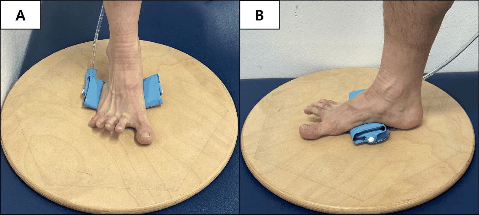
Participants were seated with the knees flexed at 90°, and weight was evenly distributed on the plantar surface. They were instructed to lift all toes while maintaining contact with the metatarsal heads and heel with the floor. Subsequently, the fifth toe was moved laterally and the great toe medially, while ensuring that the metatarsal heads and heel remained in contact with the ground.
For TSOP, the pressure biofeedback unit was positioned beneath the MLA and inflated to 30 mmHg. Participants were instructed to perform the TSO movement while drawing the first metatarsal head toward the heel to elevate the MLA, with the pressure reduced from 30 mmHg to 20 mmHg. This specific pressure reduction was targeted to ensure measurable, consistent activation of the MLA, in line with protocols established in previous studies.18 The knees remained flexed at 90°, and contact of the metatarsal heads and heel with the floor was maintained throughout the task (Figure 1-A, B).
The collected data were analyzed using IBM SPSS Statistics version 20.0 (IBM Corp., Armonk, NY, USA). The Shapiro–Wilk test was used to assess the normality of the data. (p>0.05). A paired t-test was conducted to compare the AbdH muscle activity between the two exercise conditions. The level of statistical significance was set at alpha=0.05.
RESULTS
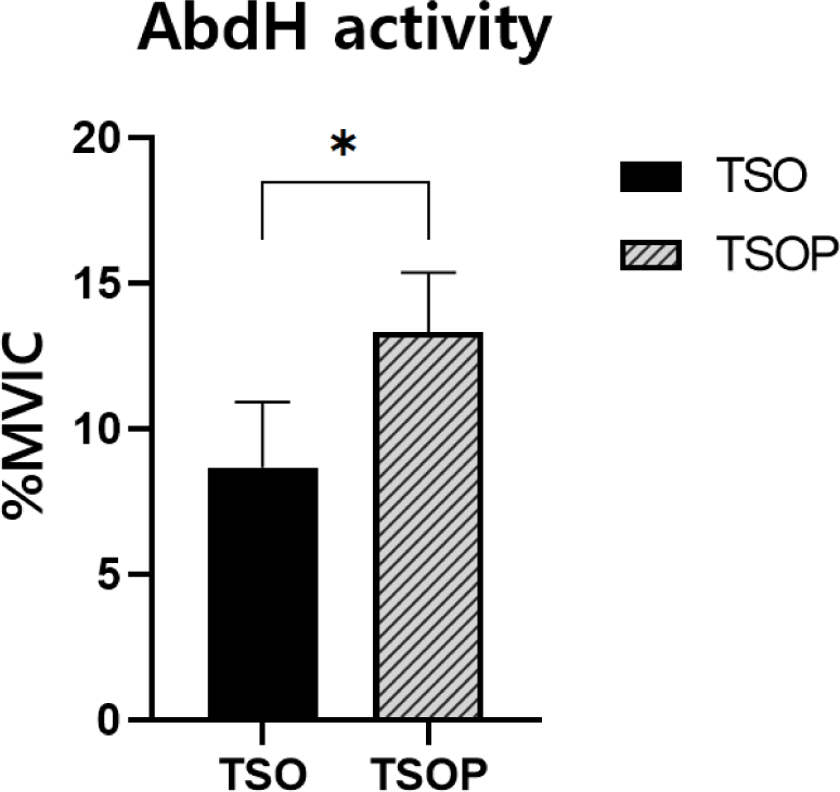
AbdH muscle activity differed significantly between the two exercise conditions. The mean AbdH activity during TSO was 8.67±2.26 %MVIC, whereas it was 13.32±2.06 %MVIC during TSOP. AbdH muscle activity during TSOP was significantly greater than during TSO (p<0.001), representing an average increase of approximately 53.6% (Table 2) (Figure 2).
| %MVIC | Mean±SD | Difference value | t | p | Effect size | |
|---|---|---|---|---|---|---|
| TSO | TSOP | |||||
| AbdH | 8.67±2.26 | 13.32±2.06 | –4.65 | –12.74 | <0.001* | 3.28 |
DISCUSSION
The present study compared AbdH muscle activity during the TSO exercise performed with and without pressure biofeedback in individuals without HV. The AbdH muscle activity was significantly greater during TSOP than during conventional TSO, with an average increase of approximately 53.6%. These findings suggest that incorporating MLA control through pressure biofeedback may enhance activation of the AbdH during the TSO exercise. Although the absolute %MVIC values in both conditions were relatively low, this result should be interpreted considering the study purpose, which was to determine whether pressure biofeedback could modify AbdH activation in individuals without structural deformity. In this context, the greater activation observed during TSOP suggests that incorporating MLA control through pressure biofeedback may provide a more effective exercise condition for facilitating AbdH activation than TSO.
The observed difference in AbdH activation between TSOP and conventional TSO may be attributable to pressure biofeedback, which influences medial longitudinal arch control. The TSO exercise is designed to promote activation of the AbdH through transverse plane toe movement.21 Because the AbdH also contributes to MLA support, the absence of active arch control during TSO may allow variability in arch posture.20-21 In contrast, TSOP incorporates a pressure biofeedback unit that provides real-time feedback regarding MLA position.18 Participants were required not only to abduct the great toe but also to maintain the arch within a predetermined pressure range. This simultaneous control of arch position and toe movement likely increased task demands compared with conventional TSO.21-22 Although arch kinematics were not directly measured in the present study, the added requirement for arch stabilization may have altered the coordination strategy of the intrinsic foot muscles, thereby contributing to the greater AbdH activity observed during TSOP.21-22 The presence of this pattern in individuals without HV suggests that the effect is more likely attributable to task structure rather than correction of pathological alignment.
Previous studies have primarily involved individuals with HV and have reported both increased AbdH activity and reductions in HV angle.18 In those investigations, improvements in muscle activity were interpreted in relation to correction of altered first-ray alignment and restoration of intrinsic muscle balance in structurally deformed feet.17,21 Because joint alignment, soft tissue tension, and mechanical loading are altered in HV, increased AbdH activity in those populations may partially reflect normalization of pathological mechanics.10,22 In contrast, the present study evaluated individuals without HV. Despite the absence of structural deformity, AbdH activity remained significantly greater during TSOP than during conventional TSO. The substantially lower %MVIC values observed in the present study, compared with those reported in previous studies involving individuals with HV, may be related to differences in baseline foot alignment and neuromuscular recruitment patterns. In individuals with HV, altered joint alignment and soft-tissue mechanics may require greater relative activation of the AbdH during corrective tasks, whereas participants without deformity may perform the same task with less compensatory muscle recruitment. Therefore, although the direction of the response to TSOP was similar across studies, the magnitude of activation may differ according to deformity status and its associated biomechanical demands. This similar pattern of results suggests that the effect of TSOP is not solely dependent on correction of structural deformity but may also be related to the additional task demands imposed by active MLA control. Because TSOP requires simultaneous maintenance of the MLA and abduction of the great toe, the task itself may impose greater motor-control demands on the intrinsic foot muscles, regardless of the presence of deformity. Further longitudinal investigations are required to determine whether repeated TSOP training produces measurable changes in arch morphology, foot alignment, or functional performance. Clinically, TSOP may represent a structured progression of the conventional TSO exercise, in which arch position is actively regulated through biofeedback. Regulation through biofeedback may enhance intrinsic muscle engagement by integrating arch stabilization with toe movement, although further evidence is required to confirm its functional relevance.
This study has several limitations. First, the participants were limited to young adults in their twenties, which restricts the generalization of the findings to other age groups. Second, because only immediate muscle activity during a single session was analyzed, the long-term training effects and potential structural adaptations were not determined. Third, the muscle activity of intrinsic foot muscles other than the AbdH was not assessed, limiting the interpretation of overall neuromuscular coordination within the foot. Fourth, baseline MLA height or arch morphology was not directly measured or controlled, and the pressure biofeedback unit was set to identical target pressures (30 mmHg baseline reduced to 20 mmHg during task performance) for all participants. Therefore, individual characteristics such as foot size, arch morphology, and intrinsic muscle capacity may have influenced the observed responses. Further studies should investigate whether pressure settings tailored to arch morphology result in distinct patterns of intrinsic foot muscle activity.
CONCLUSIONS
The TSOP exercise resulted in significantly greater AbdH activity compared to the conventional TSO exercise in individuals without HV. These findings suggest that utilizing pressure biofeedback for MLA control might serve as an effective strategy to enhance activation of the AbdH during the TSO exercise.