


INTRODUCTION
Lumbar extension rotation syndrome (L-ext & rot syn) is the most common among 5 classification category of low back pain.1 One of the characteristic of patients with L-ext & rot syn is that low back pain is increased with excessive lumbopelvic motion in sagittal and transverse plane during active or passive knee flexion.2 Repeated lumbar extension or rotation in a daily activity induces tissue microtrauma, tissue failure, and resulting the low back pain.2,3
Rectus femoris (RF) is a two joint muscle between the tibia and pelvis that is susceptible to increased passive stiffness in patients with low back pain.4 Previous research suggested that increased passive muscle stiffness of RF can be considered as possible reasons of excessive lumbar extension and rotation during active or passive knee flexion.2,5
Active muscle stretching is effective management to reduce the passive muscle stiffness.6 Previous studies demonstrated the effects of stretching exercise on passive muscle stiffness in healthy subjects.7,8 Passive muscle stiffness of hamstrings was decreased by 31% after 4-week stretching program in healthy subjects.7 The passive stiffness of gastrocnemius muscle was also immediately reduced by 47% after passive stretching program in healthy subjects.8 However, there has not been investigated the effects of RF stretching exercise on the reduction of passive muscle stiffness of RF in patients with low back pain, especially L-ext & rot syn. No studies have been investigated the effects of reduced RF stiffness by active stretching exercise on the pain intensity of low back in subjects with L-ext & rot syn.
The aim of the this study was to investigate the effects of active RF stretching exercise on the change of objective RF passive stiffness, subjective stretch sensation, and the low back pain intensity in subjects with L-ext & rot syn. We hypothesized that there would be differences in (1) the RF passive stiffness, (2) stretch sensation, (3) intensity of low back pain during daily activity after active RF stretching program.
METHODS
We recruited twenty subjects with L-ext & rot syn (14 males and 6 females). The characteristics of subjects were mean age of 23.7±3.2 years, mean height of 172.6±4.7 cm, and a mean weight of 68.9±4.2 kg. The inclusion criteria for this study were 1) the same as criteria for classifying L-ext & rot syn, proposed by Sahrmann2 and Van Dillen et al.1 (Table 1) and that duration of low back pain were over 6-week.9 An examiner in this study had 6-year experience for evaluating patients with low back pain using classification method of movement system impairments. The exclusion criteria were present radiating pain, strain injury of RF, knee joint contractures and muscle strength weakness of the hamstrings that would not be able to flex the knee actively in the prone position. Prior to this study, the principal investigator explained all procedures and signed an informed consent form to recruited subjects. This study was approved by the Yonsei University Wonju Campus Human Studies Committee.
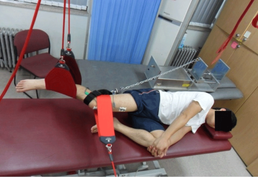
A test leg for passive RF stiffness were determined the side of greater low back pain and lumbar extension or rotation during performing active prone knee flexion.10 The subject was positioned in resting sidelying position on a treatment table, which is antigravity position to measure passive RF stiffness. In order to ensure the relaxed position throughout the all period of the measurement of passive RF stiffness, surface electromyographic monitoring (Noraxon, Scottsdale, Arizona, USA) on RF was used. Before attaching the electromyographic electrode for RF, the skin was cleaned with alcohol. The electrode for RF was attached on medial anterior surface of the thigh, approximately halfway between the hip and the knee.11 Test leg was placed above non-test leg in sidelying position and then, supported by the sling with full hip and knee joint extension without hip abduction or adduction. Subject flexed hip and knee in non-test leg to 90 degrees for providing stability (Figure 1). Strain gauge (Noraxon, Inline Force sensor 320, Scottsdale, AZ) and electrogoniometer (Noraxon, Scottsdale, Arizona, USA) were used for measurement of change in passive muscle tension and for measurement of change in passive muscle length, respectively, because the definition of passive muscle stiffness is the change in tension per unit change in length.12 Data of surface electromyography, strain gauge and electrogoniometer were recorded using a wireless telemetry system (TeleMyo 2400T, Noraxon, Scottsdale, Arizona, USA) with a sampling frequency of 1,000 Hz. A bandpass filter of surface electromyography was used between 20 and 250 Hz. An electrogoniometer was placed the lateral side of the femur and the fibula of the test leg and then attached using tape to prevent movement artifact.12 Distal femur of test leg was stabilized by non-elastic strap so that hip joint was maintained at same position during passive knee flexion in sidelying. Proximal tibia of test leg was secured by non-elastic strap, connecting the motorized pulling machine using iron chain. Before recording the data, calibration of the electrogoniometer was performed at the hip flexion at 0 degree and knee flexion at 45 degrees. Motorized pulling machine pulled the test leg into knee flexion from 45 to 90 degrees while recording data of passive tension and angle of knee flexion. The reasons for recording the data from 45 to 90 degrees of knee flexion were two as follows: 1) Range of knee flexion between 45 and 90 degrees was sensitive to measure the through-range RF stiffness in pilot study, and 2) 45 and 90 degrees of knee flexion was mostly used in daily activity.2 If increased RF muscle activity, severe pain or severe stretching sensations during the recording the data, data were discarded. Passive RF stiffness was collected at a speed of 5 degree/s.13 The mean value of the three trials of passive RF stiffness measurement was used for the data analysis. 30-second rest period were provided between measurements. Intrarater reliability of stiffness measurements was established to be high (intraclass correlation coefficient (3,1) = 0.98) in this study.
Subjective stretch sensation was measured using numerical rating scale before and after 6-week active stretching program.14 The subjects rated stretch sensation from “0” (no stretch sensation) to “10” (extremely severe stretch sensation) at end range during passive prone knee flexion with manual stabilization of pelvis using tester’s hand.
Low back pain intensity was measured using numeric rating scale before and after 6-week active stretching program.14 During daily activity in recent a week, the subjects rated pain intensity from “0” (no pain) to “10” (extremely severe pain).
Procedures of this study divided three parts; 1) pre-stretching period, 2) active stretching exercise period during 6-week, and 3) post-stretching period. All subjects were measured for passive RF stiffness, stretch sensation, and low back pain intensity in pre-stretching period and post-stretching period.
In active stretching program during 6-week, subjects performed RF stretching exercise in half-kneeling position at home as follow steps; (1) Kneel onto the subject’s knee of stretched leg on the folded towel with neutral hip joint and place opposite leg in front of subject with knee joint flexed at 90 degrees. (2) Grab subject’s ankle of the stretched leg while holding the wall using opposite hand to maintain balance for half-kneeling. (3) Pull subject’s ankle of the stretched leg to subject’s buttocks during 30 seconds until subject can feel stretching sensation of anterior thigh (4) After 30 seconds stretching, rest for 30 seconds in half-kneeling position and then repeat two time.14 Previous study demonstrated that three trials with one trial for 30 seconds stretching is enough time for muscle stretching to reduce passive muscle stiffness.6 Active RF stretching exercise during 6-week was performed one session a day on 5 consecutive days of the week.15 During intervention period, subjects were asked not to perform vigorous physical exercise and any stretching exercises. Stretching diary was given out to each subject whether the stretching exercise was completed in a day or not throughout the intervention period. Consistency of the RF stretching exercise using stretching diary was calculated as number of participating day of RF stretching / 30 days×100, representing the percentage.16
To compare the differences of RF stiffness, stretch sensation and low back pain between pre- and post-6 week stretching, the paired t-test was used. The level of statistical significance was set at p<0.05. The statistical package for SPSS version 18.0 (SPSS, Inc., Chicago, IL, USA) was used for statistical analysis.
RESULTS
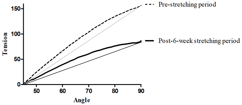
Exercise compliance was 86% in the 6-week RF stretching exercise. Passive RF stiffness was significantly decreased in Figure 2. Passive stiffness in pre-stretching period and post -stretching period was 3.29±1.36 and 1.98±1.07 (mean±SD, N/degrees), respectively. Slope of passive RF stiffness was significantly decreased after 6-week active RF stretching exercise compared to pre-stretching period (mean difference, 1.83; 95% CI, 1.15 to 2.52; p<0.05).
Stretch sensation in pre-stretching period and post -stretching period was 5.39±1.54 and 3.56±0.86 (mean±SD), respectively. Stretch sensation of RF in prone position decreased significantly after active RF stretching period (mean difference, 1.84; 95% CI, 1.15 to 2.52; p<0.05).
Low back pain intensity during daily activity in pre-stretching period and post -stretching period was 5.1±0.8 and 5.0±0.6 (mean±SD), respectively. Low back pain intensity showed no significant difference after active RF stretching period (mean difference, 0.18; 95% CI, –0.01 to 0.57; p>0.05).
DISCUSSION
Previous study suggested that increased stiffness of RF relative to lumbar spine is one of possible reasons for the compensatory lumbopelvic motion during knee flexion, resulting low back pain in patients with L-ext & rot syn.5,10 Therefore, this study demonstrate the effects of the active RF stretching exercise on the RF passive stiffness, stretch sensation, and perceived low back pain in patients with L-ext & rot syn. To our knowledge, this is the first study to demonstrate that decreased RF stiffness and stretch sensation failed to reduce low back pain intensity in subject with L-ext & rot syn.
Present study demonstrated that 6-week RF stretching program was effective way to decrease passive stiffness and stretch sensation scale of RF. However, a previous study demonstrated that 2-week RF active stretching program fail to change in passive stiffness, although stretch sensation of anterior thigh significantly decreased.14 The difference between the results of our study and previous study is stretching duration, so that 6-week stretching duration is more effective than 2-week duration.14 Additionally, in a previous study, passive knee flexion during passive stiffness measurement was performed by examiner’s hand,14 however, automatic motorized pulling machine was used for passive knee flexion in this study. Our study also measured through-range stiffness of RF than end-range stiffness in previous study.14 Through-range stiffness indicates the muscle stiffness assessed during passive lengthening at consistent slow velocities, therefore, can calculate more precisely of the viscoelastic components after stretching exercise.17,18 Thus, assessment of through-range stiffness is more meaningful than end-range stiffness after stretching. Stretching duration, using the automatic motorized pulling machine, and measurement of through-range stiffness make the difference between the results of our study and previous study.14
Previous study suggested that cause of low back pain in patients with L-ext & rot syn was compensatory lumbopelvic motions by altered motor control as well as biomechanical restriction such as relative increased stiffness of RF during prone knee flexion.5 Relative increased stiffness of RF than those of abdominal muscle and anterior supporting structure of lumbar spine, can lead to increased compensatory lumbopelvic motion pelvic during prone knee flexion.10 However, although biomechanical restriction of RF was improved after active RF stretching exercise, there were no significant differences of low back pain intensity in present study. About these results, there are two possible explanations. First, altered motor control would be more suitable factor to influence the intensity of low back pain than RF stiffness, because the results of present study showed that the reduced RF passive stiffness failed to decrease the pain intensity. Therefore, motor control exercise to facilitate deep abdominal muscle contraction may be suitable exercise selection for reducing low back pain than only active RF stretching exercise.
Second explanation is that the amount of decreased RF passive stiffness was not enough to decrease the pain intensity of low back. That is, reduced relative stiffness of RF might be still greater than that of anterior supporting structure of lumbar spine and abdominal muscle in present study. Further study would be needed to investigate the effects of intervention for increased stiffness of anterior supporting structure of lumbar spine relative to RF stiffness on the improvement of low back pain in patients with L-ext & rot syn.
The present study has several limitations. First, we could not measure the stiffness of anterior structure of lumbar spine and abdominal muscle, thus relative stiffness between the RF and anterior structure of lumbar spine could not be compared. Second, we did not compare the effects of RF stretching exercise versus control exercise. Thus, further longitudinal study would be required to compare the effect of lumbopelvic stabilization exercise plus active RF stretching exercise on the reduced pain intensity in subjects with L-ext & rot syn.
CONCLUSIONS
This study demonstrated that the 6-week active RF stretching exercise can decrease the passive RF stiffness and stretch sensation, but fail to reduce the intensity of low back pain in daily activity. Based on these results, the biomechanical restriction such as increased RF stiffness may be not one of the causative factors contributing the severity of low back pain in patients with L-ext & rot syn. Thus, we recommend for clinicians to apply lumbar stabilization exercise or motor control exercise than only stretching exercise when designing the exercise program and managing the patients with L-ext & rot syn.